www.nature.com/scientificreports
OPEN
Received: 12 May 2017 Accepted: 31 August 2017 Published: xx xx xxxx
Preferential sensing and response to microenvironment stiffness of human dermal fibroblast cultured on protein micropatterns fabricated by 3D multiphoton biofabrication Ming Hui Tong1, Nan Huang1, Alfonso Hing Wan Ngan2, Yanan Du3 & Barbara Pui Chan1 While cells are known to sense and respond to their niche including the matrix and the mechanical microenvironment, whether they preferentially sense and react to the stiffness of their microenvironment regardless of its intrinsic material properties is unknown. In this work, protein micropillar arrays with independently controllable stiffness via alterations in pillar height and elastic modulus via laser power used during photochemical cross-linking, were fabricated using a recently developed multiphoton-based 3D protein micro-patterning technology. Human dermal fibroblasts were cultured on these micropillar arrays and the specific interactions between cells and the protein micropatterns particularly on the formation and maturation of the cell-matrix adhesions, were investigated via immunofluorescence staining of the major molecular markers of the adhesions and the measurement of their cluster size, respectively. Our results showed that the cluster size of focal adhesions increased as the stiffness of the micropillar arrays increased, but it was insensitive to the elastic modulus of the protein micropillars that is one of the intrinsic material properties. This finding provides evidence to the notion that cells preferentially sense and react to the stiffness, but not the elastic modulus of their microenvironment. A great deal of recent evidence has shown that mechanical microenvironment, particularly elastic modulus and stiffness, plays a very important role in determining the cell fates, such as migration1,2, proliferation3 and differentiation4–6. Cell matrix adhesions are complex structures mediating the interaction and attachment of cells to a substrate and enabling cells to sense such mechanical microenvironment in the extracellular matrix (ECM). The primary sites of cell-matrix interactions are known as matrix adhesions such as focal adhesions7 which consist of complex sub-cellular structures, linking the cytoskeleton to the ECM and regulating crucial activities such as angiogenesis8 and wound healing9. Integrins10 are matrix receptors consisting of heterodimers with one α-chain and one β-chain and they initialize the adhesion between cells and their ECM. Different integrins usually have different preferences for different ECM ligands and are involved in different cell behaviors. Both αv and β1 chains of integrin were reported to form the nascent adhesion11 and activate FAK family kinase12. Paxillin13 as another important component of focal adhesions plays an important role in several signaling pathways that regulate cell activities such as attachment14 and migration15. Upon ECM engagement through integrin receptors, FAK was activated and recruited to the focal adhesions16,17. In some studies, the size of the focal adhesion clusters was reported to be positively related to both the elastic modulus18 and stiffness19 of the ECM. Although ample evidence has been gathered to show the importance of microenvironment stiffness on cellular fate processes20,21, it remains unclear whether the ECM stiffness is an overriding factor, compared with other factors of the ECM such as its intrinsic elastic modulus, which is known to associate with the microstructure of the 1 Tissue Engineering Laboratory, Department of Mechanical Engineering, The University of Hong Kong, Pokfulam Road, Hong Kong Special Administrative Region, China. 2Department of Mechanical Engineering, The University of Hong Kong, Pokfulam Road, Hong Kong Special Administrative Region, China. 3Department of Biomedical Engineering, School of Medicine, Tsing-hua University, Beijing, China. Correspondence and requests for materials should be addressed to B.P.C. (email:
[email protected])
Scientific Reports | 7: 12402 | DOI:10.1038/s41598-017-12604-z
1
www.nature.com/scientificreports/ material. Clarifying this issue is of high significance to the reliability of in vitro experimentation, since, firstly, it is never possible to create a microenvironment in the laboratory that can duplicate exactly the in vivo conditions. And secondly, one can legitimately query whether experiments performed on different types of microenvironment are directly comparable. If the ECM stiffness can be proven to be a dominating factor for cell fate mechanoregulation, then the need becomes a simple one which is to ensure that the microenvironment used exhibits comparable stiffness between experiments, or with in vivo conditions, regardless of its elastic modulus. Both elastic modulus and stiffness measure a substance’s resistance to deformation, but they are not totally the same. Elastic modulus is an intrinsic property of a material that is independent of geometry22, while stiffness, measured in Newton per meter, is an extrinsic property of a structure that usually depends on both material and shape conditions23. Another reason to differentiate the effects of stiffness and elastic modulus on cellular fates is the confusion of the use of terminologies related to mechanoregulation studies. For example, in a report investigating the dependence of stem cell lineage specification on substrate stiffness20, what was being manipulated was actually elastic modulus, measured in Newton per unit area, rather than stiffness. Moreover, some studies used rigidity, instead of stiffness, to denote elastic modulus21 but it usually refers to stiffness. Hydrogel assay has been used to study mechanosensing of stiffness and elastic modulus for long and it has an advantage of allowing for studying both normal and tangential traction stresses. Some studies that made use of polyacrylamide gel as the substrate to culture cells modulated elastic modulus by changing the ratio of substrate to crosslinker concentration ratio21. Other studies modulated stiffness by changing the thickness of hydrogel without altering its composition, microtopography and elastic modulus, enabling the decoupling between elastic modulus and stiffness. For tangential deformations and traction stresses, Lin YC. et al. showed theoretically and experimentally that altering substrate thickness of polyacrylamide gels modifies the substrate stiffness24. For the general 3D case, del Alamo et al. developed the theory showing how stiffness depends on substrate thickness24. More recently, Aung et al. used this idea to show experimentally that cancer cells sense substrate stiffness using matrigel substrates of constant composition but varying thickness25,26. However, the differential sensitivity of cells, in terms of cell matrix adhesion maturation, to elastic modulus and stiffness has not been studied. To study the relative importance of stiffness versus the intrinsic property of the microenvironment on focal adhesion maturation, which is a pre-requisite for important cellular fate processes including proliferation and migration, we employ arecently developed multiphoton-based microfabrication technology27 to fabricate micropillar-array substrates for cell culture that exhibits different in-plane stiffness and contact areas sensed by the cells. The technique involved is a 3D printing technology that enables the fabrication of user-defined protein micropatterns with sub-micron resolution and high aspect ratios, allowing spatial and independent control of the topology27, porosity28, mechanical proprieties28 and the type of ECM proteins used, via a wide range of independently controlled fabrication parameters including the scanning power, reagent parameters such as the photosensitizer concentration, and geometry parameters such as the shape of the protein micropatterns. The micropatterns fabricated this way can be used directly as cell culture substrates for cell niche studies particularly on the mechanical and the matrix niche. In this work, we aim to study the maturation of the cell-matrix adhesions of human dermal fibroblasts cultured on protein micropillar arrays with independently controlled stiffness and elastic modulus produced by this fabrication technology.
Mechanics Foundations
Before discussing the experiments, a clear definition of the stiffness of a substrate for cell culture needs to be given. For a flat substrate without surface patterning, a tangential stiffness may be defined as the magnitude of a force acting tangentially on the substrate surface required to produce unit displacement of the contact region along the force direction. The tangential stiffness of an elastic flat surface over a circular contact region of diameter D, as felt by a rigid stylus, is given as29 4G Sflat = D 2−ν
(1)
where ν is Poisson ratio and G the shear modulus of the surface material. For a homogeneous, flat substrate without elastic anisotropy, all locations on the surface would have the same tangential stiffness defined this way which is also a scalar rather than a tensor. For a substrate for cell culture with a micropatterned surface, however, the above definition of tangential stiffness is inconvenient as the stiffness value would no longer be location or direction independent. Also, a cell lying on top of a micropatterned substrate may not touch the trough regions of the substrate, and so for the purpose of discussing cell-matrix interactions or mechanoregulation, a more relevant definition of stiffness would be needed. For the type of micropillar-array patterned substrates studied in this work, traction-force microscopy has shown that a cell sitting on such a substrate will exert tangential forces trying to bend each of the contacting micropillars30. We therefore define the tangential stiffness of a micropillar array, for the purpose of studying cell-matrix interactions, as the tangential force a rigid cylindrical stylus of diameter D would need to exert in order to bend each of the contacting pillars by unit displacement along the force direction. For a single pillar, its bending stiffness is given as s=
3EI 3E πd4 = 3 ⋅ 3 64 L L
(2)
where E is the intrinsic Young modulus of the material, I the second moment of the cross-sectional area of the pillar (for a circular pillar), and L and d are the height and diameter of the pillar respectively. The tangential stiffness of a regular array of micropillars that would be analogous to that in eqn. (1) for a flat substrate is therefore Scientific Reports | 7: 12402 | DOI:10.1038/s41598-017-12604-z
2
www.nature.com/scientificreports/
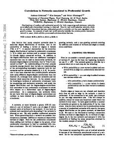
Figure 1. F-actin staining of human dermal fibroblast on micropillars with heights 4 µm (A), 8 µm (B) and 18 µm (C). Scale bar 20 µm. Cell spreading area (D) defined as the area of F-actin was quantified by Image J. (n = 10) SEM images were taken of micropillars with heights 4 µm (E), 8 µm (F) and 18 µm (G). Scale bar 50 µm.
Spillar −array = (πD 2 /4) ⋅ n ⋅ s
(3)
where n is the number of pillars per unit area in the array. In our experiments, micropillar arrays of fixed center-to-center spacing of the pillars were used for cell culture. Thus, in eqn. (3), the areal density n of the pillars was a constant, although the pillar geometry and Young modulus were varied to enable s to change. Therefore, for a given stylus dimension, i.e. only the bending stiffness of individual pillars matters. The pillar bending stiffness s in eqn. (2) is referred simply to as “stiffness” in the following. In our experiments, we aim to test a hypothesis that cells will preferentially sense and react to the stiffness defined in eqn. (2) above, which is related to both the geometry factors I and L, as well as the intrinsic Young modulus E of the protein material of the micropillars, rather than E alone, which is related to the material microstructure or physical nature. As mentioned earlier on, although eqns. (1) to (3) above would make the difference between stiffness and elastic modulus clear in the case of cell-culture substrates, such a difference is often mixed up in the tissue-engineering literature, and hence understanding the specific effects of the two quantities on cell-matrix interactions is important.
Experimental Results
Increasing micropillar stiffness increased cell spreading and enhanced focal adhesion maturation. According to eqn. (2), keeping the material elastic modulus E the same by using the same fabrication
parameters for substrate, the stiffness of the micropillars was modulated by varying the height of the micropillars while their diameter and center-to-center spacing were kept at 2 and 6 μm respectively. Figure 1 shows the F-actin expression of human dermal fibroblast after three days’ culture on protein micropillar arrays with different heights and hence stiffness. Cells were widely spread with extensive stress fibers when cultured on arrays of protein micropillars with high stiffness at 1.014 nN/μm, corresponding to a pillar height of 4 µm (Fig. 1A). On micropillars with moderate stiffness of 0.137 nN/μm, corresponding to a micropillar height of 8 µm, cells were less spread and stress fibers less extensive (Fig. 1B). On arrays of protein micropillars with extremely low stiffness at 0.012 nN/µm, corresponding to a micropillar height of 18 µm, cells were least spread with short actin fibers (Fig. 1C). The cell spreading area quantified from the F-actin staining showed an increasing trend as the substrate stiffness increased (Fig. 1D), in that the average cell spread area was 2000 μm2 on the stiffest micropillars (1.014 nN/μm). Figure 1E–G shows the SEM images of micropillars with different heights 4 µm (E), 8 µm (F) and 18 µm (G) while, as said before, their diameter and center-to-center spacing are constant at 2 μm and 6 μm respectively. Integrin alpha v, pFAK(Y397) and paxillin are important components of focal adhesions. Figure 2 shows the immunofluorescence staining of these molecular markers for focal adhesions in human dermal fibroblasts after three days’ culture. The size of the focal adhesions on micropillars increased on increasing stiffness, i.e. decreasing height of the micropillars, from small dots in micropillars with height 18 µm (Fig. 2A3) corresponding to a stiffness of 0.012 nN/µm, to small patches on micropillars with 8 and 4 μm heights (Fig. 2A2, A1) corresponding to a stiffness of 0.137 and 1.014 nN/µm, respectively. A statistically significant linear relationship exists between the stiffness of the micropillars and the cluster area of integrin alpha v expression (straight line curve fitting, R2 = 0.8526, p