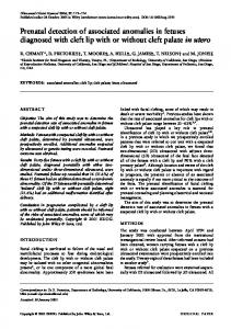
Ultrasound Obstet Gynecol 2003; 21: 564–569 Published online 9 May 2003 in Wiley InterScience (www.interscience.wiley.com). DOI: 10.1002/uog.132
Prenatal detection of velamentous insertion of the umbilical cord: a prospective color Doppler ultrasound study W. SEPULVEDA, I. ROJAS, J. A. ROBERT, C. SCHNAPP and J. L. ALCALDE Fetal Medicine Center, Department of Obstetrics and Gynecology, Clinica Las Condes, Santiago, Chile
K E Y W O R D S: color Doppler ultrasound; prenatal diagnosis; three-dimensional (3D) ultrasound; umbilical cord; vasa previa; velamentous insertion
ABSTRACT Objective Velamentous insertion of the umbilical cord, with a reported incidence of 1% in singleton pregnancies, has been associated with several obstetric complications including fetal growth restriction, prematurity, congenital anomalies, low Apgar scores, fetal bleeding and retained placenta. The aim of this study was to determine the feasibility of identifying velamentous insertion of the umbilical cord during routine obstetric ultrasound. Design This was a prospective, cross-sectional ultrasound study in 832 unselected second- and third-trimester singleton pregnancies. Color Doppler ultrasound was routinely performed to identify the placental cord insertion site. The role of three-dimensional (3D) ultrasound in evaluating the placental cord insertion site was also studied in a subset of 50 pregnancies from this population. Results The placental cord insertion site was identified in 825/832 (99%) cases. Visualization was not achieved in seven third-trimester pregnancies with a posterior placenta. A velamentous insertion was suspected prenatally in eight cases, seven of which were confirmed after delivery as velamentous and one as markedly eccentric (battledore placenta). 3D ultrasound performed poorly at evaluating placental cord insertion site, being less efficient due to poor-quality resolution and far more time-consuming than the combined use of gray-scale and color Doppler ultrasound. Conclusions Velamentous insertion of the umbilical cord can reliably be detected prenatally by gray-scale and color Doppler ultrasound. 3D imaging had limited value in the evaluation of the placental cord insertion site in our subset of patients. Systematic assessment of the placental cord insertion site at routine obstetric ultrasound has
the potential of identifying pregnancies with velamentous insertion and, therefore, those at risk for obstetric complications including vasa previa. Copyright 2003 ISUOG. Published by John Wiley & Sons, Ltd.
INTRODUCTION In approximately 99% of singleton pregnancies the umbilical cord inserts either directly into the placental tissue or marginally on the placental edge1 . In the remaining 1% of cases the umbilical cord insertion is located away from the placental mass, allowing a variable segment of umbilical vessels to run between the membranes unsupported by Wharton’s jelly. This condition, known as velamentous insertion, has been associated with several obstetric complications including fetal growth restriction (FGR), prematurity, congenital anomalies, low Apgar scores and retained placenta1 – 7 . One of the most serious, although rare, complications associated with this condition is vasa previa. In this particular circumstance, the unprotected velamentous vessels cross the lower uterine segment below the presenting part, exposing large fetal vessels to the risks of compression and rupture, especially during labor1,2 . A case of fetal exsanguination and death due to laceration of vasa previa from an undiagnosed velamentous insertion was recently seen at our institution8 . In retrospect, this tragic outcome could have only been prevented with accurate prenatal diagnosis and planned Cesarean delivery9 . As the placental cord insertion site can be readily identified prenatally by ultrasound10,11 , we undertook this study to determine prospectively the feasibility of identifying velamentous insertion of the umbilical cord in the second and third trimesters using the currently available ultrasound technology.
Correspondence to: Prof. W. Sepulveda, Centre for Fetal Care, Queen Charlotte’s and Chelsea Hospital, Hammersmith Hospitals NHS Trust, Du Cane Road, London W12 0HS, UK (e-mail:
[email protected]) Accepted: 28 February 2003
Copyright 2003 ISUOG. Published by John Wiley & Sons, Ltd.
ORIGINAL PAPER
Velamentous umbilical cord insertion
PATIENTS AND METHODS In an 8-month period from November 2001 to July 2002, women attending our center for routine obstetric ultrasound were prospectively enrolled into this study after Departmental Review Board approval. The majority of women were referred by staff obstetricians with primary indications for scanning being dating, fetal anomaly screening, placental location, evaluation of fetal growth intervals and assessment of amniotic fluid volume. For the purposes of this study, only live singleton pregnancies of at least 16 weeks’ gestation, with adequate amniotic fluid volume (deepest vertical pool measurement greater than 2 cm) and scanned by one of the authors (W.S.) were included. All examinations were performed using a Voluson 730 real-time 4D ultrasound system (General Electric Medical Systems, Milwaukee, WI, USA) equipped with color Doppler, power Doppler and threedimensional (3D) imaging capabilities. Women were allotted 20 min for standard secondand third-trimester transabdominal obstetric ultrasound. Once placental location was determined, attempts to image the placental cord insertion site were undertaken by searching the fetal surface of the placenta with highresolution gray-scale ultrasound until the umbilical cord was identified. Care was taken not to mistake the true insertion site with loops of umbilical cord overlying the placental surface by demonstrating the entry of main branches of umbilical vessels into the chorionic plate with color flow imaging in all cases (Figure 1). In pregnancies with posterior placenta, gentle manipulation of the fetus through the maternal abdomen allowed proper placental surface visualization in most cases. If this maneuver proved unsuccessful, the woman was placed in a lateral position to produce an acoustic window and then the placental surface was imaged as before. The potential role of 3D ultrasound in evaluating the placental cord insertion site was also explored in a subset of unselected pregnancies referred for 3D fetal ultrasound. In such cases, an additional 10–20 min were allocated for 3D ultrasound, which included multiplanar and surface-rendered views of the placental cord insertion site. According to the ultrasound findings, the placental cord insertion site was classified as normal, if the umbilical cord inserted on the substance of the placental mass, or marginal, if the cord insertion was found in the periphery of the placenta closer than 1 cm to the edge. Insertion of the umbilical cord on the chorioamniotic membranes away from the placental mass was considered indicative of velamentous insertion. In these cases, the relationship between the velamentous vessels and the internal cervical os was established transabdominally. If insufficient transabdominal image quality was obtained, transvaginal ultrasound was performed to search the lower uterine segment with color flow imaging and rule out vasa previa. Representative images of the placental cord insertion site were stored in a hard copy or digital format for further analysis. In women who had more
Copyright 2003 ISUOG. Published by John Wiley & Sons, Ltd.
565
a
b
c
Figure 1 (a) Gray-scale, (b) color Doppler and (c) power Doppler imaging show umbilical cord insertion into the placental mass.
than one evaluation during the study period, only the information obtained from the first scan was used for analysis. All ultrasound findings were reported to the referring obstetrician and, therefore, used for clinical management.
Ultrasound Obstet Gynecol 2003; 21: 564–569.
Sepulveda et al.
566
The placenta was routinely examined after delivery by the attending obstetrician or midwife caring for the woman during labor. The diagnosis of velamentous insertion was made macroscopically if the cord inserted into the membranes away from the placental edge. The medical records from cases of velamentous insertion were reviewed for information on maternal and perinatal demographic characteristics, as well as the mode of delivery, Apgar scores and any significant complication seen in the fetus or neonate.
a
RESULTS During the study period, 832 women fulfilled the study entry criteria. The median gestational age at the time of evaluation was 23 (range 16 to 40) weeks. In 235 cases (29%) the gestational age varied from 16 to 20 weeks, in 251 (30%) from 21 to 24 weeks, in 142 (17%) from 25 to 30 weeks, in 139 (17%) from 31 to 34 weeks and in 65 (8%) from 35 to 40 weeks. The placenta was predominantly located on the anterior uterine wall in 399 cases (48%), on the posterior wall in 417 (50%) and on the lateral wall in the remaining 16 (2%). A lowlying placenta or placenta previa was noted in 16 cases (2%) and a succenturiate-lobed or bilobed placenta was identified in four cases (0.5%). Table 1 shows the distribution of the study population according to gestational age and placental cord insertion site as determined by prenatal ultrasound. Confident identification of the placental cord insertion site was achieved in 825/832 (99%) cases. In more than 95% of these cases this task was accomplished in less than 1 min. The placental cord insertion site was not visualized in seven pregnancies (1%) in spite of repeated attempts during the scan; all these cases were scanned after 30 (median 33, range 31 to 39) weeks and the presumed reason for non-visualization was that the fetus was lying over a posteriorly located placenta. The prenatal diagnosis of velamentous insertion was made in eight cases (Figure 2), although in two of these cases there was reasonable doubt as to whether a markedly eccentric insertion (battledore placenta) was present. The overall incidence of velamentous insertion as
b
c
Table 1 Distribution of the study population according to gestational age and placental cord insertion site as determined by prenatal ultrasound Placental cord insertion site GA (weeks)
Cases ( n)
Velamentous
Marginal
Normal
Not visualized
16–20 21–24 25–30 31–34 35–40
235 251 142 139 65
2 4 1 1 0
13 16 7 5 2
220 231 134 129 60
0 0 0 4 3
Total
832
8
43
774
7
GA, gestational age.
Copyright 2003 ISUOG. Published by John Wiley & Sons, Ltd.
Figure 2 Velamentous cord insertion. (a) Gray-scale ultrasound shows insertion of the umbilical cord away from the placental mass. (b) Power Doppler imaging shows velamentous vessels. (c) Photograph of the placenta after delivery shows large, prominent velamentous vessels.
determined by prenatal ultrasound was, therefore, 1/104 or 1%. Table 2 shows the relevant ultrasound features and clinical characteristics of these eight pregnancies.
Ultrasound Obstet Gynecol 2003; 21: 564–569.
Velamentous umbilical cord insertion
567
Table 2 Prenatal diagnosis of velamentous insertion of the umbilical cord
Case 1 2 3* 4 5 6 7 8*
MA (years)/ parity
GA (weeks)
Placental location
GA at delivery (weeks)
33/2 35/2 32/1 36/1 28/0 35/0 30/1 28/0
18 19 22 22 23 23 27 34
Posterior Anterior Posterior Anterior Posterior Posterior Anterior Anterior
37 35 39 37 37 38 36 35
Cord insertion at delivery Velamentous Velamentous Velamentous Velamentous Velamentous Velamentous Velamentous Marginal
Remarks
Infant with transient tachypnea Encephalocele Trisomy 21 Bilobed placenta; infant with RDS FGR; maternal hypertension
*Markedly eccentric (battledore placenta)/velamentous insertion on prenatal ultrasound. GA, gestational age; FGR, fetal growth restriction; MA, maternal age; RDS, respiratory distress syndrome.
In one case the antenatal course was complicated with maternal hypertension and FGR requiring delivery at 35 weeks. There were no antenatal complications in the remaining seven cases. Among the eight newborn infants, three required admission to the neonatal intensive care unit. The first had trisomy 21, congenital heart defects and low birth weight, the second had an occipital encephalocele requiring early neonatal neurosurgery and the third developed respiratory distress syndrome after being delivered by Cesarean section at 36 weeks because of repeated non-reassuring cardiotocography and breech presentation. No cases of vasa previa or false-negative diagnosis of velamentous insertion were identified in this series. In a pilot study, the feasibility of 3D ultrasound in evaluating the placental cord insertion site was further studied in a subset of 50 unselected pregnancies: 30 in the second and 20 in the third trimester. Multiplanar views did not add any significant information to that already obtained by gray-scale ultrasound alone, and it was less efficient due to poor-quality resolution and was far more time-consuming than the combined use of gray-scale and color Doppler ultrasound. In addition, 3D surfacerendered ultrasound performed poorly, with the placental cord insertion site being clearly demonstrated in only 12 (40%) second-trimester pregnancies (Figure 3) and in two (10%) third-trimester pregnancies. Furthermore, even in successful cases the relationship between the umbilical cord insertion site and placenta was difficult to establish due to poor definition of 3D-rendered images in differentiating the placenta from the uterine wall. Although the acquisition time was between 3 and 5 s, the post-processing of 3D surface-rendered images also made this image modality a time-consuming technique.
DISCUSSION Early prenatal identification of velamentous insertion of the umbilical cord is a desirable clinical goal since these pregnancies are at greater risk for adverse perinatal outcome including FGR, preterm birth, congenital anomalies, fetal distress and fetal bleeding1 – 7 . However, as expected for any fetal, placental or umbilical cord anomaly which is not obvious, routine non-targeted
Copyright 2003 ISUOG. Published by John Wiley & Sons, Ltd.
a
b
Figure 3 Three-dimensional ultrasound of the placental cord insertion site. (a) Panoramic surface-rendered views in two different second-trimester pregnancies show the fetus, umbilical cord and placenta. (b) Multiplanar views and the corresponding surfacerendered ultrasound image of the placental cord insertion site. Note the poor definition in differentiating between the placenta and uterine wall.
ultrasound examination often fails to detect velamentous insertion in utero as has been demonstrated in several large retrospective studies5,6 . In spite of the obvious importance of prenatal detection of velamentous insertion of the umbilical cord in obstetric practice, only a few studies have focused on the systematic identification of the placental cord insertion site during prenatal ultrasound10 – 12 . Pretorius et al.10 studied 917 pregnancies greater than 15 weeks, and were able to identify the placental cord insertion site in 93% of cases.
Ultrasound Obstet Gynecol 2003; 21: 564–569.
568
Pathological examination of the placenta was available in 128 singleton pregnancies and six cases (4.7%) of velamentous insertion were confirmed. Among them, the prenatal diagnosis of velamentous insertion had been made in two cases and of marginal insertion in three. The placental cord insertion site was not identified in the remaining case; this fetus died during labor due to exsanguination from ruptured vasa previa10 . A similar study involving 360 pregnancies from 13 to 39 weeks was reported by Di Salvo et al.11 . Placental pathology in 38 singleton and eight twin pregnancies confirmed four cases of velamentous insertion, only one of which was prenatally diagnosed by ultrasound. A much better performance was obtained by Nomiyama et al.12 who studied the placental cord insertion site at 18–20 weeks in 555 singleton and 32 twin pregnancies. The placental cord insertion site was identified prenatally in all but one case and all five cases of velamentous insertion were correctly diagnosed prenatally. A higher visualization rate of the placental cord insertion site and detection of velamentous insertion throughout a wide range of gestational ages was achieved in our series in comparison to two previous studies10,11 . Several factors can explain these findings. First, we used color Doppler ultrasound routinely whereas Di Salvo et al.11 did only at the discretion of the operator. We noted that the use of color Doppler ultrasound enhanced umbilical cord visualization even in difficult cases and, therefore, should be the modality of choice to image the placental cord insertion site at routine obstetric ultrasound. The fact that we were able to identify the placental cord insertion site in all cases before 30 weeks, regardless of placental location, supports this observation. This is also in keeping with the findings of Nomiyama et al.12 in whose study the placental cord insertion site was identified with color Doppler ultrasound in practically 100% of cases at 18–20 weeks. However, even though the umbilical cord increases in size with advancing gestation and theoretically should be progressively easier to identify with color Doppler ultrasound, this proved not to be true later in pregnancy as visualization of the placental cord insertion site becomes more difficult in the third trimester, especially in cases with posterior placenta. Second, motivation of the operator might have also played a role. We were highly influenced by the poor pregnancy outcome seen in one of our institutional cases in which an undiagnosed velamentous insertion of the umbilical cord led to laceration of vasa previa, fetal exsanguination and intrauterine death in early labor8 . This case prompted our study and, therefore, every effort to rule out velamentous insertion was made in our prospective cases. Third, all ultrasound scans were performed by a single fetal medicine specialist, who is also trained in percutaneous umbilical cord sampling and has, therefore, been familiar with ultrasound identification of placental cord insertion site for many years. These factors may have significantly contributed to the improved visualization rate seen in this study. Nevertheless, we consider that ultrasound identification of the placental cord insertion site does
Copyright 2003 ISUOG. Published by John Wiley & Sons, Ltd.
Sepulveda et al. not require additional skills, but it should be performed in a systematic fashion in order to be useful for screening velamentous insertion. A potential criticism of our study is the fact that full pathological examination of all the placentae was not performed. However, we consider that this does not invalidate our findings as velamentous insertion can be easily diagnosed with gross examination of the placenta after delivery. We also excluded multiple gestations from our analysis as these pregnancies are considered a major risk factor for velamentous insertion, with almost a 10% incidence of abnormal umbilical cord insertion being reported in these cases1 . In singleton pregnancies, however, velamentous insertion has been largely overlooked at prenatal ultrasound. Our study suggests that velamentous insertion in singleton pregnancy is prevalent enough to warrant routine screening by ultrasound, a policy that should be further supported by the fact that this condition can lead to several obstetric complications, some of them associated with a catastrophic outcome such as vasa previa8,9 . Our preliminary experience with the use of 3D ultrasound in determining the placental cord insertion site was disappointing. In the third trimester, 3D ultrasound performed poorly at evaluating placental cord insertion site, mainly due to the presence of fetal parts, increasing fetal size and relatively reduced amniotic fluid volume surrounding the fetus in comparison to the second trimester. This is in keeping with a previous study, which showed that 3D ultrasound is often unable to identify the placental cord insertion site after 28 weeks13 . In the second trimester, however, we were able to obtain adequate 3D surface-rendered images in about 40% of cases but, even when good views were analyzed, the exact relationship between the insertion site and the placental mass was sometimes difficult to ascertain. Probably, the use of 3D color Doppler mapping with superimposed gray-scale ultrasound could be more useful in the evaluation of velamentous insertion, as it has been previously shown in cases of vasa previa14 , but this was not explored in our study. However, this technique presumably is also time consuming and therefore impractical for routine screening purposes. In conclusion, our study confirms that velamentous insertion of the umbilical cord can reliably be detected prenatally and color Doppler ultrasound can efficiently assist in this task. According to our experience, visualization of the placental cord insertion site can be easily achieved and could be incorporated into the protocol of the second-trimester anomaly scan. This policy will not result in prolongation of the allotted time for scanning and has the potential of identifying a significant number of pregnancies at risk for obstetric complications. If the placental cord insertion site is not identified initially, every effort to rule out velamentous insertion and vasa previa should be undertaken. The new 3D-ultrasound technology was not a useful tool for routine evaluation of the placental cord insertion site in our subset of patients. However, its role in the targeted
Ultrasound Obstet Gynecol 2003; 21: 564–569.
Velamentous umbilical cord insertion evaluation of previously detected cases of velamentous insertion and vasa previa should be further investigated. Conversely, gray-scale ultrasound, with the adjunctive use of color Doppler imaging, was accurate in identifying the placental cord insertion site. Therefore, this should be the technique of choice for the screening of velamentous insertion in utero.
569
6.
7.
8.
ACKNOWLEDGMENT Dr Iliana Rojas was partially supported by Hospital San Francisco de Asis de Quibdo, Choco, Colombia.
9.
10.
REFERENCES 1. Benirschke K, Kaufmann P. Pathology of the Human Placenta. New York, NY: Springer-Verlag, 2000; 353–359. 2. Sepulveda W, Sebire NJ, Harris R, Nyberg DA. The placenta, umbilical cord, and membranes. In: Diagnostic Imaging of Fetal Anomalies, Nyberg DA, McGrahan JP, Pretorius DH, Pilu G (eds). Lippincott Williams & Wilkins: Philadelphia, 2003; 85–132. 3. Robinson LK, Jones KL, Benirschke K. The nature of structural defects associated with velamentous and marginal insertion of the umbilical cord. Am J Obstet Gynecol 1983; 146: 191–193. 4. Bjoro K. Vascular anomalies of the umbilical cord. I. Obstetric implications. Early Hum Dev 1983; 8: 119–127. 5. Eddleman KA, Lockwood CJ, Berkowitz GS, Lapinski RH, Berkowitz RL. Clinical significance and sonographic diagnosis
Copyright 2003 ISUOG. Published by John Wiley & Sons, Ltd.
11.
12.
13.
14.
of velamentous umbilical cord insertion. Am J Perinatol 1992; 9: 123–126. Heinonen S, Ryynanen M, Kirkinen P, Saarikoski S. Perinatal diagnostic evaluation of velamentous umbilical cord insertion: clinical, Doppler, and ultrasonic findings. Obstet Gynecol 1996; 87: 112–117. Daly-Jones E, Hollingsworth J, Sepulveda W. Vasa praevia: second trimester diagnosis using colour flow imaging. Br J Obstet Gynaecol 1996; 103: 284–286. Robert JA, Sepulveda W. Fetal exsanguination from ruptured vasa previa: still a catastrophic event in modern obstetrics. J Obstet Gynaecol 2003; 23: (in press). Oyelese KO, Turner M, Lees C, Campbell S. Vasa previa: an avoidable obstetric tragedy. Obstet Gynecol Surv 1999; 54: 138–145. Pretorius DH, Chau C, Poeltler DM, Mendoza A, Catanzarite VA, Hollenbach KA. Placental cord insertion visualization with prenatal ultrasonography. J Ultrasound Med 1996; 15: 585–593. Di Salvo DN, Benson CB, Laing FC, Brown DL, Frates MC, Doubilet PM. Sonographic evaluation of the placental cord insertion site. AJR Am J Roentgenol 1998; 170: 1295–1298. Nomiyama M, Toyota Y, Kawano H. Antenatal diagnosis of velamentous umbilical cord insertion and vasa previa with color Doppler imaging. Ultrasound Obstet Gynecol 1998; 12: 426–429. Hata T, Aori S, Hata K, Miyazaki K. Three-dimensional ultrasonographic assessment of the umbilical cord during the 2nd and 3rd trimesters of pregnancy. Gynecol Obstet Invest 1998; 45: 159–164. Lee W, Kirk JS, Comstock CH, Romero R. Vasa previa: prenatal detection by three-dimensional ultrasonography. Ultrasound Obstet Gynecol 2000; 16: 384–387.
Ultrasound Obstet Gynecol 2003; 21: 564–569.