Preparation and Evaluation of Multiple Nanoemulsions Containing Gadolinium (III) Chelate as a Potential Magnetic Resonance Imaging (MRI) Contrast Agent Estelle Sigward, Yohann Corvis, BichThuy Doan, Kadri Kindsiko, Johanne Seguin, Daniel Scherman, Denis Brossard, Nathalie Mignet, et al. Pharmaceutical Research An Official Journal of the American Association of Pharmaceutical Scientists ISSN 0724-8741 Pharm Res DOI 10.1007/s11095-015-1680-8
1 23
Your article is protected by copyright and all rights are held exclusively by Springer Science +Business Media New York. This e-offprint is for personal use only and shall not be selfarchived in electronic repositories. If you wish to self-archive your article, please use the accepted manuscript version for posting on your own website. You may further deposit the accepted manuscript version in any repository, provided it is only made publicly available 12 months after official publication or later and provided acknowledgement is given to the original source of publication and a link is inserted to the published article on Springer's website. The link must be accompanied by the following text: "The final publication is available at link.springer.com”.
1 23
Author's personal copy Pharm Res DOI 10.1007/s11095-015-1680-8
RESEARCH PAPER
Preparation and Evaluation of Multiple Nanoemulsions Containing Gadolinium (III) Chelate as a Potential Magnetic Resonance Imaging (MRI) Contrast Agent Estelle Sigward 1 & Yohann Corvis 1 & Bich-Thuy Doan 1 & Kadri Kindsiko 1 & Johanne Seguin 1 & Daniel Scherman 1 & Denis Brossard 1 & Nathalie Mignet 1 & Philippe Espeau 1 & Sylvie Crauste-Manciet 1,2 Received: 8 December 2014 / Accepted: 18 March 2015 # Springer Science+Business Media New York 2015
ABSTRACT Purpose The objective was to develop, characterize and assess the potentiality of W1/O/W2 self-emulsifying multiple nanoemulsions to enhance signal/noise ratio for Magnetic Resonance Imaging (MRI). Methods For this purpose, a new formulation, was designed for encapsulation efficiency and stability. Various methods were used to characterize encapsulation efficiency,in particular calorimetric methods (Differential Scanning Calorimetry (DSC), thermogravimetry analysis) and ultrafiltration. MRI in vitro relaxivities were assessed on loaded DTPA-Gd multiple nanoemulsions. Results Characterization of the formulation, in particular of encapsulation efficiency was a challenge due to interactions found with ultrafiltration method. Thanks to the specifically developed DSC protocol, we were able to confirm the formation of multiple nanoemulsions, differentiate loaded from unloaded nanoemulsions and measure the encapsulation efficiency which was found to be quite high with a 68% of drug loaded. Relaxivity studies showed that the self-emulsifying W/O/W nanoemulsions were positive contrast agents, exhibiting higher relaxivities than those of the DTPA-Gd solution taken as a reference. Conclusion New self-emulsifying multiple nanoemulsions that were able to load satisfactory amounts of contrasting agent were successfully developed as potential MRI contrasting agents. A specific DSC protocol was needed to be developed to characterize these complex systems as it would be useful to develop these selfformation formulations.
* Sylvie Crauste-Manciet
[email protected] 1
U1022 INSERM, UMR8258 CNRS, Unité de Technologies Chimiques et Biologiques pour la Santé, Chimie ParisTech, Faculty of PharmacyParis Descartes University, Sorbone Paris Cité, 75006 Paris, France
2
ARNA Laboratory, ChemBioMed U869, Pharmaceutical Science Faculty University of Bordeaux, 146 rue Leo Saignat, 33000 Bordeaux, France
KEY WORDS differential scanning calorimetry . magnetic resonance imaging . multiple nanoemulsions . self-emulsifying . ultrafiltration
ABBREVIATIONS DLS DSC DTPA-Eu DTPA-Gd E.E HSA ICP-AES ICP-MS MDS MRI PDI TEM TGA
Dynamic Light Scattering Differential Scanning Calorimetry Diethylene tri-amine penta acetic acid—Europium Diethylene tri-amine penta acetic acid—Gadolinium Encapsulation efficiency Human Serum Albumin Inductively Coupled Plasma Absorption Emission Spectroscopy Inductively Coupled Plasma Mass Spectrometry Mean Droplet Size Magnetic Resonance Imaging Polydispersity Index Transmission Electron Microscopy Thermo Gravimetric Analysis
INTRODUCTION A W1/O/W2 double emulsion may be an advantageous system for drug encapsulation and controlled release of chemical species initially entrapped in the internal droplets (1). For imaging, it is of interest to develop systems capable to control the pharmacokinetics of contrasting agents (2). New selfemulsifying multiple W1/O/W2 nanoemulsions were recently developed to vectorize hydrophilic compounds, and they were found to exhibit no cytoxicity (3). The interest of the formulation is to present a granulometric profile with droplet size of the multiple droplets in the nanometric size range, thus allowing parenteral administration (4). The self-emulsifying
Author's personal copy Sigward et al.
process presents a second interest to the formulation because the emulsion is obtained without the need for high energy either mechanic or thermal, thereby limiting the risk of destruction of the W1/O droplets during the second emulsification step and allowing the inclusion of sensitive compounds. In addition, the double emulsion allows co-inclusion of both hydrophilic and lipophilic drugs (5). This property, implying the possible simultaneous inclusion of therapeutic and contrast agents (6), would be advantageous in theranostics. Self-emulsifying emulsions were mainly developed for therapeutic purposes and for oral delivery (7), and there are few examples of multiple emulsions with droplet size ranges in the nanometric scale, although it allows intravenous administration (8). Magnetic resonance imaging (MRI) is a non-invasive method whose sensitivity can be increased with contrasting agents. For this purpose, several nanosystems, such as micelles (9,10), microspheres, nanoparticles, liposomes (11–13) and, niosomes (14) have been previously developed as carriers of gadolinum chelates, such as diethylenetriaminepentaaceticGadolinum (DTPA-Gd), a positive MRI contrast agent used in clinical practice nearly for two decades. Main drawback of these formulations would be low encapsulation efficiency, complex process of formation and limited stability. The main advantages of a nanoemulsion as a drug carrier over other nanotechnologies, including liposomal formulations, are high loading capacity, formulation stability and ease of manufacture (15). To the best of our knowledge multiple (double) nanoemulsions have not yet been developed as positive MRI contrast agents bearing gadolinium chelates. In the field of imaging, multiple emulsions were more likely developed as microbubbles to improve ultrasound contrast (16). This formulation could however improve the MRI contrast signal, and allow to further develop a platform for imaging, as it has been developed for iron oxyde O/W nanoemulsions (17). The purposes for this work were to improve the inital W1/O/W2 formulation in order to efficiently encapsulate DTPA-Gd using (i) wax (hard fat) to stabilize the oil /water interface of the W1/O primary emulsion (18,19) (ii) human serum albumin (HSA) in the external water phase W2 to enhance steric stabilization against flocculation and coalescence (20). On the optimized formulations, the aim was to develop relevant methods (i.e. calorimetry and ultrafiltration) to characterize the double system and assess its encapsulation capacity. Differential scanning calorimetry (DSC) has recently been suggested as a powerful method to characterize multiple W1/O/W2 emulsion (21) but to the best of our knowledge, this method has not been applied yet to characterize a complex system in which the droplets are in within the nanometric scale and are formed by a self-emulsification process. Finally, on the characterized formulations, the aim was to assess the ability of the emulsion to increase the MRI contrast in comparison to the reference DTPA-Gd solution.
MATERIALS AND METHOD Materials Polysorbate 85, polyoxyethylene 20 sorbitan trioleate (Montanox® 85) was provided by Seppic (Paris, France). Semi-synthetic glyceride hard fat comprising saturated C8-C18 triglyceride fatty acids with hydroxyl value: 5 (Suppocire® DM) were kindly provided by Gattefossé (St Priest, France), and medium chain C8-C10 triglycerides (MCT) by SIO (St Laurent Blangy, France). Glycerol was provided by Fagron (Colombes, France). Diethylene triamine penta acetic acid (DTPA), Europium (III) and Gadolinium (III) were provided by Sigma-Aldrich (Lyon, France). Human Serum Albumin (HSA) (Vialebex® 200 mg/ml) was provided by LFB (Alès, France). All other reagents were of pharmaceutical grade. The Formulation and Preparation of Self-Emulsifying W1/O/W2 Nanoemulsions The optimization of our previous formulation (3) was performed by substituting a part of the lipid phase with semisynthetic glyceride hard fat, which solidifies at ambient temperatures and thus stabilizes the multiple oil droplets. Moreover, HSA was introduced into the water external phase in order to improve the interfacial stabilization of the droplets as it was known that protein are able to give thermodynamically and kinetically more stable emulsions (22,23). Multiple W1/O/W2 nanoemulsions were prepared using a two-step emulsification process: a first step to form the primary emulsions (W1/O) and a second one to form the multiple nanoemulsions (W1/O/W2), as previously described (3). Briefly, a blend of oil and surfactants was firstly mixed with a high shear mixer (Ultra-Turrax® T 25 basic, Ika-Werke, Staufen, Germany) at 13 500 rpm for 15 min. The W/O primary nanoemulsions were formed by admixing water with oil and surfactant blend with gentle vortex stirring to ensure thorough mixing. The previous W/O nanoemulsion was directly added to water with a weight ratio nanoemulsion/water of 1:2. Self-emulsifying W1/O/W2 Nanoemulsion Characterization and Drug Encapsulation Efficiency To ease the characterization of the W1/O/W2 nanoemulsions, a lanthanide series chelate, europium, was used as an analogue for the widely used MR contrast agent gadolinium. Eu and Gd, which are neighboring elements on the periodic table, share many fundamental properties, including ionic radius, valence, and chemical reactivity. But in contrast to Gd, Eu is easier to detect due to its fluorescence properties (24). DTPA-Eu can be detected by time resolved fluorimetry (25) which represents an excellent model of DTPA-Gd MRI contrast agent.
Author's personal copy Characterization of W1/O/W2 nanoemulsions for MRI
Particle Size, Zeta Potential and pH The mean multiple droplet size (MDS) and polydispersity index (PDI) of multiple droplets were determined by dynamic light scattering (DLS) (Zetasizer Nano-SZ®; Malvern Instruments Ltd, Malvern, UK) as previously described (3). The PDI is a dimensionless measure of the width of the size distribution, calculated from the cumulant analysis ranging from 0 to 1. A small value of PDI, usually 0.2, is indicative of a monodisperse population. The zeta potential was obtained from electrophoretic mobility measurements using a Zetasizer Nano-SZ® (Malvern Instruments Ltd) as previously described (3). The pH of the formulations was measured using Seven Compact™ pH/ion meter S220 (Mettler-Toledo International Inc., Columbus, OH, USA) at 25°C. The short term stability of the multiple nanoemulsion was assessed after 30 days of storage at room temperature (25°C) by visual observation and by measuring MDS and PDI. Transmission Electron Microscopy (TEM) The multiple W1/O/W2 emulsions were observed by TEM with negative coloration with a JEOL JEM 2100 (JEOL Ltd, Tokyo, Japan), as previously described (3). TEM was operating at 200 kV and magnifications of×20,000×40,000, and× 100,000. Images were recorded using a 2 k Ultrascan® 1000 CCD Gatan camera (Gatan Inc, Pleasanton, CA, USA). Ultrafiltration Encapsulation efficiency (E.E.) of drugs into the W1/O/W2 system was determined by using an ultrafiltration method adapted from Gan et al. (26), using centrifugal filter tube (Amicon Ultra, Ultracel-3 K, Millipore, Ireland) with a 3000 Da cut-off. Three mL of the compound-loaded nanoemulsions were introduced into the filter tube and one unique centrifugation cycle was applied for 30 min, 4500 rtpm at 25°C. The amount of encapsulated drug was calculated by measuring the difference between the total amount of the drug used to prepare the emulsion and the amount of drug that remained in the aqueous phase after isolating the external water phase and the W1/O primary emulsion, applying the equation from Gan et al. (23). The measurement of the europium signal was performed by time-resolved fluorescence at 616 nm with a spectrofluorimeter (Wallac 1420 Victor2, Perkin Elmer). Measurements were done in triplicate. Additionally, recovery experiments were performed to validate of the method. To assess the recovery of the drug by ultrafiltration, reference solutions with low concentrations of DTPA-Eu (1 and 3 mM) first, then solutions with higher concentrations (10 and 50 mM) were simultaneously filtered with an unloaded emulsion. The recovery was compared to the filtered DTPA-Eu
reference water solution. The recovery was calculated according to R=Cd/Ct×100%, where Cd is the europium concentration detected in the filtrate, and Ct is the theoretical amount added. Potential interaction with polysorbate 85 surfactant was assessed by recovering of a DTPA-solution (3 mM) when it was mixed with the surfactant after an ultrafiltration/ centrifugation cycle. Additionally, the possible formation of a polydispersed system was assessed by DLS on filtrate and upper part of the filter after an ultrafiltration/centrifugation cycle. Thermal Analysis The Differential Scanning Calorimetry (DSC) experiments were achieved using a differential scanning calorimeter 822e (Mettler-Toledo, Switzerland) calibrated beforehand with high purity indium (Tfus =156.6°C, and ΔfusH=28.45 J.g−1) and zinc (T f u s = 419.6°C, and Δ f u s H = 107.5 J.g − 1 ). Thermogravimetry analysis (TGA) was carried out with a TGA 851 (Mettler-Toledo, Switzerland). DSC and TGA runs were performed at 1 or 2°C.min−1 under an atmosphere of dry nitrogen gas. Before each DSC experiment, a fresh multiple emulsion sample was introduced in a hermetically closed aluminum pan which was then opened on the top by means of a hole of controlled size (0.7 mm). The multiple emulsions were first studied by cooling the sample from 25 to −80°C, then by heating it from −80 to 25°C. In order to understand the behavior of the multiple emulsions upon heating and in order to highlight the internal water phase DSC signal, the external water phase was evaporated from 25 to 300°C at 2°C.min−1thanks to the thermogravimetric experiments performed. The evaporation profile as a function of the temperature allowed evaporating the external water phase (W2) while monitoring the DSC endothermic signal of the evaporation of water. Once the total amount of the external water was evaporated, the pan was immediately quenched at room temperature, and then cooled from 25 to −80°C. The thermograms thus obtained present only the internal water phase (W1) and the mixture of oil and surfactants of the W1/O primary emulsion endothermic signals. The encapsulation efficiency was estimated by comparing the signals relative to the evaporated external water of the multiple emulsions with and without the DTPA-Eu in W1 phase. MRI Experiments Purification of Nanoemulsions Prior to MRI experiments, nanoemulsions were purified using ultrafiltration method to remove the potential amount of free drug (not encapsulated) in W2 phase. For validation of the purification methods, the previous ultrafiltration method was
Author's personal copy Sigward et al.
applied but with gentle conditions of centrifugation of 15 min 4600 rtpm at 25°C. At the end of each centrifugation cycle, all the W2 phase extracted was removed and analyzed for fluorimetric detection. Measurement of the europium signal was performed by time-resolved fluorescence at 616 nm with a spectrofluorimeter (Wallac 1420 Victor2, Perkin Elmer). Measurements were done in triplicate. The amount of extracted aqueous phase was compensated in the emulsion phase with an equal volume of water introduced in the nanofilter. The number of ultrafiltration/centrifugation cycles necessary to extract non-encapsulated DTAP-Eu was determined, and the same number of cycles was applied for purification of loaded DTPA-Gd W/O/W nanoemulsion for MRI experiments. MRI Protocol The MRI experiments were carried out at a magnetic field of 7T. A 7T MR imaging vertical spectrometer was fitted with an ultra shielded refrigerated magnet (300WB, Bruker, Avance II, Wissembourg, France), and equipped with a nominative 200 mT/m actively shielded gradient coil and Paravision 5 acquisition software. For in-vitro relaxivity experiments, T2 and T1 maps were recorded at 298 K. The parameters were as follows: for T2 map, multi echo SE images: hermitian pulse 2000 μs/1236 μs 90°/180°, TR/TE=15 s/11 ms, 32echos, for T1 quantitation, saturation RARE images TR=15 s, 8 s, 3 s, 1.2 s, 0.8 s, 0.6 s, 0.3 s, 0.15 s, 0.050 s, 0.033 s, RARE factor 4. We used: FOV= 4×4 cm, matrix size= 128×64, 1 slice, 1.5 mm thickness. T1 and T2 relaxation times of samples with 0.05, 0.1, 0.5 and 1 mM Gadolinium were quantified by relaxation time T1 and T2 fitting. The molar relaxivities r1 and r2 were obtained from the slope of the linear fit of the measured T1-values and T2-values as a function of the gadolinium concentration. The relaxivity r1 is given by 1/ T1 =1/T10 +ri1 [Gd3+] and r2 in mM−1.s−1 were computed using excel software. Determination of the Amount of Gadolinium in Multiple Nanoemulsion by Inductively Coupled Plasma Absorption Emission Spectroscopy (ICP-AES) The amount of Gadolinium incorporated into the multiple nanoemulsion was determined by inductively coupled plasma mass spectrometry (ICP-MS). Multiple W1/O/W2 emulsions were loaded Gadolinium (III) at 0.05, 0.1, 0.5 and 1 mM of final concentration. Control of the final concentration by ICP-AES was determined after a 1:100 dilution of the samples in 2% HN03 according to a calibration curve of GdCl3 in presence of empty emulsion treated similarly to the samples of interest. The Gd values were used to evaluate the real relaxivity.
Statistical Analysis One-way ANOVA and Dunett’s multiple comparison tests were performed to compare the characteristics and encapsulation efficiency of the formulations (GraphPad Software, Inc., La Jolla, CA 92037 USA).
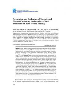
RESULTS Formulation of W1/O/W2 Nanoemulsions A first W1/O/W2 nanoemulsion was optimized and evaluated in terms of cytotoxicity (3). For this formulation, in our previous work (3) no cytotoxicity was detected by Alamar Blue (AB) test both after 1 h and 24 h of incubation with undeterminable IC50 values. Moreover apoptotic mechanisms were not evidenced by chromatin condensation and P2X7 cell death receptor activation tests. In this study, the stability of this first formulation was tentatively improved by the addition of semi-synthetic glycerides wax in the oil phase and/or Human Serum Albumin (HSA) in the external water phase (W2). Moreover, the volume of the internal phase was increased in order to improve the encapsulation efficiency. Compositions of the investigated nanoemulsions are given in Table I for the primary W1/O nanoemulsions and Table II for the multiple W1/O/W2. Characterization of the Multiple Nanoemulsions Transmission Electron Microscopy Double emulsion was identified with negative coloration transmission microscopy. Formulations showed a multiple system by inclusion of water droplets in oil vesicles (Fig. 1), Figure 1 shows TEM micrographs of multiple nanoemulsions with Fig.1a W1/ O/W2 nanoemulsions including HSA in W2 phase, Fig.1b W1/O/W2 nanoemulsions including wax in the oil phase of the primary W1/O nanoemulsion and Fig. 1c including both HSA and wax. No difference was observed between the formulations tested in terms of electron microscopic images. The encapsulation of the drug did not show any difference in the electron microscopic images. Table I
Composition (% w/w) of the Primary W1/O Nanoemulsions
Formulation
Control
(b)wax
Polysorbate 85 Glycerol Oil : (Medium Chain Triglyceride) Wax (Suppocire® DM) Water (W1)
13.3 31.1 35.6 20
13.3 31.1 17.8 17.8 20
Author's personal copy Characterization of W1/O/W2 nanoemulsions for MRI Table II Composition (% w/w) of the Multiple W1/O/W2 Nanoemulsions
Formulation
Control
(a)HSA
(b)wax
(c) HSA & wax
Primary W1/O emulsion Water (W2) Water (W2) including 5% HSA
33 67 –
33 – 67
33 67 –
33 – 67
Particle Size Distribution and Zeta Potential
Short-term Stability of W1/O/W2 Nanoemulsions
Using one-way ANOVA and Dunett’s multiple comparison test, no difference was found at D0 between the control formulation and a formulation including 5% HSA with regards to Mean Droplet Size (MDS) (Table III), whereas when wax was included, in combination or not with HSA, the MDS were significantly lower than the control p