© 2015. Published by The Company of Biologists Ltd | Disease Models & Mechanisms (2015) 8, 591-602 doi:10.1242/dmm.019570
RESEARCH ARTICLE
Presence of multiple lesion types with vastly different microenvironments in C3HeB/FeJ mice following aerosol infection with Mycobacterium tuberculosis
ABSTRACT Cost-effective animal models that accurately reflect the pathological progression of pulmonary tuberculosis are needed to screen and evaluate novel tuberculosis drugs and drug regimens. Pulmonary disease in humans is characterized by a number of heterogeneous lesion types that reflect differences in cellular composition and organization, extent of encapsulation, and degree of caseous necrosis. C3HeB/FeJ mice have been increasingly used to model tuberculosis infection because they produce hypoxic, well-defined granulomas exhibiting caseous necrosis following aerosol infection with Mycobacterium tuberculosis. A comprehensive histopathological analysis revealed that C3HeB/FeJ mice develop three morphologically distinct lesion types in the lung that differ with respect to cellular composition, degree of immunopathology and control of bacterial replication. Mice displaying predominantly the fulminant necrotizing alveolitis lesion type had significantly higher pulmonary bacterial loads and displayed rapid and severe immunopathology characterized by increased mortality, highlighting the pathological role of an uncontrolled granulocytic response in the lung. Using a highly sensitive novel fluorescent acid-fast stain, we were able to visualize the spatial distribution and location of bacteria within each lesion type. Animal models that better reflect the heterogeneity of lesion types found in humans will permit more realistic modeling of drug penetration into solid caseous necrotic lesions and drug efficacy testing against metabolically distinct bacterial subpopulations. A more thorough understanding of the pathological progression of disease in C3HeB/FeJ mice could facilitate modulation of the immune response to produce the desired pathology, increasing the utility of this animal model. KEY WORDS: C3HeB/FeJ, Tuberculosis, Mouse models, Chemotherapy, Neutrophil
INTRODUCTION
It has been estimated that approximately one third of the world is infected with Mycobacterium tuberculosis, with the vast majority of these individuals serving as a latently infected asymptomatic 1
Mycobacteria Research Laboratories, Department of Microbiology, Immunology and Pathology, Colorado State University, Fort Collins, CO 80523, 2 USA. Center for Tuberculosis Research, Department of Medicine, Johns Hopkins University School of Medicine, Baltimore, MD 21231, USA. *Author for correspondence (
[email protected]) This is an Open Access article distributed under the terms of the Creative Commons Attribution License (http://creativecommons.org/licenses/by/3.0), which permits unrestricted use, distribution and reproduction in any medium provided that the original work is properly attributed.
Received 17 December 2014; Accepted 26 March 2015
reservoir (Lin and Flynn, 2010). Since 2008, the incidence of multiple-drug-resistant (MDR) tuberculosis (TB) in Africa has almost doubled, while in southeast Asia the incidence has increased more than 11 times (World Health Organization, 2013). Globally, the incidence of MDR TB increased 42% from 2011 to 2012, with almost 10% of those cases being extensively drug-resistant (XDR) TB. With the rise in MDR and XDR TB, new drugs and especially drugs with a novel mechanism of action or drugs that can shorten the duration of treatment are desperately needed. In humans, TB presents as a diverse spectrum of disease (Laennec, 1823). Historically, it took the discovery of the tubercle bacillus and development of diagnostic staining methodologies to definitively ascertain that the diverse manifestations of this disease were the result of a single infectious agent. Histopathological studies revealed that, even within the lungs of a single individual, multiple lesion types coexist (Canetti, 1955). Through the use of 18 F-fluorodeoxyglucose positron emission tomography (PET) computed tomography (CT), it has recently become apparent that individual pulmonary lesions can change substantially over time (Barry et al., 2009). Importantly, these observations have been confirmed and extended using the nonhuman primate model, suggesting that lesions within an individual animal follow independent pathological trajectories and that the sum of these trajectories might best represent disease outcome (Lin et al., 2013, 2014). Although nonhuman primates can replicate many of the lesion types and histological features of human disease, the high cost precludes their use for drug screening. The ultimate goal of TB drug development programs is to shorten the duration of therapy needed to cure disease, prevent the emergence of drug resistance and phenotypic drug tolerance, and target difficult-to-treat persistent subpopulations of bacilli. Small animal models of TB infection are an important component of drug screening efforts and represent a cost-effective means of accelerating preclinical drug development. In order to obtain results that have predictive utility for human clinical trials, small animal models should replicate the pathophysiological conditions believed to exist within human pulmonary TB lesions. Important parameters include: (1) the production of caseous necrosis accompanied by collagen encapsulation, which can influence drug penetration and the nutrient supply available to bacilli (Dartois, 2014); (2) hypoxia, which can influence the metabolic state of M. tuberculosis bacilli (Wayne and Hayes, 1996); and (3) intracellular as well as extracellular populations of bacilli, which can impact drug efficacy (Grosset, 2003). Although BALB/c and C57BL/6 mouse strains have been extensively used for modeling aerosol infection with TB, these strains present with a single morphological lesion type following low-dose aerosol (LDA) infection with virulent M. tuberculosis (Rhoades et al., 1997), and 591
Disease Models & Mechanisms
Scott M. Irwin1, Emily Driver1, Edward Lyon1, Christopher Schrupp1, Gavin Ryan1, Mercedes Gonzalez-Juarrero1, Randall J. Basaraba1, Eric L. Nuermberger2 and Anne J. Lenaerts1,*
RESEARCH ARTICLE
TRANSLATIONAL IMPACT Clinical issue Tuberculosis (TB) is a pulmonary (lung) disease spread by aerosol transmission. Around 2-billion people worldwide are infected with Mycobacterium tuberculosis, the bacterium that causes TB. Once infected, approximately 10% of individuals rapidly develop active TB, which is characterized by fever, weight loss and productive cough. However, 90% of infected individuals contain and control the infection. Although these individuals are asymptomatic and noncontagious, they represent a reservoir of potential disease and a proportion of them subsequently present with active disease. Reactivation of TB is typically associated with an age-related decline in immunity, immunosuppression or immune-compromising diseases such as HIV. With the emergence of multi-drug-resistant TB, new drugs and chemotherapeutic regimens are desperately needed to treat the disease. Although mouse models have proven useful for TB drug discovery, many mouse strains fail to accurately reproduce the pulmonary pathology observed in human disease. Results Here, the authors investigate the histopathological response to aerosol infection in the C3HeB/FeJ mouse model, which develops caseous necrotic pulmonary granulomas, a type of lung lesion often seen in humans with TB. A comprehensive pathological evaluation reveals that three morphologically distinct lesion types emerge following infection, in contrast to the more commonly used mouse strains, which only present with a single lesion type. Notably, the bacterial load varies between lesion types and this variability reflects differences in the immunological control of bacterial replication. Finally, the authors identify a single lesion type that is responsible for the significant early mortality observed in a subpopulation of C3HeB/FeJ mice following infection.
Disease Models & Mechanisms (2015) 8, 591-602 doi:10.1242/dmm.019570
Mouse models that display more realistic pulmonary pathology might be more predictive of drug activity in human clinical trials. C3HeB/FeJ mice (also known as the Kramnik mouse model) generate hypoxic, caseous necrotic pulmonary lesions with abundant intracellular and extracellular bacteria following LDA infection with M. tuberculosis, and therefore represent an attractive model in which to study TB drug activity. In support of this, previous work from our laboratory demonstrated that clofazimine (CFZ) had significant activity in BALB/c mice and that this activity was highly attenuated in C3HeB/FeJ mice (Irwin et al., 2014). The attenuation observed for CFZ was shown to be dependent upon the pathological progression of disease, because CFZ had significant activity in C3HeB/FeJ mice when treatment was initiated prior to the formation of well-defined pulmonary granulomas. The differential activity of CFZ in a mouse model that reproduces the complex pathological manifestation of granulomatous disease underscores the central role of the pathological process in assessing in vivo drug efficacy. Owing to the crucial role of the pathological response in the lungs in TB, a more comprehensive understanding of the histopathological response to aerosol infection in C3HeB/FeJ mice was warranted. In these experiments, we characterized the pathological progression of lesion development over time following an LDA infection and examined the cellular composition and distribution of cell types in the resulting granulomas. We identified three distinct lesion types that arose following LDA infection. Importantly, the heterogeneity of lesion types represented differing
therefore do not represent the full range of disease manifestations observed in affected humans. The cellular, inflammatory granulomas in these strains of mice fail to become hypoxic (Driver et al., 2012; Harper et al., 2012) and might not produce the microenvironmental conditions needed to generate metabolically distinct subpopulations of bacteria, especially persistent organisms that are difficult to treat. In addition, the majority of the bacilli are located intracellularly (Hoff et al., 2011), which could potentially bias drug screens by overestimating the efficacy of compounds that accumulate within macrophages. It has become apparent that the caseum of mature granulomas might substantially impede drug penetration, limiting in vivo drug efficacy (Prideaux et al., 2011). Mouse strains that do not exhibit this aspect of the granulomatous process might fail to accurately model the effects of drug penetration and activity under specific microenvironments, aspects that are crucial components of in vivo drug efficacy (Lanoix et al., 2015; Dartois, 2014). 592
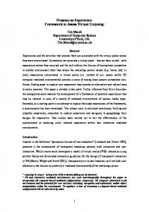
Fig. 1. Kinetics of bacterial replication and effects on morbidity and mortality of C3HeB/FeJ mice. (A) Bacterial growth curve in the lungs of C3HeB/FeJ mice exposed to an LDA of M. tuberculosis Erdman. Data points represent mean log10 CFU±s.e.m. of five animals per time point from one representative experiment. (B) A separate experiment showing the survival curve and characteristic window of mortality (arrows) of C3HeB/FeJ mice (n=85) following aerosol infection.
Disease Models & Mechanisms
Implications and future directions Cost-effective small animal models that represent the full spectrum of human TB are needed to augment drug development efforts, evaluate novel multi-drug combination regimens and prevent the emergence of drug resistance. Although the C3HeB/FeJ mouse model has shown promise, variability in pulmonary colony-forming unit (CFU) counts and early mortality have complicated its usage. The realization that three distinct lesion types with different bacterial loads can emerge following aerosol infection of C3HeB/FeJ mice will allow researchers to understand and properly interpret the variability of pulmonary bacterial counts typically observed when using C3HeB/FeJ mice as a model for TB. Moreover, a more complete understanding of the pathological process that underlies lesion development in the lungs will increase the reproducibility, and therefore the utility, of this animal model. Immunomodulatory and chemotherapeutic strategies are presently being investigated to reduce early mortalities while preserving the caseous necrotic lesions that make this animal model relevant to human TB.
levels of host control of bacterial replication and of the progressive inflammatory response as evidenced by the number of bacteria and morphometric analysis of lesion size over time. The exceptionally large number of bacilli characteristic of C3HeB/FeJ mice coupled with the highly sensitive SYBR Gold staining methodology allowed us to easily visualize the location of the bacilli, which is normally quite challenging in most animal models (Hoff et al., 2011; Manabe et al., 2008; Rayner et al., 2013; Turner et al., 2003) and in humans (Canetti, 1955) owing to the low number of bacilli present in the lungs. Finally, we correlated rapid pulmonary neutrophilia with progressive consolidation of lung parenchyma that led to the early mortality observed in a proportion of C3HeB/FeJ mice, underscoring the damaging role of an excessive neutrophilic response in the lungs. RESULTS Bacterial replication and C3HeB/FeJ morbidity/mortality following aerosol infection
Following LDA infection of C3HeB/FeJ mice using 50-75 colonyforming units (CFU) of M. tuberculosis Erdman, bacterial numbers within the lung rapidly increased to more than 7 log10 CFU by 30 days post-infection (Fig. 1A). The increase in bacterial numbers in the lung slowed after 30 days, culminating in an additional 0.6 log10 CFU increase between 30 and 90 days. In a series of experiments, we measured mouse survival over time. A consistent pattern emerged whereby significant mortality was observed between 28-45 days after LDA; however, mice that survived beyond 45 days generally survived at least 14 weeks with only minimal mortality observed in the intervening time (Fig. 1B). Of importance, although the timing of this window of mortality remained fairly constant across multiple experiments, the percentage of mortality within this window varied significantly between multiple experiments, ranging from 10 to 40% (data not shown).
Disease Models & Mechanisms (2015) 8, 591-602 doi:10.1242/dmm.019570
C3HeB/FeJ mice developed three distinct pulmonary lesion types following aerosol infection
We next performed a comprehensive pathological examination of the pulmonary lesions that arose following LDA infection in the C3HeB/FeJ mice. Interestingly, upon histological examination we were able to identify three distinct lesion types by 5 weeks following aerosol infection that differed by cellular composition and organization. For descriptive efficiency, we arbitrarily identified these lesions as Type I, Type II or Type III. Type I lesions (Fig. 2A) most closely resembled classical human TB granulomas in that they were solid, encapsulated caseous necrotic lesions. These granulomas were initially identified by Igor Kramnik’s group (Pichugin et al., 2009) and have been described previously (Driver et al., 2012). These lesions became evident 35-45 days following LDA, and originated as a focal accumulation of foamy macrophages interspersed with neutrophils, often proximal to a bronchus. The peripheral margins contained abundant, loosely aggregated epithelioid macrophages interspersed with a small number of scattered lymphocytes (Fig. 2A). As this lesion morphology progressed, the number of neutrophils increased rapidly, and the beginning of a dense central neutrophilic core was evident, variably surrounded by smaller regions of loosely packed neutrophils and foamy macrophages (Figs 2B and 3A,B). The epithelioid macrophages immediately adjacent to the neutrophilic core stained less intensely with eosin, and it appeared that these cells were transforming into foamy macrophage cells. Loosely packed epithelioid macrophages, activated macrophages and a small number of lymphocytes composed the peripheral extremity of the lesion. By 7-10 weeks following infection, the Type I granulomas took on a highly organized appearance, composed of a densely packed neutrophilic core, with or without central caseation (Fig. 2C). This core region was surrounded by a rim of diffusely stained foamy macrophages, encapsulated by a fibrotic rim (Fig. 3C). The
Fig. 2. Progression of Type I, II and III lesions over time. Each panel represents a single lesion (H&E-stained) obtained from an individual representative animal (n=5) euthanized at the time point defined here. Progression of Type I lesions from individual animals at 45 (A), 50 (B), 55 (C) and 61 (D) days following aerosol infection. Progression of Type II lesions from individual animals at 20 (E), 35 (F), 38 (G) and 40 (H) days following aerosol infection. Progression of Type III lesions from individual animals at 30 (I), 35 (J), 61 (K) and 75 (L) days following aerosol infection. CN, caseous necrosis.
593
Disease Models & Mechanisms
RESEARCH ARTICLE
RESEARCH ARTICLE
Disease Models & Mechanisms (2015) 8, 591-602 doi:10.1242/dmm.019570
peripheral margin of the granuloma was composed of fibroblasts, epithelioid and activated macrophages, and a small number of scattered lymphocytes. At this time, multiple coalescing Type I granulomas were occasionally observed. After 8-10 weeks, Type I granulomas continued to progressively enlarge, although the cellular composition did not change. However, the central region of the neutrophilic core progressively degenerated into an acellular homogeneous caseum diffusely stained by eosin (Fig. 2D). Initially, alveolar septae were clearly visible and retained the structural appearance of the lung. Gradually, as neutrophils within individual alveoli degenerated as evidenced by punctate karyorrhectic debris, the structure of the interalveolar septae within the central region of the granuloma began to degenerate into isolated islands as previously described (Driver et al., 2012). Ultimately, the karyorrhectic debris and even the septal wall fragments completely degenerated until no histologically identifiable lung structure was evident (Fig. 3D). This degeneration typically originated within the centermost region and progressed outward as the granuloma enlarged. The periphery of the core region still retained a distinct band of intact neutrophils within the collagen rim. Type II lesions resembled a rapidly progressive, granulocytic pneumonia composed almost entirely of neutrophils. By 20 days following LDA, small cellular aggregates composed of activated 594
and epithelioid macrophages and aggregates of neutrophils that had completely impacted the alveoli were evident (Fig. 2E). By day 25, the beginning stage of rapidly progressive neutrophilia was evident as fulminant necrotizing alveolitis (Fig. 2F). A distinct central region of epithelioid macrophages was surrounded by a large rim of neutrophils that appeared to be expanding outward by progressively infiltrating adjacent alveoli. Small aggregates of lymphocytes were occasionally observed, but were confined to the extreme periphery of the lesion boundary. The initial stages of cellular necrosis were evident at this time, particularly within the central region. By 30 days post-infection, the majority of Type II lesions had substantially increased in size and contained a central region of caseous necrosis (Figs 2G and 3E). The central region had evidence of cellular necrosis, karyorrhectic debris, and fragmentation and degeneration of septal walls, similar to the caseous necrotic response observed in the center of Type I lesions (although at a much earlier time point than the Type I lesions). Notably, few if any lymphocytes were observed in the vicinity of this lesion type and, in mice in which this lesion type predominated, lymphocytes were notably absent in other areas of the lung. Inflammatory exudate, primarily composed of edematous fluid, neutrophilic debris and foamy macrophages, frequently consolidated terminal bronchioles, often leading to complete occlusion. By 40 days after infection,
Disease Models & Mechanisms
Fig. 3. Type I, II and III lesions vary with respect to their cellular composition, organization and collagen encapsulation. (A) Type I granuloma showing the acellular caseum, distinct band of darkly stained intact neutrophils (arrows delineate margins), primarily neutrophil-derived zone of karyorrhectic debris, lightly stained foamy macrophage rim, and collagen encapsulation. (B) Higher-magnification image of A illustrating a zone of intact neutrophils (black arrows), foamy macrophage rim and collagen encapsulation (white arrows). (C) Masson’s Trichrome stain for collagen along the peripheral margin of a Type I lesion. (D) Interior region of a Type I granuloma, showing the acellular caseum and karyorrhectic debris. (E) Type II lesion with a caseous necrotic interior, and extensive consolidation of peripheral alveoli. (F) Pulmonary acinus (*) completely occluded with neutrophils and cellular debris. (G) Type III lesion with distinct lymphocytic clusters (arrows). (H) Higher-magnification image of G showing foamy macrophages (black arrows) and a lymphocytic cluster (L). Small isolated pockets containing small numbers of neutrophils (white arrows) are occasionally present, especially at later time points as foamy macrophages undergo necrosis. AC, acellular caseum; K, karyorrhectic debris; FM, foamy macrophages; CN, central necrosis. H&E staining unless otherwise noted.
rapidly progressive neutrophilia resulted in large areas of consolidation of lung parenchyma that radiated outwards over time (Figs 2H and 3F). The central areas of these lesions exhibited the various stages of caseous necrosis as described above, with large areas of amorphous eosin-stained material with no visible cellular structure. This lesion type was rapidly progressive, and culminated in complete consolidation of large areas of lung and collapse of the lung parenchyma along the exterior edge of the lesion. Complete consolidation of entire lung lobes was frequently observed. Although Type II lesions were similar to the Type I granulomas in that they were primarily neutrophil-dominated, they lacked the highly organized fibrotic structure and collagen deposition diagnostic of encapsulated Type I granulomas. Type III lesions in C3HeB/FeJ mice were cellular, inflammatory lesions that were indistinguishable from pulmonary lesions typically observed in BALB/c (Hoff et al., 2011) and C57BL/6 (Rhoades et al., 1997) mice. Briefly, Type III lesions were initially composed primarily of mononuclear phagocytes and activated macrophages, located proximally to a blood vessel with mild alveolitis (Fig. 2I). By 35 days post-infection, large numbers of epithelioid macrophages were evident, admixed with activated macrophages and large numbers of lymphocytes typically arranged in perivascular and peribronchiolar cuffs that exhibited mild to moderate interstitial fibrosis and alveolar thickening (Figs 2J and 3G). Small isolated pockets of neutrophils were occasionally present (Fig. 3H), usually confined to localized regions within a lesion. Inflammatory exudate was sometimes apparent within bronchioles, but rarely resulted in complete occlusion. By 55 days, many of the epithelioid-like macrophages had transformed into foamy macrophages (Figs 2K and 3H). Large numbers of lymphocytes were found both in large aggregates and interspersed throughout the lesion, often in association with macrophages. By 75 days following infection, large numbers of foamy macrophages (Figs 2L and 3H) containing abundant lipid vesicles were present. Localized individual cellular necrosis with associated punctate karyorrhectic debris occurred primarily within foamy macrophages, and resulted in microcavities containing necrotic debris and variably small numbers of neutrophils within otherwise densely packed cellular lesions. C3HeB/FeJ mice with predominantly Type II lesions had decreased survival
We next examined the mice that had to be euthanized prior to experimental endpoints to adhere to Colorado State University’s Institutional Animal Care and Use Committee (IACUC) guidelines (loss of greater than 20% body weight, unthrifty appearance, etc.). This group of mice exhibited significant morbidity in multiple experiments within a highly reproducible window (28-45 days after infection). Morphometric analysis of the total lesion area occupied by each of the three lesion types in comparison to the total lung area was performed on hematoxylin and eosin (H&E)-stained whole lung sections to quantify disease severity. Analysis of the earlymortality mice (mice humanely euthanized between days 30 and 41 in this particular experiment) indicated that 100% of these animals had predominantly Type II lesions composed of fulminant neutrophilia (Fig. 4A) that occupied large areas of the lung parenchyma and resulted in observable weight loss, compromised respiratory function and a decline in overall health of the affected animals. In contrast, of the mice that survived to 8 weeks, all had a mixture of Type I and Type III lesions, with little evidence of Type II lesion involvement. Of the three surviving mice that had detectable Type II lesions, only one mouse (Fig. 4A, mouse 16) had a significant percentage of lung area occupied by Type II
Disease Models & Mechanisms (2015) 8, 591-602 doi:10.1242/dmm.019570
lesions, although it should be noted that the area occupied by Type II lesions in this mouse was approximately half that observed in the early-mortality mice. Pulmonary bacterial load was assessed by plating serial dilutions of whole-lung homogenates on 7H11 agar. Seventeen mice displaying signs of morbidity substantial enough to warrant humane euthanasia (early-mortality mice) were selected for CFU determination. All of these mice were euthanized between 3041 days post-infection in this experiment. Fourteen surviving mice were euthanized 8 weeks post-infection (Fig. 4B). The earlymortality mice had 1.3 log10 CFU more bacteria than the mice that survived 8 weeks, even though the infection in the surviving mice had progressed approximately 3 weeks longer. We next compared pulmonary bacterial load with the terminal body weight at the time of euthanasia for individual mice (Fig. 4C). Mice were euthanized at 25 (n=10), 35 (n=15) and 45 (n=14) days post-infection. Early-mortality mice (n=17) were euthanized between 30-41 days post-infection based upon IACUC morbidity guidelines. Pulmonary bacterial load inversely correlated with mouse body weight, which proved to be a reliable indicator of disease severity in conjunction with physical parameters (e.g. dyspnea, ruffled fur) and behavioral characteristics (e.g. hunched appearance, lethargy). Initial deviations in individual animal body weights could be observed by 20 days following LDA, and were predictive of disease severity and early mortality (data not shown). Lesion burden stabilized by 45 days following infection
The progression of pulmonary disease in C3HeB/FeJ mice following LDA infection was characterized by collecting mice for histological examination at 5-day intervals through 85 days of infection (Fig. 5). The first evidence of inflammation related to M. tuberculosis infection occurred at 15 days following infection and pulmonary lesion burden increased with each subsequent time point through 45 days of infection, with a particularly abrupt transition between 35 and 40 days of infection. After 45 days, the pathology in the lungs of C3HeB/FeJ mice as measured by total lesion burden scoring began to stabilize. The 35- to 40-day time period coincided with the 28- to 45-day window of mortality observed in the survival experiments (Fig. 1B). Beyond 45 days of infection, there was no significant increase in the number of lung lesions and it seemed that the pathology in the lungs of C3HeB/FeJ mice began to stabilize, although individual lesions continued to increase in size and severity over time. Intra-mouse lesion heterogeneity was observed throughout the time course of infection
Owing to the presence of three lesion types, a pathological assessment was performed to determine the spatial distribution of each lesion type within each lung lobe of individual animals following LDA infection. Type I lesions were non-uniformly distributed between the five lung lobes of individual animals (Fig. 6A,C-E). Type III lesions were more numerous, generally smaller in size, and more evenly distributed between lung lobes. In the mice that predominantly had Type II lesions, nearly all of the lung lobes had evidence of such lesions (Fig. 6B). M. tuberculosis number and location varied by lesion type as visualized by SYBR Gold staining
Using the SYBR Gold acid-fast stain, we next examined the distribution of M. tuberculosis bacilli to determine the spatial location of the bacilli within pulmonary lesions, whether they were 595
Disease Models & Mechanisms
RESEARCH ARTICLE
RESEARCH ARTICLE
Disease Models & Mechanisms (2015) 8, 591-602 doi:10.1242/dmm.019570
Fig. 4. Early-mortality mice displayed primarily Type II lesions, and had higher pulmonary bacterial loads and lower body weights. (A) Early-mortality mice primarily exhibited Type II lesions, whereas the surviving mice exhibited primarily Type I and Type III lesions. Lesion area analysis was performed on H&Estained histological sections of all five lung lobes from individual mice (n=9 for early mortality mice and n=10 for surviving mice). Early-mortality mice represent mice euthanized between day 30 and 41 post-infection owing to morbidity. Surviving mice were euthanized 8 weeks following infection. Data are expressed as the percentage of the total lung area (all five lobes) for each lesion type compared to the total lung area for individual mice. (B) Early-mortality mice (n=17) had a higher pulmonary bacterial load compared to surviving mice (n=14). Data represent the logarithms of serial dilutions of lung homogenates obtained from whole lungs of individual mice. Statistical comparison performed using Student’s t-test. *P