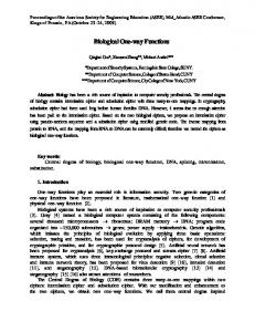
have now determined whether the yeast MTase can prevent ." -.4 A. 1 kb. LacZ. 0. Fig. 1. ..... to killing by the UV-mimetic agent 4-nitroquinoline-1-oxide. (4NQO) ...
The EMBO Journal vol. 1 0 no.8 pp.2179 - 2186, 1991
Primary sequence and biological functions of a Saccharomyces cerevisiae 06-methylguanine/ 04-methylthymine DNA repair methyltransferase Wei Xiao, Bruce Derfier, Jin Chen and Leona Samson Laboratory of Toxicology, Harvard School of Public Health, Boston, MA 02115, USA
Communicated by J.Cairns
We previously identified and characterized biochemically an 06-methylguanine (06MeG) DNA repair methyltransferase (MTase) in the yeast Saccharomyces cerevisiae and showed that it recognizes both 06MeG and 04-methylthymine (04MeT) in vitro. Here we characterize the cloned S.cerevisiae 06MeG DNA MTase gene (MGTJ) and determine its in vivo role in protecting yeast from DNA alkylation damage. We isolated a yeast DNA fragment that suppressed alkylation-induced killing and mutation in Escherichia coli ada ogt MTase deficient mutants and produced in these cells a protein similar to the yeast MTase. The cloned yeast fragment was mapped to chromosome IV and DNA sequencing identified an open reading frame, designated MGT], which encodes a 188 amino acid protein with a molecular weight of 21 500 daltons. An 88 amino acid stretch of the MGT1 protein displays remarkable homology with four bacterial MTases and the human DNA MTase. S.cerevisiae mutants bearing an insertion in the MGT] gene lacked DNA MTase activity and were very sensitive to alkylation induced killing and mutation. MGT] transcript levels are not increased in response to DNA alkylation damage, nor is the MGT1 MITase involved in the regulation of the yeast 3-methyladenine DNA glycosylase gene (MAG). Expression of the MGT] gene in E.coli prevented the induction by alkylating agents of both G:C to A:T and A:T to G:C transition mutations indicating that this eukaryotic MTase repairs both 06MeG and 04MeT in vivo. Key words: alkylation/DNA repair/methyltransferase/yeast
Introduction Agents like N-methyl-N'-nitro-N-nitrosoguanidine (MNNG) and methylmethanesulphonate (MMS) cause at least a dozen different kinds of DNA lesions, and most cells express a number of DNA repair enzymes to protect against the mutation and cell death that can be caused by some of these lesions. In both prokaryotic and eukaryotic cells, 06-methylguanine (0 MeG) and 04-methylthymine (O4MeT) cause transition mutations by mispairing when replicated (Loveless, 1969; Preston et al., 1986; Lindahl et al., 1988). In Escherichia coli, these lesions are repaired by DNA methyltransferases (MTases) and by the nucleotide excision repair pathway (Lindahl et al., 1988; Rebeck et al., 1988; Samson et al., 1988). Two E. coli MTases remove methyl groups from alkylated DNA. The 39 kd Ada MTase Oxford University Press
gene
removes methyl groups from 06MeG, 04MeT and methylphosphotriester (MePT) lesions, and is induced as part of the adaptive response to alkylating agents (Samson and Cairns, 1977; Lindahl et al., 1988). The Ada protein also acts to regulate transcription; once a methyl group is transferred from a MePT lesion to Cys69, the Ada MTase becomes a positive activator of transcription for a group of genes which include alkA, aidB and the ada-alkB operon (Nakabeppu and Sekiguchi, 1986; Teo et al., 1986; Lindahl et al., 1988). A second MTase, known as DNA MTase II or the Ogt MTase, is constitutively expressed and repairs only 06MeG and 04MeT (Potter et al., 1987; Rebeck et al., 1988, 1989). The Ada and Ogt MTases protect E. coli against mutation, and to some extent against killing, by alkylating agents (Rebeck and Samson, 1991). Bacillus subtilis has three MTases, namely Dati, AdaA and AdaB. Dat], like ogt, is non-inducible (Morohoshi et al., 1989), but adaA and adaB overlap by 11 bp and form an alkylation inducible operon that encodes two MTases. AdaA is a MePT MTase as well as a regulatory protein, and AdaB is an 06MeG MTase (Morohoshi et al., 1990). Recently, the human 06MeG MTase cDNA was isolated (Hayakawa et al., 1990; Rydberg et al., 1990; Tano et al., 1990) and the enzyme characterized (Koike et al., 1990; von Wronski et al., 1991). It appears that, at least in some cell lines, transcription of the human MTase gene is not increased in response to alkylating agents (Fornace et al., 1990). 06MeG DNA MTase activity has been found in a variety of organisms including several species of bacteria (Lindahl et al., 1988; Vaughan, 1991), insect (Green and Deutsch, 1983), fish (Nakatsuru et al., 1987) and mammalian cells (Harris et al., 1983; Pegg and Dolan, 1987). Despite the existence of DNA repair MTases in prokaryotes and higher eukaryotes, it was commonly believed that the yeast Saccharomyces cerevisiae does not have a MTase and that 06MeG is therefore removed by other DNA repair pathways (Hadden et al., 1983; Maga and McEntee, 1983; Friedberg, 1998; Goth-Goldstein and Johnson, 1990). However we have identified a 25 kd protein in crude yeast cell extracts that accepts methyl groups from 06MeG to form S-methylcysteine (Sassanfar and Samson, 1990). More recently, we found that this acceptor protein and the human MTase, have a weak affinity for 0 MeT in DNA (Sassanfar et al., 1991). Here we describe the cloning of the S.cerevisiae DNA repair MTase gene (by its ability to functionally complement MTase deficient ada- ogt- E. coli) and we determine its role in the response of yeast cells to DNA alkylation damage.
Results Cloning a yeast gene that functionally complements ada- ogt- E.coli E. coli strain GWR1 11 (ada- ogt -) totally lacks O6MeG DNA MTase activity and is very sensitive to MNNG-induced 2179
W.Xiao et al.
killing and mutation (Rebeck and Samson, 1991). A yeast genomic library cloned into pUC 19 was introduced into GWR1 11, and the transformants were challenged with MNNG (100 Itg/ml). The survivors were then challenged a second time (150 ,tg/ml MNNG) and the survivors from the second challenge were screened for MTase activity (see Materials and methods for details). In this way, we isolated a clone, pUC 11-3, that produced 06MeG DNA MTase activity in ada- ogt - E. coli. This MTase activity was like the yeast MTase (Sassanfar and Samson, 1990) having a molecular weight of -25 kd (Figure lA) and a relatively short half life at 37°C (data not shown). The pUC 11-3 clone had a 5.3 kb insert that hybridized with yeast DNA but not with E. coli or human DNA (Figure ID), and made GWR1 11 cells very resistant to the killing and the mutagenic effects of MNNG (Figure lB and C). We have called the yeast gene which encodes this 06MeG DNA MTase MGTJ, for 06MeG methyltransferase.
Localization of the yeast 06MeG DNA MTase gene
(MGT1) A restriction map of the pUC 1 1-3 insert was used as a guide to make a series of pUC 1-3 deletions and subclones to localize the MGT] gene (Figure 2). The subclones were tested for their ability to produce MTase activity in adaogt- E.coli. Since subclone A44 (pMGT1-44) was the smallest subclone to retain MTase activity, the MGT] gene was localized to the left end of the insert, within a 1.9 kb HincII fragment (Figure 2). Further deletion of either a 0.15 kb HindIll fragment from the left end (A117), or a 0.75 kb Bgll fragment from the middle (Al 14) of the 1.9 kb HincII fragment abolished MTase activity (Figure 2), suggesting that the MGT] gene was located at the extreme left end of the yeast DNA insert, just downstream from the bacterial lacZ promoter (Figure 2). When the 1.9 kb insert orientation was inverted with respect to the lacZ promoter, MTase activity was abolished (data not shown). Further, in the appropriate strains, MTase expression was induced by the lac operon inducer IPTG (data not shown). We therefore believe that MGTJ expression from pUC 1 1-3 and pMGT1-44 is driven by the lacZ promoter. The yeast 06MeG DNA MTase also repairs 04MeT in vivo
We recently showed that the MTase in crude yeast cell extracts is specifically inactivated upon incubation with oligonucleotides containing a single 06MeG or 04MeT, indicating that the eukaryotic MTase is able to repair both of these DNA lesions in vitro (Sassanfar et al., 1991).
."
-.4
However, because the affinity of the MTase for 04MeT was much lower than for 06MeG, its ability to repair 04MeT might not be significant in vivo. To test this we
A
have now determined whether the yeast MTase can prevent LacZ
1 kb Plasmid
flTase
Activitg
pUC 1 1-3 &
4
I
38 o22
0
&
6
,&
19
,&4 & & & &
35 21
2180
m
4
+
34 44
_117 Fig. 1. Characterization of GWR 1 1 /pUC 1 1-3 transformant. (A) Gel electrophoretic assay of MTase activity in bacterial extracts from GWR1 1 l/pUC19 (open circle) and GWR1 1 l/pUCl 1-3 (closed circle). Slice 1 is the top of a 12% SDS-polyacrylamide gel. (B) Killing of GWRl11 (open circle) and GWR1 1 1/pUC1 1-3 (closed circle) after treatment with MNNG (5 ig/ml) for the indicated times. (C) MNNGinduced mutation in GWRl1 I/pUC19 (open circle) and GWRl 1 I/pUC1 1-3 (closed circle) after treatment with 0.01 Ag/ml MNNG for different times. (D) Southern hybridization of HindIII (H) and EcoRI (E) digested total E.coli DNA (2 ag), yeast DNA (5 yg) and human DNA (10 yg) using the 1.9 kb HincII MGT] fragment from pUC 11-3 (see Figure 2) as probe.
+ +
_
_114_ Fig. 2. Localization of MGT] in pUCl 1-3. Known restriction sites were used to generate subclones that delete a DNA segment from the left end (Al, A38, A22, A6, A19 and A4), the right end (A35, A21 and A44) or the interal part (A34) of the insert. The bar represents the DNA that remains after deletion. The presence (+) or absence (-) of yeast MTase activity is indicated in the right panel. A1 17 and A1 14 were constructed by HindIlI and BglII digestion, respectively, of A44 containing the 1.9 kb insert. Restriction sites: B, BamHI; Bg, BglII; E, EcoRI; H, HindlIl; Hc, HincII.; K, Kpnl; N, NdeI; P, PstI; S,
SmaI; Sa, Sall; Sc, SacI; Sp, SphI; Xb, XbaI.
(-,X.
Yeast DNA methyltransferase sequence and function
A:T to G:C transition mutations which are known to be induced by 04MeT (Preston et al., 1986). The yeast MGTJ MTase gene in pUC 1 1-3 was introduced into two E. coli strains, each containing a different lacZ- allele; one can only revert via a G:C to A:T transition, the other only by an A:T to G:C transition (Cupples and Miller, 1989). As controls, we introduced into the same strains pUC19ogt containing the E. coli ogt gene (since Ogt efficiently repairs both 06MeG and 04MeT) (Rebeck et al., 1989; Sassanfar et al., 1991) and pUC 19 alone. All six strains were streaked onto agar with and without MNNG and the induction of lacZ+ mutants was scored by the appearance of dark blue papillae. Figure 3 shows, as expected, that MNNG induces lacZ+ papillae via G:C to A:T and A:T to G:C transitions and that both kinds of transition were prevented by the E. coli Ogt MTase. As the figure shows, the S. cerevisiae MGT1 MTase prevented both G:C to A:T and A:T to G:C transition mutations suggesting that this MTase similarly repairs both 06MeG and 04MeT in vivo. This result was also obtained using the much smaller DNA fragment in pMGT1-44 as the source of yeast MTase in E.coli (data not shown). MGT1 maps to yeast chromosome IV The 1.9 kb yeast fragment containing MGTJ was radioactively labelled and used to probe an agarose gel containing electrophoretically separated yeast chromosomes. A single band appeared in the chromosome IV/VII area (Figure 4, lane 1). In order to determine which of these chromosomes contains MGTJ, a 1.5 kb BamHI-EcoRI fragment containing part of the ADHI gene, known to reside in chromosome XV (Mortimer and Schild, 1985) which runs close to IV and VII in the gel, was included as a marker. Hybridization with mixed ADHJ -MGTi probes indicates that MGT] is most likely located on chromosome IV rather than on chromosome VII (Figure 4, lane 2). Basepair Substitution
G[C to AT
blt
to Al
AT to GC
MNN'm/7n,)
(
p ill
Shenk, 1981). The carboxy-terminal half of the deduced MGT1 amino acid sequence has significant homology with every other sequenced 06MeG MTase, namely Ada and Ogt from E.coli, Datl and AdaB from B.subtilis and MGMT from human cells. Over an 88 amino acid stretch, the identity between MGT1 and the bacterial MTases is 42-43.8%, and that between MGT1 and the human MGMT is 34.1%, suggesting that they may have had a common origin (Figure
i
--
.X:..'v
i.
t. .
0X
AT to BC
I I 1j
Nucleotide and amino acid sequences of MGT1 We determined the nucleotide sequences for the 0.9 kb downstream from the lacZ-MGTI junction to the downstream NdeI site (Figure 2) and found an open reading frame that is in-frame with the lacZ coding region (Figure 5). The possible LacZ-MGTl fusion protein would have a molecular weight of 23 873 daltons. We have tentatively marked the first in-frame ATG in the insert (Figure 5) as the start codon for the MGT] gene because the predicted 188 amino acid protein coded from this AUG to the first stop codon has a calculated molecular weight of 21 502 daltons, which is consistent with previous studies (Sassanfar and Samson, 1990) and shows that the yeast 06MeG MTase has a molecular weight that is close to the new value of 21 700 daltons for the human MTase (Hayakawa et al., 1990; Rydberg et al., 1990; Tano et al., 1990). However, it remains possible that this ATG may not be the authentic translation initiation site in yeast. Sequences downstream of the translation stop codon (Figure 5) show similarity to yeast transcription termination signals (Zaret and Sherman, 1982) and the eukaryotic polyadenylation signal (Fitzgerald and
.
...
.
Fig. 3. The prevention of MNNG-induced transition mutations by the MGT1 and Ogt MTases. pUC19, pUCogt (producing the Ogt MTase) and pUC11-3 (producing the yeast MTase) were introduced into Ecoli CC 102 ada- ogt - which carries a lacZ - allele revertible by a G:C to A:T transition and E. coli CC 106 ada- ogt - which carries a lacZ allele revertible by a T:A to C:G transition. Colonies of each strain were streaked on minimal plates + MNNG as indicated, and the mutant lacZ- papillae were visible after 3-4 days at 30°C.
Fig. 4. Chromosomal mapping of the MGT] gene. Yeast chromosomes separated by pulse-field electrophoresis (Clontech) were hybridized with an [cs-32P]dCTP-labelled 1.9 kb HincH fragment from pUC1 1-3 containing MGT] (lane 1) and the mixture of MGT] probe with an ADHI probe (lane 2). ADHI probe was prepared by EcoRI-BamHI digestion of plasmid pMA561 (McKnight and McConaughy, 1983) followed by isolation of the 1.5 kb fragment containing ADHI upstream sequences (Bennetzen and Hall, 1982). The positions of yeast chromosomes are indicated.
2181
W.Xiao et al. -97 agcggataacaatttcacacaggaaacagct atg acc Met Thr > PstI XbaI SalI .SphI -45 ttg cat gcc tgc agg tcg act cta gag GAT Leu Hi. Ala Cys Arg Ser Thr Lou Glu Asp
HindIII atg att acg cca agc Met Ile Thr Pro Ser CTA AAT GGA CCA ACG Leu Asn Gly Pro Thr
+1 ATG AAG GAA CTG CTT TAC TAT ACA TTC ATT GAA ACT GAA GTG ACT Met Lys Glu Lou Lou Tyr Tyr Thr Phe Ile Glu Thr Glu Val Thr 46 GGT GCA TTT TTG GTG TTT AGG GAA AAG ACT CAA AAC CTT GTT TTT Gly Ala Ph. Lou Val Ph. Arg Glu Lys Thr Gln Asn Lou Val Phe 91 GCC TCG TTA GGT AAT GAT AAG CTT TTT TTA TTG GGA AAG GTG GAA Ala Sor Leu Gly Asn Asp Lys Lou Pho Lou Leu Gly Lys Val Glu 136 GGC TTC TTG AAG AAA CAT GAG AAA CAG GAT ACA ATG TAC GAT TTA Gly Phe Leu Lys Lys His Glu Lys Gln Asp Thr Met Tyr Asp Leu
181 CAG GAA CTA AAA GAG GCA GAA ACA TAT AAG AAA TCA ATC GAA AAT Gln Glu Leu Lys Glu Ala Glu Thr Tyr Lys Lys Ser Ile Glu Asn 226 TAT ACA ATA TGT TTA GAA AAC AAA ATG CCA TTA CCA TCG GGC GCT Tyr Thr Ile Cys Leu Glu Asn Lys Met Pro Lou Pro Ser Gly Ala 271 ATT CCC TTT GAG TTC CTG TTT GGA ACA GAT TTT CAA CGT AAA GTT Ile Pro Phe Glu Phe Leu Phe Gly Thr Asp Phe Gln Arg Lys Val
316 TGG AAT GAG CTT TTA AAC GTG GAA CAC GGC CAC GTC GTA ACA TAT Trp Asn Glu Leu Lou Asn Val Glu His Gly His Val Val Thr Tyr 361 GGT GAT ATT GCA AAG AGA ATA GGG AAG CCA ACT GCC GCA AGA TCT Gly Asp Ile Ala Lys Arg Ile Gly Lys Pro Thr Ala Ala Arg Ser 406 GTC GGA AGA GCT TGC GGC TCA AAT AAC CTG GCA TTG TTA GTA CCT Val Gly Arg Ala Cys Gly Sor Asn Asn Lou Ala Leu Leu Val Pro 451 TGC CAT AGA ATC GTT GGT AGC AAT AGA AAA TTA ACC GGA TAT AAA
Cys His Arg Ile Val Gly Ser Asn Arg Lys Leu Thr Gly Tyr Lys
496 TGG AGC TGT AAA CTG AAA GAA CAG TTA TTA AAT AAT GAA AAG GAA Trp Ser Cys Lys Leu Lys Glu Gln Leu Leu Asn Asn Glu Lys Glu 541 AAT AGC TTA AGC CTT AGT AGA TTG TAG TTGGATAAAGGATTTTAGGAAAA Asn Ser Leu Ser Leu Ser Arg Leu ***
MGMT 85 MGT1 91 Ada 261 79 Ogt Datl 70 AdaB 81 MGMT 111 MGT1 117 Ada 287 Ogt 105 Datl 96 AdaB 107
MGMT MGT1 Ada Ogt Datl
137
143
313 131 122 AdaB 133
MGMT MGT1 Ada Ogt Datl AdaB
162
168 338 156 148 158
H[
K L K V VK F G H v FQ Q E S F T R V W NEL L N H G D K I P F E F LF W Q L R T I P C G L D I R G T A FQQ T L L R T I P C G1 WK T L P T A T G G T PFQ E K G L P L S Q E R I P G T P Q V E Y A GTQ CE IYJ AVWNA
iY
Q VY G
A L ANP XKAARAV GI A M R G AARMVGRIAICGS R AKR GrKAPRG SACAA ARAV ANa X G A A NGS A E QLG RIPLA AR AV
A
TVV yQ YjG YjA DAIAAAV G S
EM SR TK
S
N D
PKXArVIR A V GQ AA IN GK AR
GA PrA X V R A V Y Sn A VC
N P V PWL I PC it S V LA L P C H RVGS R KL T G Y K W S N G SLG Y RWGN KLAIVIP C IV VG T G Y AF SIV N P C VIIGRT HR PCIRVIGKIN| A TGYA G K nPIF S T G Y G GLVTl P C H R VG KG
NC
L A VK EWLA HEG H R L GK P G L G G.. E N S L S L S R L C K L K Q L LN N E
207
V S R KAQ L LR R E A E N E E R R HE G Y L L L L V Q
188 354 171
T E I AFL N IER I S Y K E K F E MT LLD LK R A S S E M D V P H
165 179
RKW
Fig. 6. Amino acid alignment of MGT1 with other DNA repair MTases. An 88 amino acid region of deduced MGTI sequence is compared with five 06MeG MTases, Ada (Demple et al., 1985) and Ogt (Potter et al., 1987) from Ecoli, Datl (Morohoshi et al., 1989) and AdaB (Morohoshi et al., 1990) from B.subtilis and MGMT from human cells (Hayakawa et al., 1990; Rydberg et al., 1990; Tano et al., 1990). Alignment was by a Doolittle multi-sequence comparison program (Eugene, Houston, TX, USA) and identical residues shared by more than three MTases are boxed.
591 TAAACATAAGAAATAGTTATGTATATGTGGTAAATTGTTCTAGTTATACATCTATGTTA 650 CAATATGGCTGGCGTTGGTAATCTTGTGAAACAAGCGACGAACTTGTCACCCTTCTTTC
709 TTTCCACTTTTTTACCTTCTTCTGTGGCATTTCTCATTATTTTCACACATTCATCGTTT 768
TCTTCCCATTCTTTGCTCAACCGTTCXTMGGATGCTCTTCTAAATGTTTTACCAT
827 CCACTCATGCAGGTCTTTGACGTCTGTAATTGTGTACACGACACCGCCCTCTTTCAAAA 886 CATATG
Fig. 5. Nucleotide and amino acid sequences of MGT]. The nucleotide sequence was determined by the chain termination method (Sanger et al., 1977) for both strands. The upstream pUC19 sequences are in lower case letters. The first in-frame ATG in the yeast insert is marked + 1 and the putative upstream yeast sequences which might be translated from lacZ-MTGI fusion gene are in capital italics. The potential fusion protein contains 210 amino acid residues with a molecular weight of 23.9 kd, whereas the tentative yeast MGTJ (if translated from + 1) encodes 188 amino acid residues with a molecular weight of 21.5 kd. Potential transcription termination signals are underlined and the tentative eukaryotic polyadenylation signal is sandwiched.
6). The yeast MGTl MTase contains four Cys residues, but CyslSl is likely to be the methyl acceptor because it is located within a well-defined domain, namely b-b-Pro-CysHis-Arg-b-b (where b is the branched chain amino acid Ile, Leu or Val), that is found in all 06MeG DNA repair MTases, and it is this Cys residue that is the methyl acceptor in the Ada MTase (Demple et al., 1985) and the human MGMT MTase (von Wronski et al., 1991). MGT1 transcription is not inducible by alkylating agents The MGT1 MTase is homologous to both the alkylation inducible Ada MTase and the constitutively expressed Ogt MTase. However, the MGT] gene, like ogt, is constitutively expressed at a very low level and appears not to be induced in response to alkylating agents. Using the 1.9 kb HincIl or the 0.5 kb NdeI MGT] fragments (Figure 7A) as a probe, Northern hybridization detected two transcripts of 1.2 kb and 0.8 kb. However, when the 0.4 kb HinclI-NdeI
2182
fragment from the left end of pMGT1-44 (Figure 7A) was used as a probe, only the 0.8 kb transcript was observed (Figure 7A), suggesting that the 0.8 kb band represents MGT] mRNA. We therefore used the 0.4 kb MGTI-specific probe to study MGT] expression in cells that had been treated with various doses of alkylating agents previously shown to cause the induced transcription of the yeast 3-methyl-adenine (3-MeA) DNA glycosylase MAG gene (Chen et al., 1990). Figure 7B shows that sublethal doses of MNNG (2-10 IOtg/ml) failed to increase the MGTJ mRNA levels; under the same conditions, the MAG mRNA level increased up to 7-fold (Figure 7C). Similar results were obtained when cells were treated with MMS (data not shown). Characterization of a MTase deficient yeast mutant To investigate the in vivo role of the MGTJ MTase in yeast cells, we generated a mgtl mutant. The plasmid pmgtl::LEU2 was constructed by replacing an internal 0.75 kb BglII fragment containing one-third of the MGT] gene with a 2.7 kb Bgll fragment containing the LEU2 gene (Figure 8A). A 4.7 kb HindIII fragment carrying the disrupted MGT] gene was isolated from pmgtl ::LEU2 and used to replace the endogenous MGT] gene in the yeast strain DBY747; the integrant structure (Figure 8A) was confirmed by Southern hybridization (Figure 8B). As expected, the wild type 6.8 kb XAoI MGT] fragment was replaced by 7.4 kb and 1.6 kb fragments (Figure 8B, lanes 1 and 2). This mgtl deletion mutant lost MTase activity (Figure 8C), showing that MGTJ does indeed encode the previously described yeast O6MeG MTase (Sassanfar and Samson, 1990). The Amgtl ::LEU2 mutant was extremely sensitive to MNNGand slightly sensitive to MMS-induced killing compared with the parental cells (Figure 9A and C), but it was not sensitive to killing by the UV-mimetic agent 4-nitroquinoline-1 -oxide (4NQO) (Figure 9D) suggesting that, as expected, the MGT1 MTase specifically repairs DNA alkylation damage and that mgtl is not allelic to any of the mutant rad genes that confer
Yeast DNA methyltransferase sequence and function
A. I.
NGT I
1 2
:j
0-8 ..
etIt
11
I
:..
4
1--
*ih*
'7' .-
.4W~~~~~~~~~~~~~~~~~~~~~~~~~~~~~~~
Fig. 7. Northern blot analysis of MGT] expression. In each lane in A and B, 50 Ag of total yeast RNA was separated in a formaldehyde/l % agarose gel and transferred to a nylon membrane. Panel A. Lane I was probed with the 0.55 kb NdeI fragment; lane 2 was probed with the 0.4 kb Hincll-NdeI fragment. Molecular sizes of two transcripts are indicated. Panel B. Northern hybridization using a MGTI-specific probe (upper panel) and actin probe (lower panel) after cells were treated with MNNG at 0 (lane 1), 2.5 (lane 2), 5 (lane 3) and 10 (lane 4) ytg/ml for 30 min. Panel C. Dose response of MAG (open circle) and MGT] (closed circle) mRNA level to MNNG treatment.
4-NQO sensitivity (Friedberg, 1988). The Amgtl::LEU2 mutant also became very sensitive to MNNG-induced mutation (Figure 9B). We previously found that the internal 0.75 kb BglII fragment used to generate the mgtl mutant spans two genes because it hybridized to both MGT] and a 1.2 kb transcript (Figure 7A). To ensure that the MGT] gene alone is responsible for DNA MTase activity and for the alkylation resistant phenotype in wild type cells, we disrupted MGT] in vitro at its 5' end (Figure 8A and B) by TnJOlacZ-URA3-kanR (Tn]O-LUK) transposon mutagenesis (Huisman et al., 1987) and used this construct to replace the wild type MGT] in vivo in the yeast strain DBY745. This mgtl mutant was deficient in MTase activity (Figure 8C) and was sensitive to MNNG-induced killing and mutation (data not shown). The MGT1 MTase does not regulate transcription of the MAG gene The Ada MTase plays a central role in protecting E.coli against alkylation-induced killing because the methylated Ada protein activates transcription of the alkA 3MeA DNA glycosylase gene. The S. cerevisiae MAG glycosylase displays homology to AlkA and it too is induced in response to DNA alkylation damage (Chen et al., 1990; Figure 7C). It thus seemed possible that the alkylated yeast MTase might induce MAG transcription and that the alkylation sensitivity of mgtl cells may be due, in part, to an inability to induce the MAG gene in response to DNA alkylation damage. However, the MAG transcript is just as efficiently induced in mgtl cells as in wild type cells, by both MNNG (Figure
-~~~~~~~~~~~~~~~
'.!e
!iumber
Fig. 8. Generation of mgtl mutants. (A) Expected wild type and mutant structures at MGT] locus of chromosome IV. A plasmid pmgtl::LEU2 was constructed by replacing the 0.75 kb BglII fragment in pUCI1-3 with a 2.7 kb BglII fragment from YEpl3 containing LEU2 gene (Broach et al., 1979). The plasmid was digested by HindlIl and the 4.5 kb fragment containing mgtl::LEU2 cassette was used to transform yeast strain DBY747. The TnJO-LUK triangle shows the location of the in vivo transposon mutagenesis in pUCI1-3(9-8). pUCI1-3(9-8) was cleaved by XbaI and HincIl and used to transform strain DBY745. (B) Southern hybridization of XhoI digested total yeast DNA from DBY747, DBY747(mgtl ::LEU2) and DBY745(mgtl::URA3) with the 1.9 kb MGTJ probe. Molecular sizes of radioactive bands are indicated in kb. (C) Yeast MTase assay. Yeast extracts were prepared from early log phase cells of DBY747 (open circle), DBY747(mgtl ::LEU2) (close circle) and DBY745(mgtl::URA3) (open square).
10) and MMS (data not shown); it appears therefore that the yeast MGTJ MTase is not involved in the regulation of MAG gene expression.
Discussion We recently developed a powerful method for cloning eukaryotic DNA repair genes which exploits both the abundant information about DNA repair in E.coli and the existence of well characterized E. coli DNA repair mutants (Chen et al., 1989). Eukaryotic DNA repair genes can be cloned by their ability to rescue the appropriate E. coli mutant from the biological effects of DNA damage. So far this approach has been used to clone a yeast 3MeA DNA glycosylase gene (MAG), a rat 3MeA DNA glycosylase cDNA and a human 06MeG DNA MTase cDNA (Chen et al., 1989; Berdal et al., 1990; Tano et al., 1990; O'Connor and Laval, 1991). In this report we describe the
2183
W.Xiao et al. 1000
M1NNG (pg/mi) IN
c
Im
-
'a
a
t n
ACT IN
0
80
Minutes In MNNG (30 Itg/mI)
Dose of MNNG (jg/mi)
4) .5 50
.5 Co
2
4 MNNG
fI
8
14
(jig/ml)
Co
at at
Fig. 10. Induction of MAG transcripts in Amgtl ::LEU2 mutants. Amgtl ::LEU2 mutants were exposed to the indicated concentrations of MNNG for 30 min. 25 ytg of total RNA was analysed by Northern hybridization using a MAG-specific probe and an actin probe (top panel); the relative increase in MAG transcripts was calculated using the actin transcript levels as an internal standard (bottom panel). 0
Minutes In MMS (0.3%)
10 20 30 40 Dose of 4NQO (gg/ml)
Fig. 9. Yeast mgtl mutant phenotypes. MNNG (30 ptg/ml, A) and MMS (0.3%, C) induced killing to DBY747 (open circle) and DBY747(mgtl::LEU2) (closed circle). (B) MNNG (15 min at 30°C) induced mutation on DBY747 (open circle) and DBY747(mgtl ::LEU2) (closed circle) cells. (D) Killing effect of 4NQO (15 min at 30°C) on DBY747 (open circle) and DBY747(mgtl ::LEU2) (closed circle).
isolation and characterization of an S. cerevisiae 06MeG/ 04MeT DNA repair MTase gene and its role in protecting cells against alkylating agents. Genes from S.cerevisiae may be expressed in E.coli in one of two ways. About one-third of S. cerevisiae genes so far cloned contain yeast sequences which serve as bacterial promoters; these yeast sequences are positioned just upstream of the coding sequence and allow efficient transcription and translation in E. coli. When we cloned the MAG 3MeA DNA glycosylase gene from a yeast library borne in the YEpl3 shuttle vector (Chen et al., 1989) we relied upon the existence of such sequences in the cloned yeast gene because the YEp13 plasmid does not provide a bona fide bacterial promoter positioned next to the cloned DNA fragment. But the majority of yeast genes must be cloned into bacterial expression vectors for their expression in E. coli. Our initial attempt to clone the S. cerevisiae 06MeG DNA MTase gene from the YEp13 library failed. We therefore put yeast genomic fragments under the E. coli lacZ promoter in the pUC 19 plasmid, and from this library we isolated a lacZ-yeast-gene fusion that rescued ada ogt MTase deficient E. coli mutants from killing by MNNG and produced active 06MeG DNA MTase. We called this yeast gene MGT] and have confirmed that it encodes the previously identified
2184
yeast 06MeG DNA MTase because it produces an MTase characteristic of the yeast MTase (Sassanfar and Samson, 1990) and, moreover, because mutations engineered into MGTJ result in the loss of MTase activity. Judging from the sizes of the yeast MTase mRNA and protein and the sizes of the lacZ-MTGI gene fusion and its product, the cloned MGTI coding sequence is virtually full length. Recent in vitro enzyme kinetic evidence showed that the S. cerevisiae DNA repair MTase recognizes 04MeT as well as 06MeG, albeit with a much lower affinity (Sassanfar et al., 1991). Here we show that it seems to be able to repair 04MeT in vivo (at least in the E. coli milieu) because it protects against the induction of A:T to G:C transition mutations by MNNG. Judging from the behaviour of the MTase deficient mutant, it seems that S. cerevisiae has only one MTase and this protein provides great resistance to the killing and mutation induced by alkylating agents. The protection against mutation was expected because 06MeG and 04MeT pair with thymine and guanine, respectively, during replication (Loveless, 1969; Preston et al., 1986; Lindahl et al., 1988). However, the mechanism by which the repair of 06MeG and 04MeT prevents cell death is less clear. Lesions like 3MeA are lethal if unrepaired because they block replication (Larson et al., 1985; Lindahl et al., 1988), 06MeG and 04MeT do not inhibit replication and so their repair must prevent cell death via another mechanism. In E. coli the Ada MTase protects against killing in part because when it is methylated at one of its two active sites it becomes an activator of transcription for the induction of the AlkA 3MeA DNA glycosylase (Lindahl et al., 1988). However, since the methylated MGT1 MTase does not mediate the induction
Yeast DNA methyltransferase sequence and function
of the MAG 3MeA glycosylase gene in S. cerevisiae, the prevention of cell killing might be a direct consequence of O6MeG and 04MeT repair. We speculate that 0-alkylationinduced cell death results either from lethal mutations produced by 06MeG and 04MeT, or from the inhibition of DNA replication when these lesions occur in certain parts of the genome such as replication origins (Bignami and Lane, 1990), or both. In support of the direct role of 0-alkyl repair in preventing cell death in S. cerevisiae is the fact that mgtl mutants are much more sensitive to killing by MNNG than by MMS; MNNG produces at least 20-fold more 0-alkyl DNA damage than MMS (Beranek et al., 1980). However, we cannot yet eliminate the possibility that the methylated MGTl MTase (which would be more readily methylated after MNNG than MMS) regulates the expression of some other gene whose product repairs a DNA lesion that inhibits replication. DNA repair MTases are expressed constitutively in some cell types but are inducible in others. For instance, of the 18 bacterial strains studied so far, 11 express inducible MTase activity and seven express only constitutive, noninducible MTase activities (Vaughan et al., 1991). Similarly, MTase activity was found to be inducible in three mammalian tissue culture cell lines and in rat liver in vivo, but was found to be non-inducible in several other mammalian cell lines and in hamster liver in vivo (Montesano et al., 1979, 1980; Waldstein et al., 1982; Laval and Laval, 1984; Frosina and Abbondandolo, 1985; von Hofe and Kennedy, 1988; Hall, 1990; Laval, 1990). We have shown that, in at least one strain of yeast, the 06MeG/04MeT DNA MTase is not alkylation inducible at either the enzymatic (Sassanfar and Samson, 1990) or the transcriptional level. It now appears that all organisms examined express MTases for the repair of 0-alkyl DNA damage. This suggests that most organisms commonly encounter 0-alkyl DNA damage and that the pressure to maintain an efficient means of 0-alkyl repair is very high. We recently found that MTase deficient ada- ogt- E.coli suffer an elevated spontaneous mutation rate indicating that E.coli contains a natural endogenous source of DNA alkylation (Rebeck and Samson, 1991). Apparently, one of the consequences of normal metabolism in prokaryotes and eukaryotes is the production of alkylated lesions in the genome. Thus, DNA repair MTases can be considered to act, in part, as housekeeping enzymes and the regulation of their cellular concentration may play a role in limiting the rate of spontaneous mutation.
Materials and methods Strains, plasmids and transposons E. coli strain GWRl11 was constructed in this laboratory (Rebeck and Samson, 1991) and is an ogt::kanr derivative of GW7101 (AB1157, ada-25, camr, Shevell et al., 1988). DHF15 and JM101 (Gibco BRL, Gaithersburg, MD, USA) were used for plasmid transformation and NM522 (Pharmacia LKB, Uppsala, Sweden) was used for single-stranded DNA isolation. E.coli strains CC 102 and CC 106 (Cupples and Miller, 1989) were kindly given by J.Miller (UCLA). ada and ogt mutations were introduced into these strains by P1 transduction. Yeast strains DBY745 (matcm, adel-100, ura3-52, leu2-3,112) and DBY747 (mata, his3-A1, leu2-3,112, trpl-289, ura3-52) were obtained from E.Eisenstadt (Office of Naval Research, Arlington, VA, USA), and were used as wild type strains for MGTJ disruption. Plasmid pUC19 was purchased from New England Biolabs (NEB, Beverly, MA, USA); yeast vector YEpl3 (Broach et al., 1979) was used as LEU2 donor; plasmid pMA561 (McKnight and McConaughy, 1983) was
obtained from B.D.Hall (Washington University, Seattle, WA, USA) as source of ADHI chromosome XV probe; and transposon TnJO-lacZURA3-kanR along with the necessary bacterial and yeast strains (Huisman et al., 1987) were generously given by N.Kleckner (Harvard University, Cambridge, MA, USA).
Molecular biological analyses Restriction and other enzymes were purchased from NEB and Gibco BRL and used as instructed. Standard techniques for bacterial and yeast transformation, DNA cloning, Southern and Northern hybridization were followed (Maniatis et al., 1982; Sherman et al., 1983). The yeast chromosomal gel was purchased from Clontech (Palo Alto, CA, USA) and used according to the manufacturer's instructions. Isolation of the yeast MTase gene Total yeast DNA was isolated (Sherman et al., 1983) from strain DBY747, partially digested by Sau3A and DNA fragments of 5-10 kb were isolated from a sucrose gradient. Plasmid pUCl9 was digested by BamHI , treated with calf intestine phosphatase (Boehringer Mannheim, Indianapolis, IN, USA) and served as cloning vector. This library was used to screen for DNA repair MTase clones in an E. coli ada ogt mutant strain GWR1 11. Transformants that represented 99% of the yeast genome were incubated in 1 ml of LB broth for 70 min at 25'C in the presence of 100 jtg/ml MNNG, resulting in 66% cell death. Surviving transformants (5 x 10⁶) were collected and treated with 150 ,g/ml of MNNG under the same conditions, resulting in 80% cell death. One hundred and fifty surviving colonies were pooled into 15 groups of 10 each. Crude cell extracts from each group were tested for MTase activity and individual colonies from MTase-positive groups were further analysed.
MTase assay Unless specified otherwise, E.coli and yeast cells were cultured at 250C since the yeast MTase is temperature sensitive. Bacterial and yeast crude cell extracts were prepared as previously described (Rebeck et al., 1988; Sassanfar and Samson, 1990). Protein concentrations were determined by the Bradford method (BioRad Protein Assay Reagent, Richmond, CA, USA). For a gel activity assay, cell extracts were incubated with 06-[3H]MeGlabelled Micrococcus luteus DNA substrate (Rebeck et al., 1988) under specified conditions (normally 25°C, 1 h) and proteins were separated in a 12% polyacrylamide-SDS gel. The gel slices (2 mm wide) that contain different molecular size proteins were incubated in ScintiLene (Fisher, Pittsburg, PA, USA) plus 5% Protosol (NEN, Wilmington, DE, USA) at 55'C overnight and the radioactivity was measured by scintillation counting. For a rapid assay, bacterial cell extract was incubated with 06-[3H]MeG DNA substrate (25'C, 1 h unless specified otherwise) in MTase assay buffer (50 mM HEPES-KOH, pH 8.0, 10 mM dithiothreitol and 1 mM EDTA), after 1 h at 25'C perchloric acid (PCA) was added to a final concentration of 1 M and incubated at 70'C for 1 h to hydrolyse DNA. Total protein was precipitated by 5 min microcentrifugation, washed twice with 1 M PCA, resuspended in 0.2 ml of 10 mM NaOH and the radioactivity was scintillation counted in 10 ml of Hydrofluor (National Diagnostics, Manville, NJ, USA). DNA sequencing DNA fragments to be sequenced were cloned into the plasmid pTZ 1 8R or pTZ19R (Pharmacia LKB), and the insert orientation was determined either by distinct restriction ends or by asymmetrical internal restriction sites. The recombinant clones were used to transform E.coli strain NM522, superinfected by a helper phage M13kO7 and single-stranded DNA was isolated. Standard reverse primer (5'-d[CAGGAAACAGCTATGAC]-3') and T7 DNA Polymerase Sequencing Kit were purchased from Pharmacia LKB and used as instructed.
Cell killing and mutagenesis assay MNNG stock solution (1 mg/ml in 100 mM acetate buffer, pH 5.0) was aliquoted, frozen and thawed only once. MMS (Sigma, St Louis, MO, USA) was supplied as liquid. 4NQO solutions (10 mg/ml in acetone) were made fresh each time. Bacterial cells were grown at 25'C in LB or LB+50 jLg/ml ampicillin (Amp) to 2 x l08 cells/ml. For the killing assay, 5 iLg/ml of MNNG was added and, at the indicated times, samples removed, washed, diluted, plated on LB plates and incubated at 25'C for 2-3 days before colonies were counted for survival. For the mutagenesis assay, 0.1 ig/ml MNNG was added, samples removed at the indicated times to plate on both LB medium and M9 medium deficient in arginine to monitor the frequency of revertants in GWR1 11. To determine the specificity of the mutations prevented by the yeast MTase we introduced the cloned MGTJ gene into ada- ogt - E.coli CC102 and CC106 which bear lacZ- alleles that can only revert via a G:C to A:T
2185
W.Xiao et at. or a T:A to C:G transition event, respectively (Cupples and Miller, 1989). As controls we introduced pUC19 and the pUC19 containing the cloned ogt gene into these strains. Colonies of each strain were streaked onto minimal plates (as described by Cupples and Miller) containing MNNG, and incubated at 30°C for 3-4 days until the mutant papillae were visible. MNNG was added to the cooled but still molten agar to the indicated concentrations, poured immediately and the plates used within 2 h. Yeast cells were cultured at 30°C in YPD to a density of 2 x l07 cells/mil. For the killing assay, cells were treated with 30 Ag/ml MNNG or 0.3% MMS for various times and then diluted and plated onto YPD medium. For killing by 4NQO, cells were incubated for 15 min in various doses of 4NQO then diluted and plated onto YPD medium. For the mutagenesis assay, cells were incubated for 15 min in various doses of MNNG, washed twice, resuspended in sterile distilled water and plated onto both YPD medium and SD minimal medium without arginine plus 40 itg/ml canavanine to monitor the frequency of canavanine resistant mutants (Broach et al., 1979).
Acknowledgements We thank John Cairns for his critical comments. This work was supported by American Cancer Research Society Grant NP448 and National Institute of Environmental Health Science Grant ES03926. L.S. was supported by an American Cancer Society Faculty Research Award, W.X. by a Markey Foundation Toxicology Postdoctoral Fellowship, and J.C. by a Pharmaceutical Manufacturers Association Foundation Advanced Predoctoral Fellowship in Pharmacology/Toxicology.
References Bennetzen,J.L. and Hall,.D. (1982) J. Biol. Chem., 257, 3018-3025. Beranek,R.D.T., Weiss,C.C. and Swensen,D.H. (1980) Carcinogenesis, 1, 595-606. Berdal,K.G., Bjoras,M., Bjelland,S. and Seeberg,E. (1990) EMBO J., 9, 4563 -4568. Bignami,M. and Lane,D.P. (1990) Nucleic Acids Res., 18, 3785. Broach,J.R., Strathern,J.N. and Hicks,J.B. (1979) Gene, 8, 121-133. Chen,J., Derfler,B., Maskati,A. and Samson,L. (1989) Proc. Natl. Acad. Sci. USA, 86, 7961-7965. Chen,J., Derfler,B. and Samson,L. (1990) EMBO J., 9, 4569-4575. Cupples,C.G. and Miller,J. (1989) Proc. Natl. Acad. Sci. USA, 86, 5345-5349. Demple,B., Sedgwick,B., Robins,P., Totty,N., Waterfield,M. and Lindahl,T. (1985) Proc. Natl. Acad. Sci. USA, 82, 2688-2692. Fitzgerald,M. and Shenk,T. (1981) Cell, 24, 251-260. Fornace,A.J., Jr, Papathanasiou,M.A., Hollander,M.C. and Yarosh,D.B. (1990) Cancer Res., 50, 7908-7911. Friedberg,E.C. (1988) Microbiol. Rev., 52, 70-102. Frosina,G. and Abboondandolo,A. (1985) Mutat. Res., 154, 85-100. Goth-Goldstein,R. and Johnson,P.L. (1990) Mol. Gen. Genet., 221, 353 -357. Green,D.A. and Deutsch,W.A. (1983) Mol. Gen. Genet., 192, 322-325. Hadden,C.T., Foote,R.S. and Mitra,S. (1983) J. Bacteriol., 153, 756-762. Hall,J., Bresil,H., Serres,M., Martel-Planche,G., Wild,C.P. and Montesano,R. (1990) Cancer Res., 50, 5426-5430. Harris,A.L., Karran,P. and Lindahl,T. (1983) Cancer Res., 43, 3247-3252. Hayakawa,H., Koike,G. and Sekiguchi,M. (1990) J. Mol. Biol., 213, 739-747. Huisman,O., Raymond,W., Froehlich,K.-U., Errada,P., Kleckner,N., Botstein,D. and Hoyt,M.A. (1987) Genetics, 116, 191-199. Koike,G., Maki,H., Takeya,H., Hayakawa,H. and Sekiguchi,M. (1990) J. Biol. Chem., 265, 14754-14762. Larson,K., Sham,J., Shenkar,R. and Strauss,B. (1985) Mutat. Res., 150, 77-84. Laval,F. (1990) Mutat. Res., 233, 211-218. Laval,F. and Laval,J. (1984) Proc. Natl. Acad. Sci. USA, 81, 1062 - 1066. Lindahl,T., Sedgwick,B., Sekiguchi,M. and Nakabeppu,Y. (1988) Annu. Rev. Biochem., 57, 133-157. Loveless,A. (1969) Nature, 233, 206-207. Maga,J.A. and McEntee,K. (1985) Mol. Gen. Genet., 200, 313-321. Maniatis,T., Fritsch,E.F. and Sambrook,J. (1982) Molecular Cloning-A Laboratory Manual. Cold Spring Harbor Laboratory Press, Cold Spring Harbor, NY. McKnight,G.L. and McConaughy,B.L. (1983) Proc. Natl. Acad. Sci. USA, 80, 4412-4416.
2186
Montesano,R., Bresil,H. and Margison,G.P. (1979) Cancer Res., 39, 1798-1802. Montesano,R., Bresil,H., Planche-Martel,G., Margison,G.P. and Pegg,A.E. (1980) Cancer Res., 40, 452-458. Morohoshi,F., Hayashi,K. and Munakata,N. (1989) Nucleic Acids Res., 17, 6531-6543. Morohoshi,F., Hayashi,K. and Munakata,N. (1990) Nucleic Acids Res., 18, 5473-5480. Mortimer,R.K. and Schild,D. (1985) Microbiol. Rev., 49, 181-212. Nakabeppu,Y. and Sekiguchi,M. (1986) Proc. Natl. Acad. Sci. USA, 83, 6297-6301. Nakatsuru,Y., Nemoto,N., Nakagawa,K., Masahito,P. and Ishikawa,T. (1987) Carcinogenesis, 8, 1123-1127. O'Connor,T. and Laval,F. (1990) EMBO J., 9, 3337-3342. Pegg,A.E. and Dolan,M.E. (1987) Pharmacol. Ther., 34, 167-179. Potter,P.M., Wilkinson,M.C., Fitton,J., Carr,F.J., Brennand,J., Cooper,D.P. and Margison,G.P. (1987) Nucleic Acids Res., 15, 9177-9193. Preston,B.D., Singer,B. and Loeb,L.A. (1986) Proc. Natl. Acad. Sci. USA, 83, 8501-8505. Rebeck,G.W. and Samson,L. (1991) J. Bacteriol., 173, 2068-2076. Rebeck,G.W., Coons,S., Carroil,P. and Samson,L. (1988) Proc. Natl. Acad. Sci. USA, 85, 3093-3043. Rebeck,G.W., Smith,C.M., Goad,D.L. and Samson,L. (1989) J. Bacteriol., 171, 4563-4568. Rydberg,B., Spurr,N. and Karran,P. (1990) J. Biol. Chem., 265, 9563-9569. Samson,L. and Cairns,J. (1977) Nature, 267, 281-283. Samson,L., Thomale,J. and Rajewsky,M.F. (1988) EMBO J., 7, 2261 -2267. Sanger,F., Nicklen,S. and Coulson,A.R. (1977) Proc. Natl. Acad. Sci. USA, 74, 5463-5467. Sassanfar,M. and Samson,L. (1990) J. Biol. Chem., 265, 20-25. Sassanfar,M., Dosanjh,M.K., Essignmann,J.M. and Samson,L. (1991) J. Biol. Chem., 266, 2767-2771. Sherman,F., Fink,G.R. and Hicks,J.B. (1983) Methods in Yeast Genetics, A Laboratory Manual. Cold Spring Harbor Laboratory Press, Cold Spring Harbor, NY. Shevell,D.E., Abou-Zamzam,A.M., Demple,B. and Walker,G.C. (1988) J. Bacteriol., 170, 3294-3296. Tano,K., Shiota,S., Collier,J., Foote,R.S. and Mitra,S. (1990) Proc. Natl. Acad. Sci. USA, 87, 686-690. Teo,I., Sedgwick,B., Kilpatrick,M.W., McCarthy,T.V. and Lindahl,T. (1986) Cell, 45, 315-324. Vaughan,P., Sedgwick,B., Hall,J., Gannon,J. and Lindahl,T. (1991) Carcinogenesis, 12, 263-268. von Hofe,E. and Kennedy,A.R. (1988) Carcinogenesis, 9, 679-681. von Wronski,M.A., Shiota,S., Tano,K., Mitra,S., Bigner,D.D. and Brent,T.P. (1991) J. Biol. Chem., 266, 1064-1070. Waldstein,E.A., Cao,E.H. and Setlow,R.B. (1982) Proc. Natl. Acad. Sci. USA, 79, 5117-5121. Zaret,K.S. and Sherman,F. (1982) Cell, 28, 563-573.
Received on April 2, 1991