with nick-translated gephyrin. cDNA. (B) Hydrization with a nick-translated chick B-actin ..... Nitkin, R. M., Smith, M. A., Magill, C., Fallon, J. R., Yao, Y. M.,. Wallace ...
Neuron,
Vol. 8, 1161-1170,
June, 1992, Copyright
0 1992 by Cell Press
Open access under CC BY-NC-ND license.
Primary Structure and Alternative Splice Variants of Gephyrin, a Putative Clycine Receptor-Tubulin Linker Protein P. Prior,* B. Schmitt,* C. Crenningloh,+* G. Multhaup,+ K. Beyreuther,+ Y. Maulet,+ll P. Werner,+ D. tangosch,* J. Kirsch,* and H. Betz’ *Abteilung Neurochemie Max-Planck-lnstitut fur Hirnforschung DeutschordenstraBe 46 D-6000 Frankfurt/M. 71 +Zentrum fur Molekulare Biologie Universitat Heidelberg Im Neuenheimer Feld 282 D-6900 Heidelberg Germany
I. Pribilla,+§
Summary A 93 kd polypeptide associated with the mammalian inhibitory glycine receptor (GlyR) is localized at central synapses and binds with high affinity to polymerized tubulin. This protein, named gephyrin (from the Greek y&pupa, bridge), is thought to anchor the GlyR to subsynaptic microtubules. Here we report its primary structure deduced from cDNA and show that corresponding transcripts are found in all rat tissues examined. In brain, at least five different gephyrin mRNAs are generated by alternative splicing. Expression of gephyrin cDNAs in 293 kidney cells yields polypeptides reactive with a gephyrin-specific antibody, which coprecipitate with polymerized tubulin. Thus, gephyrin may define a novel type of microtubule-associated protein involved in membrane protein-cytoskeleton interactions. Introduction Each neuron in the mammalian brain receives, on average, IOX-IO4 synaptic inputs from other nerve cells. Correspondingly, thousands of highly differentiated postsynaptic sites are found on the neuronal plasma membrane (Peters et al., 1976). These are precisely matched to the apposed nerve terminals in morphology, size, and receptor composition. The mechanisms regulating the specialization of postsynaptic plasma membrane areas are currently poorly understood. Different lines of evidence, however, indicate that both extraand intracellular components determine postsynaptic differentiation and topography. First, most synapses are characterized by dense accumula-
* Present address: Howard Hughes Berkeley, California 5 Present address: Germany. 11Present address: 23 rue Becquerel,
Department of Molecular and Cell Biology, Medical Institute, University of California, 94720. Schering AC, Postfach 65 03 11,lOOO Berlin 65, Centre National 6087 Strasboug,
de la Recherche France.
Scientifique,
tions of subsynaptic cytoskeletal elements (Heuser and Reese, 1977). Second, neurally derived factors and basal lamina components induce receptor aggregation in cultured myotubes (Olek et al., 1983; Nitkin et al., 1987). Third, a peripheral membrane protein of 43 kd is thought to be implicated in the localization and/ or maintenance of nicotinic acetylcholine receptors at the motor endplate (reviewed in Froehner, 1991). A similar function in postsynaptic receptor organization has been proposed for a peripheral membrane protein of 93 kd (Triller et al., 1985; Schmitt et al., 1987), which copurifies with the mammalian inhibitory glytine receptor (GlyR) (Pfeiffer et al., 1982; Graham et al., 1985; Langosch et al., 1990). By immunoelectron microscopy, this polypeptide has been localized at thecytoplasmic faceof glycinergic postsynaptic membranes (Triller et al., 1985, 1987, 1990; Altschuler et al., 1986; Naas et al., 1991). Moreover, during rodent spinal cord development, its accumulation correlates with the appearanceof the adult GlyR isoform (Becker et al., 1988). Recently, we have shown that the 93 kd polypeptide binds with high affinity to polymerized tubulin, suggesting that it may serve as a receptormicrotubule linker (Kirsch et al., 1991). Based on its putative function, we propose to name the 93 kd protein gephyrin (from the Greek @pupa, bridge). In this study, we describe the isolation of gephyrin cDNAs and the deduced primary structure of this polypeptide. Gephyrin exists in different alternative splice variants in rat brain, and its carboxy-terminal region displays significant homology with a bacterial protein. Expression of gephyrin cDNAs in mammalian cells generates polypeptides that bind to microtubules and react with monoclonal antibody (MAb) 5a (Pfeiffer et al., 1984), which stains gephyrin at glycinergic synapses (Triller et al., 1985). We propose that gephyrin representsa novel typeof receptor-microtubule linker protein that may have a general role in membrane protein-microtubule interactions. Results Isolation of Gephyrin cDNAs Gephyrin was isolated from affinity-purified GlyR preparations by SDS-polyacrylamide gel electrophoresis (SDS-PAGE) and subjected to tryptic or Staphylococcus aureus V8 protease digestion. After reversephase high-pressure liquid chromatography (HPLC), partial sequences were obtained from eight selected peptides (see Figure 1). The sequence VFMKPGLPTTFATLDIDGVRwas used to design four synthetic oligonucleotide probes, two degenerate IQmers, one 17-mer, and a 32-mer of low redundancy, by considering codon usage and nucleotide frequencies (Lathe, 1985). A XgtlO cDNA library of rat spinal cord (Grenningloh et al., 1987) was initially screened with the 32-mer
NeUrOfl 1162
and both 14mer oligonucleotides. Only a single clone (~7; see Figure 4) was obtained; its insert of about 1.6 kb hybridized to all four oligonucleotide probes. Subcloning and DNA sequencing identified an open readingframethatencodedtheaminoacidsequences of three of the selected gephyrin peptides. Since most of the 5’ part of the cDNA was missing, several other rat brain, brain stem, and spinal cord cDNA libraries were subsequently screened (see Experimental Procedures). This led to the isolation of six additional gephyrin cDNAs, of which one (pl) covered the entire gephyrin coding sequence (Figure 1). Structure of Cephyrin mRNA and Protein The sequences of the pl, p5, and p7 gephyrin cDNAs established a contiguous open reading frame of 2208 nucleotides, which was flanked by273 nucleotides of 5’ untranslated sequence and 911 nucleotides of 3 untranslated sequence.The latter contained a3’terminal poly(A) tail region, suggesting that most of the ~3.5 kb (see below) gephyrin mRNA was represented by our clones. The assigned initiation codon at nucleotide positions 274-276 is preceded by a sequence that fulfills the criteria of a eu karyotic translation start site (Kozak, 1989) following an upstream stop codon at nucleotides 7-9. After the in-frame stop codon at positions 2482-2484, three consensus polyadenylation signals(Figurel)arefound infrontoftheterminal poly(A) sequence. The protein predicted from the open reading frame consists of 736 amino acid residues with a calculated molecular mass of 79.833 daltons and a theoretical isoelectric point of 5.09. The amino acid sequences of the eight selected peptides were all found within the deduced protein (see Figure I), thus confirming its identity with gephyrin. Its amino acid composition is unusual: the hydroxylated amino acids serine and threonine constitute 13.6%, the charged residues aspartate, glutamate, lysine, arginine, and histidine 25.6%, and proline 7.7% of the polypeptide sequence. Most amino acids are equally distributed through the protein; however, some accumulation of hydroxylated, charged, and in particular proline residues is seen between amino acids 160 and 237. In accordance with the high overall content of charged and hydrophilic amino acids, hydropathy analysis according to Kyte and Doolittle (1982) revealed a hydrophilic amino acid sequence without any major hydrophobic regions (data not shown). Sequence inspection revealed that a potential myristylation site may be generated at the amino terminus of the gephyrin sequence by proteolytic cleavage of the first 4 residues (Figure 1). Three putative membraneassociated helices between amino acids 108 and 128, 482 and 502, and 620 and 640 were predicted by the algorithm of Eisenberg et al. (1984). Two potential consensus phosphorylation sequences for cyclic nucleotide-dependent protein kinases and nine for protein kinase C are located along the polypeptide chain. Two consensus PEST regions suggestive of rapid protein
turnover 181 and Cephyrin
(Dice, 1987) were found 201 and 259 and 271. IS a Homolog
of the
ChiE
between
residues
Protein
of Escherichia coli A computer search in the EMBLlGenBank and SwissProt/PIR sequence libraries did not reveal any eukaryotic protein showing significant homologywith gephyrin. However, the ChlE protein of E. coli displayed highly significant amino acid identity (36%) over the entire carboxy-terminal half of gephyrin (Figure 2). This high degree of conservation suggests an ancient evolutionary origin of gephyrin and points to a specific function of this part of the polypeptide. The precise role of the ChlE protein is not known; genetic evidence implicates ChlE together with other gene products in bacterial molybdopterin biosynthesis (Nohno et al., 1988). Gephyrin Transcripts Are Widely Expressed in Many Rat Tissues To examine the tissue distribution of gephyrin mRNA, Northern blots of poly(A)+ RNA isolated from different rat organs were hybridized to a gephyrin cDNA probe (Figure 3). Unexpectedly, a gephyrin transcript of 3.5 kb was revealed not only in spinal cord, cortex (data not shown), and cerebellum, but also, to a lesser extent, in kidney and lung. For liver, a strong hybridization signal was also obtained; the size of the hybridizing mRNA was, however, larger (about 3.7 kb). Control experiments showed that the same RNA bands were detected regardless of whether 5’ or 3’ probes of the gephyrin cDNA were used for hybridization (data not shown). In conclusion, the gephyrin gene is transcribed in many rat tissues. Moreover, in spinal cord its expression appears to be developmentally regulated; relative hybridization signals estimated by scanningof the p-actin and gephyrin bands increased from postnatal day 3 (P3) to PI6 and decreased to adult levels (P40) thereafter (Figure 3). Alternative Splicing Generates Cephyrin Diversity Our Northern blot analysis suggested tissue-specific differences among gephyrin mRNAs. Comparison of the nucleotide sequences of several gephyrin cDNAs indeed revealed considerable variations in their 5’ regions (Figures 4A and 4B). In clones pl to p3, the contiguous open reading frame was conserved; differences occurred within three short regions, referred to as cassettes C2-C4, of the 5’ terminal coding sequence. Another cDNA, clone p4, contained an additional sequence cassette, Cl, in front of the C2 sequence. This resulted in the generation of a novel predicted initiation codon within Cl, which is preceded by two in-frame stop triplets (Figure 4). Inspection of the cassette flanking regions indicated that these variations are likely to originate from alternative splicing events, as no suitable splice donor and acceptor consensus sequences are found at cassettes Cl, C2, and C3. Thus, these sequences are predicted
Primary
Structure
and Expression
of Gephyrin
1163
Figure 1. Sequences of Gephyrin cDNA and Protein The deduced amino acid sequence is indicated in single-letter code below the codons of the gephyrin open reading frame. Peptides identified by gas-phase microsequencing of gephyrin peptides are underlined; the peptide sequence closest to the carboxyl terminus was used for construction of oligonucleotide probes. The peptide sequence between amino acid residues 528 and 553 was derived from two overlapping peptides obtained by V8 protease and trypsin digestion, respectively. Serine and threonine residues constituting potential sites of phosphorylation by cyclic nucleotide-dependent protein kinases and protein kinase C are boxed and circled, respectively. An amino-terminal consensus site for protein myristylation is marked by an arrow. An asterisk denotes the 3’terminal stop codon, and polyadenylation signals in the 3’ untranslated region are given in bold. Numbering of nucleotides and amino acid residues is indicated on the left. The first 113 nucleotides, shown in italics, are derived from clone p5, and the remaining sequence is from the pl and p7 cDNAs.
to represent transcripts of separate exons, but not copies of incompletely processed RNA. In the case of cassette C4, a proper splice acceptor site is located at the 3’ end of the insertion; this variation thus may originate from alternate selection of two splice acceptor sites within the same exonic region. Insertion of the different cassettes significantly alters the structure of the deduced protein. Use of cassette Cl changes the amino-terminal region of gephyrin into a more hydrophobic sequence. Deletion of cassettes Cl and C2 was found only in a single clone (~5) and results in a new potential initiation codon at nucleotides 616-618 of the gephyrin cDNA. Cassette
C3 enlarges the protein by 36 amino acids, sette C4creates a novel potential phosphorylation for casein kinase II (Figure 4B).
and
cassite
To analyze the expression of the different cassettes in various rat tissues, we amplified corresponding regions of the gephyrin mRNA by polymerase chain reaction (PCR). After reverse transcription of poly(A)’ RNA into cDNA, primers flanking the Cl and C2 regions gave a prominent product (257 bp) corresponding in size to the C2 cassette in all tissues examined (Figure 4C, left). Hybridization of a blot of this gel to a 5’ gephyrin cDNA probe revealed low levels (90% of the expressed pl, p2, and p3 polypeptides were recovered from cytosof, but not from membrane fractions, of the transfected cells (Figure 5; and data not shown). These differences may reflect the absence of GlyR polypeptides and/or the lack of covalent modification, e.g., phosphorylation (Langosch et al., 1992), myristylation, etc., in the heterologous expression system. Indeed, two-dimensional SDS-PAGE of affinity-purified GlyR isolated from adult rat spinal cord according to Pfeiffer et al. (1982) reveals considerable heterogeneity of the associated gephyrin (Rienitz et al., 1989) with respect to both apparent molecular mass (see Figure 5) and isoelectric point (P. Prior and 6. Schmitt, unpublished data). Alternatively, the pl, p2, and p3 cDNAs may not correspond to the GlyR-associated gephyrin variant.
Expression of Cephyrin in 293 Cells For heterologous expression of gephyrin in a mammalian cell line, the inserts of clones pl, p2, and p3 (after completing the 5’sequences with the pl cDNA) were subcloned into theeukaryotic expression vector pClS 2 (Gorman et al., 1989) and transfected into293 human kidney cells. As shown in Figure 5, the transfected cells contained polypeptide species reacting with
Cephyrin Binds to Polymerized Tubulin in 293 Cell Extracts Previous work has shown that gephyrin copurifying with ClyR a and @ subunits binds with high affinity to polymerized tubulin (Kirsch et al., 1991). To examine the tubulin binding capacity of recombinant gephyrin, 293 cells were transfected with the pl cDNA expression construct. Most (~80% as estimated by den-
corresponding to clone p5 in all tissues, whereas a larger fragment of 315 bp representing a combination of cassettes Cl and C2 was weakly visible in spinal cord and liver (data not shown). Amplification of the C3 and C4 regions produced at least three differently sized DNA fragments (Figure 4C, right). The smallest (610 bp) indicates the absence of both insertions, whereas the 654 bp product corresponds to the C4 cassette and the 718 bp product to the C3 cassettes. The C3 cassette was predominantly expressed in liver, heart, and, to a lesser extent, lung, whereas brain, cortex, spinal cord, and cerebellum contained C3 or C4 cassettes in addition to the prominent insert-less amplification product. Control amplifications of rat genomic DNA using
Primary
Structure
and Expression
of Cephyrin
1165
12
A
3
4
5
6
7
kb 7.4 + I
5.3 + 2.8 + 1.9 + 1.6 +
B 1.9 +
Figure3. Tissues
: Detection
of Cephyrin
Transcripts
in Different
Rat
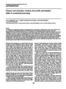
Northern blot analysis of poly(A)+ RNA samples isolated from cDNA probe different rat tissues using a 3zP-labeled gephyrin was performed as described in Experimental Procedures. (A) Hybridization with nick-translated gephyrin cDNA. (B) Hydrization with a nick-translated chick B-actin cDNA. RNA fractions applied were as follows: lane 1, kidney (P40); lane 2, lung (P40); lane 3, liver (P40); lane 4, cerebellum (P16); lane 5, spinal cord (P40); lane 6, spinal cord (P16); lane 7, spinal cord fP3). The positions of size markers are indicated. Note the relatively strong hybridization signal seen in liver, compared with the very faint B-actin band.
evaluation) of the gephyrin found in the cytosolic fraction of the transfected cells cosedimented with microtubules (Figure 6, lanes 2,4, and 5) after initiating their polymerization by GTP and taxol at37°Cunderconditionsdescribedpreviously(Kirsch et al., 1991). Control experiments showed that this could not be attributed to temperature-dependent proteolysis or aggregation of gephyrin (Figure 6, lane 3). Moreover, most of the expressed gephyrin was also precipitated by directly adding polymerized microtubules to a cytosol fraction of the transfected cells (data not shown). We therefore conclude that the pl gephyrin cDNA encodes a protein displaying tubulin binding properties similar to those previously reported for the GlyR-associated gephyrin from spinal cord (Kirsch et al., 1991).
sitometric
Discussion The
biochemically
defined
93 kd GlyR-associated
pro-
tein gephyrin binds with high affinity to tubulin and exhibits properties that qualify it as a novel type of tubulin-binding protein associated with transmembrane receptors (Kirsch et al., 1991). Here, we report the isolation of several gephyrin cDNAs. The identity of the deduced gene product with gephyrin found in affinity-purified GlyR preparations was established by several criteria: eight peptide sequences obtained by microsequencing of the 93 kd protein were all found in the amino acid sequence predicted from our cDNAs; the proteins expressed from pl, p2, and p3 cDNAs were recognized by MAb 5a, an antibody whose specificity for the 93 kd GlyR-associated protein is well documented (Pfeiffer et al., 1984); and heterologously expressed gephyrin bound to microtubules. Analysis of the predicted gephyrin amino acid sequences revealed several structural characteristics of this protein. Its amino acid composition, e.g, the high content in charged, hydroxylated, and proline residues, is unexpected for a peripheral membrane protein, but is similar to that of some structural polypeptides (for discussion see Rienitz et al., 1989). A potential myristylation site is localized at the amino terminus; myristylation at this site after polypeptide processing may contribute to a direct interaction of different gephyrins with the plasma membrane in addition to binding to cytoplasmic receptor domains (Triller et al., 1985; Schmitt et al., 1987). Nineconsensussequencesfor protein kinasecand two potential sites for cyclic nucleotide-dependent phosphorylation are located in the central portion of the polypeptide. Recent data (Langosch et al., 1992) indicate that gephyrin is indeed phosphorylated by an endogenous protein kinase in vitro. This observation is of particular interest since the state of phosphorylation appears to be crucial for the interaction of other microtubule-associated proteins, like MAP2 and tau, with microtubules in living cells (Brugg and Matus, 1991; Lindwall and Cole, 1984). A puzzling feature of the gephyrin sequence is its comparatively high homology with the ChlE gene product of E. coli. As sequence identities are found over the entire length of the ChlE open reading frame, a common evolutionary origin of both proteins must be envisaged. In view of the yet unknown function of ChlE, speculations about the functional significance of this homology are, however, premature. Northern blot hybridization revealed gephyrin transcripts in all rat tissues examined, including spinal cord, brain, liver, kidney, and lung. Thus, gephyrin is specific for neither the ClyR nor the central nervous system. Rather, a more general role of this putative membrane protein-tubulin linker has to be postulated. This interpretation is supported by the discovery of several variants of the gephyrin mRNA. Although the structure of the gephyrin gene remains to be elucidated, the four sequence cassettes identified in various cDNAs may be classified as bona fide alternative splice regions for the following reasons: no
NeUlWl 1166
A ”
x
P
5’ a
ClQ
,...**
”
E
B
PS I I
n Y C4
,..”
3’
I
?2
I
P? PC
I+ W
P5
clI I
l-l ”
200
H
P6
bp
B
bp
bp
f 298 234
f
653
Primary 1167
Structure
and Expression
1
of Cephyrin
2
93kD-W
Figure
5. Expression
of Gephyrin
cDNAs
in 293 Ceils
Cells were transfected with gephyrin cDNAs subcloned into pCIS2, and cytosolic fractions of the transfected cells were analyzed by Western blots with MAb 5a as described in Experimental Procedures. Lane 1, control lane containing affinity-purified GlyR. Lanes 2,3, and 4, cytosolic fractions of 293 cells transfected with gephyrin p2, p3, and pl cDNA, respectively.
consensussplicedonorandacceptorsiteswerefound in the cassette flanking regions of the corresponding cDNAs; PCR amplification with flanking primers failed to generate products on genomic DNA (this implies the existence of intervening sequences between our primer locations); and the remaining nucleotide sequences were identical in all clones analyzed. Thus, the cassette-containing variants do not represent incompletely processed transcripts, but constitute mature mRNAs. Interestingly, all four alternatively spliced cassettes are located in the amino-terminal half of the polypeptide, thus generating a variable region of gephyrin. One might speculate that the constant carboxy-terminal region mediates tubulin binding, whereas the variable part of the protein interacts with specific membrane proteins. Preliminary deletion experiments (J. Kirsch, unpublished data) are
Figure
4. Alternative
Splicing
of Cephyrin
Figure fected
6. Gephyrin Cells
Binds
to Microtubules
in Extracts
of Trans-
The human kidneycell line293was transfected with thegephyrin pl cDNA, and a cytosolic fraction was prepared. From this fraction, microtubules were polymerized by the addition of GTP and taxol as detailed in Experimental Procedures. Aliquots of the different fractions and 50 ng of purified GlyR were separated by SDS-PAGE and analyzed by Western blots using MAb 5a. Lane 1, affinity-purified ClyR. Lanes 2-5: extracts from transfected cell; lane 2, cytosolic fraction; lane 3, cytosolic fraction after incubation at 37°C; lane 4, supernatant of cytosolic fraction after microtubule precipitation at 37OC; lane 5, microtubule precipitate from cytosolic fraction.
consistent with this interpretation. Presently, it is unknown which variant(s) corresponds to ClyR-associated gephyrin. Indeed, none of the cDNAs expressed in 293 cells generated a polypeptide identical in size to the protein found in purified GlyR preparations. This may, however, reflect a lack of covalent modification. Moreover, only 5 (Figure 4A, clones plp5) out of 16 theoretically possible splice combinations have been isolated as cDNAs; it remains to be seen whether other splice variants are also expressed. Gephyrin diversity by alternative splicing may gener-
mRNA
(A) Physical map and alignment of the different spinal cord and brain gephyrin cDNAs. The coding region is represented as a bold line in the physical map. Restriction sites are indicated: B, BamHI; E, EcoRI; P, Pstl; S, Spel; V, EcoRV; X, Xbal. Four regions of variation (open boxes, numbered as cassettes Cl-C4) between the sequence shown in Figure 1 and the other cDNAs isolated are thought to result from alternative splicing of the gephyrin pre-mRNA. Schematic maps of all clones sequenced (pl-~7) are aligned to the combined open reading frame (a shorter version of clone pl also analyzed is not displayed). Deletions are indicated by V-shaped interuptions. Arrows symbolize potential translation start sites, and vertical bars show the positions of stop codons. All predicted translation start sites fulfill the criteria of eukaryotic initiation codons (Kozak, 1989) and are preceded by in-frame upstream stop triplets (not indicated). A dotted line at the 5’ end of clone p2 denotes recombined sequence. (B) cDNA and deduced amino acid sequences of the putative alternative splicing cassettes Cl-C4. Use of cassette Cl alters the amino terminus of gephyrin, whereas the other splice variants (with the exception of clone p5; see text) are contiguous with the open reading frame defined in Figure 1. The flanking residues and the amino acid positions of the gephyrin sequence shown in Figure 1 are also indicated. (C)Amplification of gephyrin variant mRNAs. Poly(A)+ RNA isolated from different adult rat tissues was reverse transcribed into cDNA and amplified by PCR as described in Experimental Procedures. Left, PCR with primer oligonucleotides flanking the first two variant regions (Cl and C2); right, PCR with primer oligonucleotides flanking the third and fourth variant region (C3 and C4). The following RNA fractions were used: lane 1, spinal cord; lane 2, liver; lane 3, lung; lane 4, heart; lane 7, spinal cord; lane 8, liver; lane 9, spinal cord; lane 10, liver; lane 11, lung; lane 12, heart; lane 13, cerebral cortex; lane 14, cerebellum; lane 16, cerebral cortex. Lanes 5 and 15 contain PCR reactions performed on a mixture of the gephyrin pl, p2, p3, and p4, and p5 cDNAs, and lanes 6 and 17 contain DNA size markers. The amplification products shown in lanes 7,8, and 16 were obtained by using B-actin mRNA-specific primers and represent internal controls for the integrity of the RNAs. Sizes of amplification products and marker DNAs are indicated by arrowheads and arrows, respectively.
NWKW
1168
ate tissue-specific sites for different
isoforms receptors
and/or selective and membrane
binding proteins.
Thewidespread distribution of gephyrin transcripts in various tissues of the rat suggests a general role of this protein in many membrane-microtubule interactions. Gephyrin variants may be involved not only in the synaptic organization of neurotransmitter receptors, but also in mediating the anchoring of other membrane proteins in specialized regions of the plasmalemma. Future immunohistochemical studies of cells and tissues expressing gephyrins should help to elucidate the physiological role and functional diversity of this widely expressed protein. Experimental
Procedures
Digestion of Gephyrin and Peptide Microsequencing GlyR was isolated from rat spinal cord by affinity chromatography according to Pfeiffer et al. (1982). Purified receptor (~500 kg) was electrophoresed in a preparative 10% SDS-polyacrylamide gel, and the 93 kd gephyrin band was isolated. After digestion with trypsin or S. aureus V8 protease, peptides were dissolved in70% (v/v)formicacidandpurified byHPLCasdescribed(Leube et al., 1988). A nucleosil C8 RP column was used for separation of tryptic peptides, and a nucleosil C4 RP column for that of V8 peptides. Peptide-containing peaks were processed and subjected to gas-phase microsequencing as described previously (Grenningloh et al., 1990). Isolation of cDNAs A 32.mer mixed oligonucleotide (Nut 93132: S’-AAGGTGGT(A/ C)CCCAG(G/T)CC(A/G)CGClTCATCAACAC-3’) corresponding to amino acids I-II of the tryptic peptide VFMKPCLPTTFATLDIDGVR, a 17-mer oligonucleotide (Nut 93/17: 5’-CCXGG(T/ C)TTCAT(A/C)AAXAC-3’) corresponding to amino acids l-6, and two 14mer oligonucleotides corresponding to amino acids 8-12 (Nut 93/14A: 5’CC(A/C)AAXGTXCTXCG-3’) and amino acids 1519 (Nut 93/14B: 5’-ACXCC(A/G)TC(A/C/T)AT(G/A)TC-3’) of the same peptide were synthesized by phosphoramidate chemistry (X stands for all four nucleotides). A rat spinal cord hgtl0 cDNA library (1.2 x IO6 pfu; see Grenningloh et al., 1987) was screened with the32P-labeled oligonucleotides. Hybridization with Nut 931 32 was performed at 37OC in 5x SET (1 x SET = 150 mM NaCI, 1 mM EDTA, 30 mM Tris-HCI [pH S.O]), 0.1% (w/v) SDS, 0.1% (wi v) sodium pyrophosphate, 10x Denhardt’s solution, 100 ug/ml denatured herring sperm DNA, 20% (w/v) formamide, and filters were washed in 2x SET, 0.1% (w/v) SDS, 0.1% sodium pyrophosphate at 52OC (three 15 min washes). The 17.mer oligonucleotide was hybridized at 42’C and washed at 48OC, and the ICmer oligonucleotides were hybridized at 37OC and washed at 42’C in tetramethylammonium chloride buffer as described (Crenningloh et al., 1987). After purification and subcloning into M13, pSPT18 or Bluescript vectors, the DNA sequence of an ml.6 kb cDNA insert hybridizing to all four oligonucleotides was determined by the chain termination method (Sanger et al., 1977). Since the corresponding clone (named ~7) contained only the 3 coding part and 3’untranslated sequenceof the gephyrin cDNA, several additional screening procedures were performed. Basically, a 5’cDNA fragment and an oligonucleotide corresponding tothevery5’endofthisfragmentwereused repeatedlyasprobes for sequential screening of the following cDNAs libraries: hgtl0 rat brain (Ymeret al., 1989); ilgtl0 rat brain stem (Clontech); hgtll rat brain stem (Clontech); hZAP rat brain (Werner etal., 1991); and ZAP neonatal rat spinal cord (Kuhseet al., 1991). This resulted in the isolation of six additional gephyrin cDNAs (pl-~6). Homology Searches and Sequence Comparison Homology searches against GenBank (release 70.0), EMBL (release 29.01, SwissProt (release 20.0), and PIR-Protein (release 31.0) data bases of either nucleotide or deduced amino acid se-
quences were done using the FASTA algorithm (Pearson and Lipman, 1988). Sequence comparison between gephyrin and the ChlE protein and randomized sequences thereof were performed with the GAP program based on the method of Needleman and Wunsch (1970). The HUSAR program package of the Deutsche Krebsforschungszentrum (Heidelberg), which includes the University of Wisconsin Genetics Computer Group software, was employed for computer-assisted analysis. Northern Blot Analysis Poly(A)+ RNA from different rat tissues was extracted according to Cathala et al. (1983), purified by oligo(dT) chromatography (Aviv and Leder, 1972), and electrophoresed (13-15 up per lane) on a 1% agarose gel containing 6% (w/v) formaldehyde. Following transfer to Hybond N (Amersham), the filter was hybridized in 5x SET, 0.1% (w/v) SDS, 0.1% (w/v) sodium pyrophosphate, 10x Denhardt’s solution, 100 pglml denatured herring sperm DNA, 50% (v/v) formamide at 42OC to the 5’ EcoRl fragment of clone p5 labeled by nick translation (specific activity = l x IO8 cpm/ug). In other experiments, the 3*P-labeled clone p7was also hybridized. After washing in 0.1% (w/v) SDS, 0.1% sodium pyrophosphate containing 2x SET, 1 x SET, and 0.2x SET, respectively, at 6S°C, the blot was exposed to Kodak X-Omat film for 1 week. To examine the integrity of the RNA samples, the blot was rehybridized to a nick-translated chick B-actin cDNA under the same conditions (Grenningloh et al., 1990). Relative hybridization signals were estimated by scanning the autoradiograms with a Hirschmann Elscript 400 densitometer. PCR Amplification PCR amplification (Saiki et al., 1988) of gephyrin transcripts was performed after first strand cDNA synthesis (First Strand Synthesis Kit, Stratagene) with different poly(A)+ RNA preparations by using unique oligonucleotide primers corresponding to nucleotides surrounding the sequence region of interest. Foramplification of transcripts differing in the first variant region (Cl and 62 cassettes), oligonucleotides corresponding to nucleotide positions 315-334 (sense) and 552-577 (antisense) were used as primers, and for amplification of gephyrin transcripts differing in the C3 and C4 region, oligonucleotides corresponding to position 615-634 (sense) and 1204-1223 (antisense) of the gephyrin cDNA wereused.Annealingwasdoneforl minat61°Cor580C,elongation for 1 min at 72OC, and denaturation for 1 min at 94%. The reaction was performed with 2.5 U of AmpliTaq polymerase (Perkin Elmer Cetus) for 33 cycles. Amplification products and DNA size marker VI (Boehringer) were separated in 2% or 1.5% agarose gels and stained with ethidium bromide. In some experiments, their identitywith gephyrin sequences was confirmed by Southern hybridization with the 5’ EcoRl fragments of clones pl and p5. Heterologous Expression The human embryonic kidney cell line 293 (ATCC #CRC 1573) was transfected with the gephyrin pl, p2, and p3 DNAs inserted in the eukaryotic expression vector pCIS2 as described (Puia et al., 1990). (To provide identical upstream sequences, the p2 and p3 cDNAs were digested with Bglll and their 5’ regions were exchanged with the 5’ Bgll I fragment of cione pl.) Two days after transfection, the cells were harvested, washed in 150 mM NaCI, 5 mM KCI, 0.1 mM CaC12, 3 mM NaHCOI, 5 mM glucose, and homogenized in 2 ml of homogenization buffer (200 mM mannose, 70 mM sucrose, 4 mM HEPES-NaOH [pH 7.51‘1 mM EDTA, and protease inhibitors as detailed by Becker et a!. [l988]). Nuclei and cell debris were removed by centrifugation (1000 x g for 3 min), and a cytosolic fraction was separated from membranes by subsequent centrifugation at 10,000 x g for 15 min. Western Blot Analysis Cytosolicand membranefractionsofcelis transfected with gephyrin cDNA (100 pg of protein per lane) or affinity-purified ClyR (~100 ng per lane) were subjected to 7.5% SDS-PAGE (Laemmli, 1970). Proteins were blotted onto nitrocellulose (Kyhse-Andersen, 1984) and stained after transfer with Ponceau S. Immunode-
Primary 1169
Structure
and Expression
of Gephyrin
tection scribed
with MAb 5a (Pfeiffer et al., 1984) was performed (Schmitt et al., 1987; Kirsch et al., 1991).
as de-
Coprecipitation of Gephyrin with Microtubules of Transfected Cells From the cytosolic fraction of cells transfected with pl DNA, microtubules were isolated by polymerization induced by the addition of GTP (Boehringer) and taxol (National Cancer Institute) at final concentrations of 2 mM and 20 PM, respectively. After 10 min at 37”C, the newly formed microtubules were collected by centrifugation at 20,000 x g for 10 min through a 5% sucrose cushion. Cephyrin contents of the different samples were revealed by SDS-PAGE followed by Western blotting as described above.
Mountcastle, eds. (Bethesda, Society), pp. 261-294.
Maryland:
American
Kirsch, J., Langosch, D., Prior, P., Littauer, U. Z., Schmitt, Betz, H. (1991). The 93 kDa glycine receptor-associated binds to tubulin. J. Biol. Chem. 266, 22242-22245. Kozak, M. (1989). Context effects and inefficient AUG codons in eucaryoticcell-freetranslation Biol. 9, 5073-5080.
Kyhse-Andersen, them. Biophys.
J. (1984). Electroblotting Meth. 70,203-209.
We thank H. Krischkeand C. Udri for expert technical assistance, H. Rohrer, A. Ultsch, and E. Sawruk for critical reading of the manuscript, and S. Wartha for help with its preparation. This work was supported by the Deutsche Forschungsgemeinschaft (Leibniz-Programm and Schwerpunkt Funktionelle Domlnen), the Bundesministerium fur Forschung und Technologie (BCT 365/l), and Fonds der Chemischen Industrie.
Laemmli, assembly
December
31, 1991; revised
April
6, 1992.
of structural T4. Nature
Altschuler, R. A., Betz, H., Parakkal, M. H., Reeks, K. A., and Wenthold, R. J. (1986). Identification of glycinergic synapses in the cochlear nucleus through immunocytochemical localization of the postsynaptic receptor. Brain Res. 369, 316-320. Aviv, H., and Leder, P. (1972). Purification of biologically active globin messenger RNA by chromatography on oligothymidylic acid-cellulose. Proc. Natl. Acad. Sci. USA 69, 1408-1412.
Brugg, B., and Matus, A. (1991). Phosphorylation determines the binding of microtubule-associated protein 2 (MAP2) to microtubules in living cells. J. Cell Biol. 174, 735-743. Cathala, G., Savouret, J.-F., Mendex, B., West, B. L., Karin, M., Martial, J. A., and Baxter, J. D. (1983). A method for isolation of intact, translationally active ribonucleic acid. DNA 2, 329-335. of protein
half-lives
E., Komaraomy, M., and Wall, and surface protein sequences plot. J. Mol. Biol. 779, 125-142.
FEBS Lett. 283, gels. J. Bio-
for displaying the Biol. 757, 105-132. proteins during 227, 680-685.
the
Langosch, D., Hoch, W., and Betz, H. (1992). The 93 kDa protein gephyrin and tubulin associated with the inhibitory glycine receptor are phosphorylated by an endogenous protein kinase. FEBS Lett. 298, 113-117. probes deduced from and practical considera-
Leube, R. E., Kaiser, P., Seiter,A., Zimbelmann, R., Franke, W. W., Rehm, H., Knaus, P., Prior, P., Betz, H., Reinke, H., Beyreuther, K., and Wiedenmann, B. (1988). Synaptophysin: molecularorganization and mRNA expression as determined from cloned cDNA. EMBO J. 77, 3261-3268. Lindwall, G., and Cole, R. D. (1984). Phosphorylation affects the ability of tau protein to promote microtubule assembly. J. Biol. Chem. 259, 5301-5305.
Becker, C.-M., Hoch, W., and Betz, H. (1988). Clycine receptor heterogeneity in rat spinal cord during postnatal development. EMBO J., 7, 3717-3726.
Dice, J. F. (1987). Molecular determinants eukaryotic cells. FASEB J. 7, 349-357.
V.,
Langosch, D., Becker, C.-M., and Betz, H. (1990). The inhibitory glycine receptor: a ligand-gated chloride channel of the central nervous system. Eur. J. Biochem. 794, l-8.
Lathe, R. (1985). Synthetic oligonucleotide amino acid sequence data. Theoretical tions. J. Mol. Biol. 783, 1-12.
References
Eisenberg, D., Schwarz, Analysis of membrane hydrophobic moment
U. (1970). Cleavage of the bacteriophage
Schmieden,
of multiple
Kyte, J., and Dolittle, R. F. (1982). A method hydrophobic character of a protein. J. Mol.
B., and protein
initiation at nonsystems. Mol. Cell
Kuhse,J., Kuryatov,A., Maulet, Y., Malosio, M.-L., and Betz, H. (1991).Alternativesplicinggeneratestwoisoformsof the a2 subunit of the inhibitory glycine receptors. 73-77.
Acknowledgments
Received
Physiological
in
R. (1984). with the
Froehner, S. (1991). The submembrane machinery for nicotinic acetylcholine receptor clustering. J. Cell Biol. 774, l-7. Corman, C. M., Cies, D., McCray, C., and Huang, M. (1989). Products of adenovirus early regions, Ela and Elb, increase expression from the human cytomegalovirus major intermediate early promoter. Virology 777, 377-385. Graham, D., Pfeiffer, F., Simler, R., and Betz, H. (1985). Purification and characterization of the glycine receptor of pig spinal cord. Biochemistry 24, 990-994. Grenningloh, C., Rienitz,A., Schmitt, B., Methfessel, C., Zensen, M., Beyreuther, K., Cundelfinger, E. D., and Betz, H. (1987). The strychnine-binding subunit of the glycine receptor shows homology with nicotinic acetylcholine receptors. Nature 328,215220. Crenningloh, G., Pribilla, I., Prior, P., Multhaup, G., Beyreuther, K., Taleb, O., and Betz, H. (1990). Cloning and expression of the 58 kd B subunit of the inhibitory glycine receptor. Neuron 4,963970. Heuser, J. E., and Reese, T. S. (1977). Structure of the synapse. In Handbook of Physiology, Vol. I, J. M. Brookhart and V. M.
Naas, E., Zilles, K., Cnahn, Schroder, H. (1991). Clycine and human cerebral cortex.
H., Betz, H., Becker, C.-M., and receptor immunoreactivity in rat Brain Res. 567, 139-146.
Needleman, S. B., and Wunsch, C. D. (1970). A general applicable to the search for similarities in the amino quence of two proteins. J. Mol. Biol. 48, 443-453.
method acid se-
Nitkin, R. M., Smith, M. A., Magill, C., Fallon, J. R., Yao, Y. M., Wallace, B. C., and McMahan, U. 1. (1987). Identification of agrin, a synapticorganizing protein from Torpedoelectricorgan. J. Cell Biol. 705, 2471-2478. Nohno, T., Kasai, Y., and Saito,T. (1988). Cloning and sequencing of the E. coli ChlE operon involved in molybdopterin biosynthesis. J. Bacterial. 770, 4097-4102. Olek,A. J., Pudimat, P. A.,and Daniels, M. P. (1983). Directobservation of the rapid aggregation of acetylcholine receptors on identified cultured myotubes after exposure to embryonic brain extract. Cell 34, 255-264. Pearson, W. R., and Lipman, logical sequence comparison. 24442448.
D. J. (1988). Improved Proc. Natl. Acad.
Peters, A., Palay, S. L., and Webster, ofthe Nervous System: The Neurons delphia, Pennsylvania: Saunders). Pfeiffer, F., Graham, D., and Betz, ity chromatography of the glycine Biol. Chem. 257, 9389-9393. Pfeiffer, F., Simler, R., Monoclonal antibodies similarities between the spinal cord. Proc. Natl.
tools for bioSci. USA 85,
H. de F. (1976). Fine Structure and Supporting Cells (PhilaH. (1982). receptor
Purification by affinof rat spinal cord. J.
Grenningloh, G., and Betz, H. (1984). and peptide mapping reveal structural subunits of the glycine receptor of rat Acad. Sci. USA 81, 7224-7227.
Puia, G., Santi, M., Vicini, S., Pritchett, D. B., Purdy, R. H., Paul, S. M., Seeburg, P. H., and Costa, E. (1990). Neurosteroids act on recombinant human CABAA receptors. Neuron 4, 759-765.
NWNXl 1170
Rienitz, A., Grenningloh, G., Hermans-Borgmeyer, I., Kirsch, Littauer, U. Z., Cundelfinger, E. D., Schmitt, B., and Betz, (1989). Neuraxin, a novel putative structural protein of the central nervous system that is immunologically related microtubule-associated protein 5. EMBO J. 8, 2879-2888.
J., H. rat to
Saiki, R. K., Gelfand, D. H., Stoffel, S., Scharf, S. J., Higuchi, R., Horn, G. T., Mullis, K. B., and Erlich, H. A. (1988). Primer-directed enzymatic amplification of DNA with a thermostable DNA polymerase. Science 239,487-491. Sanger, F., Nicklen, S., and Coulson, ing with chain-terminating inhibitors. 74, 5463-5467.
A. R. (1977). DNA sequencProc. Natl. Acad. Sci. USA
Schmitt, B., Knaus, P., Becker, C.-M., and Betz, H. (1987). M, 93,000 polypeptide of the postsynaptic glycine receptor peripheral membrane protein. Biochemistry 26, 8055811.
The is a
Triller, A., Cluzeaud, F., Pfeiffer, F., Betz, H., and Korn, H. (1985). Distribution of glycine receptors at central synapses: an immunoelectron microscopy study. J. Cell Biol. 701, 683-688. Triller, butyric ceptors
A., Cluzeaud, F., and Korn, H. (1987). Gamma-aminoacid-containing terminals can be apposed to glycine at central synapses. J. Cell Biol. 704, 947-956.
re-
Triller, A., Seitanidou, T., Franksson, O., and Korn, H. (1990). Size and shapeof glycine receptor clusters in a central neuron exhibit a somato-dendritic gradient. New Biol. 2, 637-641. Werner, W., Voigt, M., Keinanen, K., Wisden, W., and Seeburg, P. H. (1991). Cloning of a putative high-affinity kainate receptor expressed predominantly in hippocampal CA3 cells. Nature 357, 742-744. Ymer, S., Schofield, P. R., Draguhn, A., Werner, P., Kohler, M., and Seeburg, P. H. (1989). CABA, receptor B subunit heterogeneity: functional expression of cloned cDNAs. EMBO J. 8, 16651670. EMBUGenBank
Accession
The EMBUGenBank ported in this paper
Number
accession is X66366.
number
for
the
sequence
re-