966
IEEE TRANSACTIONS ON BIOMEDICAL ENGINEERING, VOL. 51, NO. 6, JUNE 2004
Principles of a Brain-Computer Interface (BCI) Based on Real-Time Functional Magnetic Resonance Imaging (fMRI) Nikolaus Weiskopf*, Klaus Mathiak, Simon W. Bock, Frank Scharnowski, Ralf Veit, Wolfgang Grodd, Rainer Goebel, and Niels Birbaumer
Abstract—A brain-computer interface (BCI) based on functional magnetic resonance imaging (fMRI) records noninvasively activity of the entire brain with a high spatial resolution. We present a fMRI-based BCI which performs data processing and feedback of the hemodynamic brain activity within 1.3 s. Using this technique, differential feedback and self-regulation is feasible as exemplified by the supplementary motor area (SMA) and parahippocampal place area (PPA). Technical and experimental aspects are discussed with respect to neurofeedback. The methodology now allows for studying behavioral effects and strategies of local self-regulation in healthy and diseased subjects. Index Terms—Brain-computer interface, blood oxygen level-dependent, neurofeedback, self-regulation.
I. INTRODUCTION
B
RAIN-COMPUTER interfaces based on electroencephalography (EEG) have been used to train voluntary regulation of electrical brain activity [1], [2]. Specific behavioral effects dependent upon the functional role of the Manuscript received July 11, 2003; revised February 6, 2004. This work was supported in part by the WIN-Kolleg of the Heidelberg Academy of Sciences and Humanities (Germany), in part by the Deutsche Forschungsgemeinschaft (DFG) under Grant SFB 550/B5+B1 and Grant Th 812/1-1, and in part by the National Institute of Health (NIH). Asterisk indicates corresponding author. *N. Weiskopf is with the Institute of Medical Psychology and Behavioral Neurobiology, University of Tübingen, Tübingen, Germany and also with the Section of Experimental MR of the CNS, Department of Neuroradiology, University of Tübingen, Tübingen, Germany (e-mail:
[email protected]). K. Mathiak is with the Center for Neurology, University of Tübingen, Tübingen, Germany (e-mail:
[email protected]). S. W. Bock is enrolled in the Graduate School of Neural and Behavioral Sciences and the International Max Planck Research School, University of Tübingen, Tübingen, Germany (e-mail:
[email protected]). F. Scharnowski is enrolled in the Graduate School of Neural and Behavioral Sciences and the International Max Planck Research School, University of Tübingen, Tübingen, Germany (e-mail:
[email protected]). R. Veit is with the Institute of Medical Psychology and Behavioral Neurobiology, University of Tübingen, Tübingen, Germany (e-mail:
[email protected]). W. Grodd is with the Section of Experimental MR of the CNS, University of Tübingen, Tübingen, Germany (e-mail:
[email protected]). R. Goebel is with the Department of Cognitive Neuroscience, Faculty of Psychology, University of Maastricht, Maastricht, The Netherlands (e-mail:
[email protected]). N. Birbaumer is with the Institute of Medical Psychology and Behavioral Neurobiology, University of Tübingen, Tübingen, Germany, and also with the Center for Cognitive Neuroscience, University of Trento, Trento, Italy (e-mail:
[email protected]). Digital Object Identifier 10.1109/TBME.2004.827063
regulated brain area have been shown [2]. However, the regional specificity of the feedback is restricted by the relatively low spatial resolution of the EEG. In contrast, functional magnetic resonance imaging (fMRI) measures the blood oxygen level-dependent (BOLD) signal with high spatial resolution and can access the whole brain (for a review cf. [3]). Electric brain activity as measured by EEG and the BOLD signal seem to be highly correlated (e.g., [3]). In order to increase the spatial specificity of neurofeedback training, different studies investigated the feasibility to use fMRI for operant training of the BOLD-response with feedback of the BOLD signal as reward. Two previous studies applied delayed feedback (by approx. 60 s) of BOLD-responses by visual feedback of functional brain activation maps [4] or verbal ratings of the experimenter based on the local BOLD signal [5]. In two other studies, immediate feedback of local BOLD signals was provided to further facilitate learning [6], [7]. We will discuss the technical and experimental basis of a brain-computer interface (BCI) based on real-time fMRI and present an implementation for neurofeedback. II. TECHNICAL AND EXPERIMENTAL ASPECTS In this section, we consider the demands on the fMRI-BCI as concerns speed, signal-to-noise ratio (SNR), artifact suppression, and usability of the feedback signal. A. Temporal Properties of BOLD-Response Feedback It has been shown that self-regulation of electric brain activity is facilitated by minimum delays between physiologic activity and feedback [2]. However, the BOLD signal reflects vascular effects and only indirectly the neuronal signal. The hemodynamic response function (see inset in Fig. 1) introduces a physiological delay of 3 to 6 s before signal changes can be observed. Finally, the cognitive processing of the feedback signal introduces an additional delay into the regulatory loop. We suggest to reduce the data, e.g., from an appropriate combination of regions of interest (ROI’s), to one dimension for easy and unequivocal design of a fMRI-BCI and to facilitate cognitive processing (see “BCI” in Fig. 1). B. Signal- and Contrast-to-Noise Ratio 1) Field Strength: Contrast-to-noise ratio (CNR), i.e., the sensitivity to the BOLD effect, and SNR determine the reliability of the feedback signal and indirectly how fast changes in
0018-9294/04$20.00 © 2004 IEEE
WEISKOPF et al.: PRINCIPLES OF A BCI BASED ON REAL-TIME fMRI
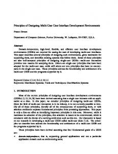
Fig. 1. Setup and data-flow of the fMRI brain-computer interface. Local brain activity is measured by fMRI using the blood oxygen level-dependent (BOLD) effect which resembles the neurovascular response to electric brain activity (red curve superimposed over the rendered brain). The measured hemodynamic response is delayed by approx. 3–6 s from the neuronal activity. Images are reconstructed, undistorted, and averaged on the “Trio” scanner console. Turbo-BrainVoyager retrieves the data via local area network (LAN), and performs data preprocessing (including 3D motion correction) and statistical analysis. The signal time-series of interactively selectable regions of interest are exported via LAN to the custom-made visualization software “BCI” which provides feedback to the subject using video projection. Feedback is presented with a delay of less than 1.3 s from time of image acquisition.
the feedback signal can be observed. At higher field strengths, the magnetization of the probe increases, and, thus, the MR signal and SNR might increase linearly with magnetic field strength. However, higher field strengths lead to more distortions and signal dropouts in the image due to larger frequency offsets in the object. Moreover, a shorter apparent transversal and a longer longitudinal relaxation time relaxation time ameliorate the theoretical gain as well as safety problems such as radio frequency (RF) deposition. In general, the range of 3–4 T seems to be an appropriate compromise for BOLD sensitive imaging [8]. 2) Single-Shot Multi-Echo Acquisition: The BOLD signal depends upon the echo time (TE). For the apparent transversal relaxation time , the expected contrast is proportional to and maximal for TE close to [9]. The sampling of multiple images after a single RF excitation pulse and their appropriate combination, thus, increases CNR without the disadvantageous effects of increased readout bandwidth [10]. C. Real-Time Artifact Control 1) Head Motion: Even the movement of a minor fraction of a resolution element, i.e., below 1 mm, can induce signal changes comparable to the one achieved by maximal brain activation. Efficient real-time motion correction should be robust and should not add significant noise and artifact sources [11]. Mathiak et al. [11] showed that at least 3 slices should be acquired in order to reduce movement noise to less than 1% of the voxel size in case of a 64 64 image matrix. 2) Image Distortions: Single-shot image encoding as used for echo-planar imaging (EPI), i.e., one RF excitation pulse per image, suffers from significant image distortions in case of off-resonances caused by inhomogeneities of the static
967
magnetic field which can be observed close to air-tissue interfaces. These distortions depend upon motion and, thus, could simulate BOLD signal changes by head motions which cannot be corrected by motion correction. We recently developed a multi-echo EPI technique that dynamically reduces image distortions in a single-shot [12]. This technique is particularly useful for real-time and feedback application as the correction is immediate and does not depend on previous (time demanding) reference scans (e.g., [13]). 3) Signal Dropout: Inhomogeneities of the magnetic field may also cause signal dropouts and lead to a reduced BOLD sensitivity [9]. It is possible to compensate these effects partially by adapted MR pulse sequences [9], but, in general, speed or SNR is traded for signal recovery. We focused on areas that were unaffected by large signal dropouts such as supplementary motor area (SMA) and parahippocampal place area (PPA).
III. PROCEDURE A. Participants and Experimental Paradigms Four volunteers (3 male, age 27–30 yr.) were trained to differentially control the BOLD response of the supplementary motor area (SMA) and the parahippocampal place area (PPA) using the fMRI-BCI. Three of the subjects were members of the research group and experienced in fMRI and neurofeedback experiments. Subject 2 was naïve with respect to fMRI and the task. Written informed consent was obtained prior to the study in accordance with the local ethics committee. Each experiment began with two functional localizer sessions followed by feedback sessions. The first localizer session was run to delineate the SMA. It consisted of five baseline blocks separated by three blocks of bimanual finger tapping (50 s each; e.g., [14]). The second localizer session determined the location of the PPA and the fusiform face area (FFA). During PPA blocks outdoor scenes with houses, and during FFA blocks human faces were presented to the subject [15]. Beginning and ending with a baseline block, three PPA blocks alternated with three FFA blocks (37.5 s each) and were separated by baseline blocks (25 s each). During the baseline blocks, the subject counted from 100 downwards. During feedback sessions, the difference of the mean BOLD signal of two regions of interest (ROI) approximating SMA and PPA was presented to the subject by video projection [Fig. 2(b)]. Each rectangular ROI [Fig. 2(a)] was based on the preceding functional localizers and enclosed in average about 32 voxels for the SMA and 29 voxels for the PPA. During up-regulation blocks, the subjects should raise the curve, and during downregulation blocks, they had to lower the curve. Three up-regulation blocks alternated with three down-regulation blocks (50 s each) and were separated by baseline blocks [31 s; see also Fig. 2(b)]. Subjects counted down from 100 during baseline blocks in order to suppress rehearsal of mental strategies. In subjects 1 and 2, sessions were acquired on two consecutive days (3 and 4 feedback sessions on the first day, respectively). Subjects 2 and 3 saw inverted curves to rule out effects of the direction. The participants should use an individual strategy to control the differential BOLD signal. However, visual imagery (e.g.,
968
IEEE TRANSACTIONS ON BIOMEDICAL ENGINEERING, VOL. 51, NO. 6, JUNE 2004
Fig. 3. Differential regulation of activity in supplementary motor area (SMA) and parahippocampal place area (PPA) across feedback sessions. In all subjects significant task correlated changes in the differential feedback signal were observed (t-value >1:63 is equivalent to p < 0:05). In subjects 1 and 2, the effect increased across feedback sessions (p < 0:05; one-sided Pearson).
Fig. 2. On-line statistical analysis and neurofeedback. (a) Display presented to the experimenter. On the left panel statistical maps were superimposed over one oblique echo-planar image. Green spots indicated areas which were activated during up-regulation blocks, and blue spots indicated areas which were activated during down-regulation blocks. Regions of interest were marked as rectangles which approximated the supplementary motor area (red), and parahippocampal place area (green). On the right upper and middle panel time courses of SMA and PPA, respectively, were plotted as white curves superimposed over the experimental block design which was represented by grey (baseline), green (up-regulation), and blue (down-regulation) stripes. The right lower panel displayed the estimated and corrected head motion. Statistical maps and time courses were updated continuously within 1.3 s after image acquisition. (b) Feedback screen presented to the subject. The differential BOLD signal was presented as a constantly updated yellow curve on a color-coded background (signal of SMA minus PPA). The tasks were presented as colored stripes: grey indicated the baseline, during green blocks subjects had to raise the curve (up-regulation), and during blue blocks subjects had to decrease the curve (down-regulation). The red curve is the low-pass filtered (Gaussian FWHM = 31 stime points) time-series. Arrows and filtered curve were not presented during on-line feedback. Mean and standard deviation (std)were estimated based on the data of the first baseline block.
pictures of buildings; [15]), spatial navigation (e.g., routing, orienteering) or motor imagery (e.g., fist clenching, dancing; [14]) served as a general starting point. Moreover, subjects were instructed that the feedback signal was delayed by approx. 6 s and that they should not move any body parts and should breathe regularly.
were recorded with alternating phase encoding direction, corrected for geometric distortions [12] and averaged to improve BOLD sensitivity in real-time [10], [12]. The first ten volumes equilibration of each session were discarded to suppress effects. To improve BOLD sensitivity, the experiments were performed at a high static magnetic field (3 T) on a whole body scanner equipped with a volume head coil (Magnetom Trio, Siemens, Erlangen, Germany). C. On-Line Statistical Analysis and Visual Feedback Statistical analysis was performed by Turbo-BrainVoyager (Brain Innovation, Maastricht, The Netherlands; [16]) on a separate personal computer retrieving the image files as soon as they were created by the image reconstruction system via local area network (LAN). The program performed data preprocessing, statistical analysis, and export of ROI time-courses to hard disk in real-time [Fig. 2(a)]. Preprocessing of the data included incremental linear detrending of the time-series and 3D motion correction. Statistical analysis based on a general linear model (GLM) was performed cumulatively using the recursive least squares regression algorithm. For separate treatment of visual feedback of brain activity from statistical analysis we developed custom-made software (“BCI”; Fig. 1) based on Matlab 6.5 (The MathWorks, Natick, MA). This visualization software was run on a separate personal computer and generated configuration files for the Turbo-BrainVoyager which included information about the paradigm and imaging parameters. During the fMRI session, it accessed the ROI time courses provided by the Turbo-BrainVoyager via LAN and displayed the feedback curve [Fig. 2(b)].
B. fMRI Data Acquisition and Image Reconstruction For all functional scans, 10 oblique transversal-coronal slices were acquired using a single-shot multi-echo EPI s, (echo-planar imaging) sequence (repetition time , effective echo times ms, flip angle , voxel mm matrix mm, mm, partial fourier phase encoding, kHz/pixel). The echoes
IV. RESULTS AND DISCUSSION All subjects achieved significant differential BOLD amplitudes between SMA and PPA (Fig. 3) as determined by off-line ROI analysis [6]. This analysis of the differential feedback signal was performed with custom-made software based on SPM99 (Wellcome Department of Imaging Neuroscience, Queens Square, London, U.K.); time-series were
WEISKOPF et al.: PRINCIPLES OF A BCI BASED ON REAL-TIME fMRI
high-pass ( s) and low-pass filtered s), and motion parameters (3 translation, 3 ro( tation) were included into the GLM [17]. In subjects 1 and 2, control of the feedback signal increased significantly across ; one-sided Pearson; Fig. 3). training sessions ( Taken together, the present study showed the feasibility of on-line differential feedback and regulation of local BOLD activity using a fMRI-BCI. However, potential learning effects have to be further assessed in a larger sample size across more sessions to allow for conclusions on the efficiency of this neurofeedback approach. V. OUTLOOK Several extensions of the current technical and experimental approach are possible. Recently, fast imaging sequences reducing signal dropouts in areas with large magnetic field inhomogeneities became available [5], [9]. Thus, activity of brain regions such as amygdala and orbitofrontal cortex can be measured. Moreover, prospective motion correction techniques can further reduce artifactual feedback signals by continuously tracking the brain with the measured volume [18]. Real-time cortex-based statistical analysis focuses on the grey matter of the cortex with high spatial precision and will possibly improve the spatial specificity and sensitivity even further [16]. Subcortical and orbitofrontal structures which are involved in emotional regulation could be a target for a fMRI based neurofeedback training. In anxiety disorders or other mood disorders, dysregulation in areas not accessible to other neurofeedback techniques such as EEG-BCI were reported. The training itself might be integrated in a broader therapeutical approach. Moreover, the methodology can be used to study and presumably improve the self-regulation of brain potentials, such as slow cortical potentials (SCP), which are successfully used by patients to communicate [1]. Feedback of specific areas of the thalamus and basal ganglia may improve the control of SCP [19]. Moreover, spatially specific feedback might reveal optimized strategies in the individual and in general for SCP self-regulation. In general, subjects might be trained by fMRI feedback to control local brain activity. This offers the opportunity to study behavior, e.g., mental strategies, physiological measures, affective state, dependent on self-regulated activity in circumscribed brain regions. This approach adds to the current neuroimaging methodology which usually measures brain activity dependent on behavior. ACKNOWLEDGMENT The authors would like to thank M. Erb, B. Kardatzki, and U. Klose for technical support and helpful discussions, and B. Newport for technical assistance. They would also like to thank Siemens Medical (Erlangen, Germany) and especially S. Thesen for their support with sequence programming. REFERENCES [1] N. Birbaumer, N. Ghanayim, T. Hinterberger, I. Iversen, B. Kotchoubey, A. Kubler, J. Perelmouter, E. Taub, and H. Flor, “A spelling device for the paralyzed,” Nature, vol. 398, pp. 297–298, 1999.
969
[2] B. Rockstroh, T. Elbert, N. Birbaumer, and W. Lutzenberger, “Biofeedback-produced hemispheric asymmetry of slow cortical potentials and its behavioral effects,” Int. J. Psychophysiol., vol. 9, pp. 151–165, 1990. [3] N. K. Logothetis, “MR-imaging in the nonhuman primate: Studies of function and of dynamic connectivity,” Curr. Opinion Neurobiol., vol. 13, pp. 630–642, 2003. [4] S. S. Yoo and F. A. Jolesz, “Functional MRI for neurofeedback: Feasibility study on a hand motor task,” NeuroReport, vol. 13, pp. 1377–1381, 2002. [5] S. Posse, D. Fitzgerald, K. Gao, U. Habel, D. Rosenberg, G. J. Moore, and F. Schneider, “Real-time fMRI of temporolimbic regions detects amygdala activation during single-trial self-induced sadness,” NeuroImage, vol. 18, pp. 760–768, 2003. [6] N. Weiskopf, R. Veit, M. Erb, K. Mathiak, W. Grodd, R. Goebel, and N. Birbaumer, “Physiological self-regulation of regional brain activity using real-time functional magnetic resonance imaging (fMRI): Methodology and exemplary data,” NeuroImage, vol. 19, pp. 577–586, 2003. [7] R. C. deCharms, K. Christoff, G. H. Glover, J. M. Pauly, S. Whitfield, and J. D. E. Gabrieli, “Learned regulation of spatially localized brain activation using real-time fMRI,” NeuroImage, vol. 21, pp. 436–443, 2004. [8] G. Kruger, A. Kastrup, and G. H. Glover, “Neuroimaging at 1.5 T and 3.0 T: Comparison of oxygenation-sensitive magnetic resonance imaging,” Magn. Reson. Med., vol. 45, pp. 595–604, 2001. [9] R. Deichmann, O. Josephs, C. Hutton, D. R. Corfield, and R. Turner, “Compensation of susceptibility-induced BOLD sensitivity losses in echo-planar fMRI imaging,” NeuroImage, vol. 15, pp. 120–135, 2002. [10] K. Mathiak, A. Rapp, T. T. J. Kircher, W. Grodd, I. Hertrich, N. Weiskopf, W. Lutzenberger, and H. Ackermann, “Mismatch responses to randomized gradient switching noise as reflected by fMRI and whole-head magnetoencephalography,” Human Brain Map., vol. 16, pp. 190–195, 2002. [11] K. Mathiak and S. Posse, “Evaluation of motion and realignment for functional magnetic resonance imaging in real time,” Magn. Reson. Med., vol. 45, pp. 167–171, 2001. [12] N. Weiskopf, U. Klose, M. Erb, W. Grodd, N. Birbaumer, and K. Mathiak. Distortion correction in fMRI using single-shot multi-echo EPI. presented at 9th Int. Conf. Functional Mapping of the Human Brain. [CD-ROM] [abstract]. Available in NeuroImage [13] P. Jezzard and R. S. Balaban, “Correction for geometric distortion in echo planar images from B0 field variations,” Magn. Reson. Med., vol. 34, pp. 65–73, 1995. [14] M. Lotze, P. Montoya, M. Erb, E. Hülsmann, H. Flor, U. Klose, N. Birbaumer, and W. Grodd, “Activation of cortical and cerebellar motor areas during executed and imagined hand movements: An fMRI study,” J. Cogn. Neurosci., vol. 11, pp. 491–501, 1999. [15] K. M. O’Craven and N. Kanwisher, “Mental imagery of faces and places activates corresponding stimulus-specific brain regions,” J. Cogn. Neurosci., vol. 12, pp. 1013–1023, 2000. [16] R. Goebel, “Cortex-based real-time fMRI,” NeuroImage, vol. 13, p. S129, 2001. [17] K. J. Worsley and K. J. Friston, “Analysis of fMRI time-series revisited–Again,” NeuroImage, vol. 2, pp. 173–181, 1995. [18] S. Thesen, O. Heid, E. Mueller, and L. R. Schad, “Prospective acquisition correction for head motion with image-based tracking for real-time fMRI,” Magn. Reson. Med., vol. 44, pp. 457–463, 2000. [19] T. Hinterberger, R. Veit, U. Strehl, T. Trevorrow, M. Erb, B. Kotchoubey, H. Flor, and N. Birbaumer, “Brain areas activated in fMRI during selfregulation of slow cortical potentials (SCP),” Exp. Brain Res., vol. 152, pp. 113–122, 2003.
Nikolaus Weiskopf studied physics at the University of Miami, Coral Gables, FL, and University of Tübingen, Tübingen, Germany. He received the diploma degree in physics in 2000. He developed techniques to measure slowly varying magnetic fields and to analyze epileptic activity at the Magnetoencephalography Center at the University of Tübingen. Since 2000, he is working towards the Ph.D. degree at the Institute of Medical Psychology and Behavioral Neurobiology and is enrolled in the Graduate School of Neural and Behavioral Sciences and the International Max Planck Research School, University of Tübingen. His research is focused on the development of magnetic resonance imaging (MRI) techniques, real-time analysis of functional MRI data, and brain-computer interfaces.
970
IEEE TRANSACTIONS ON BIOMEDICAL ENGINEERING, VOL. 51, NO. 6, JUNE 2004
Klaus Mathiak studied medicine and mathematics at the Universities of Berlin, Berlin, Germany. He received the diploma in applied mathematics in 1993 and the M.D. degree in 1996. He did his thesis on medical statistics in 1997 and received the Ph.D. degree and habilitation in behavioral neuroscience in 2003. Since 1998, he is with the Center for Neurology, University of Tübingen, Tübingen, Germany. He developed techniques for real-time artefact handling in fMRI, nonparametric statistics in neuroimaging, and neurophysiological-based warning systems. His research is focused on the neurophysiology of the auditory system and the development of neuroimaging methods.
Wolfgang Grodd studied biology and medicine at the University of Tübingen, Tübingen, Germany. He received his diploma in biology in 1976 and the M.D. degree in medicine in 1984. He did an internship at the Department of Medical Radiology University of Tübingen (Chairman: Prof. Dr. W. Frommhold) and was 1984-1985 Postdoctoral Fellow at the contrast media laboratory of R.C. Brasch at the Department of Radiology at the University of California at San Francisco (Chairman: Prof. Dr. A. Margulis). Since 1995, he is Professor of Neuroradiology and Head of the section on experimental magnetic resonance of the CNS at the University of Tübingen. His research interest focuses on proton spectroscopy and functional imaging of the brain with special emphasis on brain development and cerebellar functions. Dr.Grodd certified as a radiologist in 1986 and as a neuroradiologist in 1990.
Simon W. Bock received the B.Sc. degree in physics. He finished medical studies at the University of Tübingen Medical School, Tübingen, Germany, in 2002. He is enrolled in the Graduate School of Neural and Behavioral Sciences and International Max Planck Research School, University of Tübingen. He received medical training at the University Hospital in Tübingen, at Brown University, Providence, RI, and at the Universidad Mayor de San Simón, Cochabamba, Bolivia. In 2003, he completed research projects at the Institute of Medical Psychology and Behavioral Neurobiology, University of Tübingen, and at the Institute of Pharmacology and Toxicology, University of Zürich, Zürich, Switzerland. Since 2004, he is with the Center for Neurology, Department of Cognitive Neurology, University of Tübingen, focusing on awake behaving monkey fMRI and social attention.
Rainer Goebel studied psychology and computer science at the University of Marburg. He received the Ph.D. degree in cognitive psychology from the Technical University of Braunschweig, Braunschweig,Germany, in 1995. From 1995-1999, he was Postdoctoral Fellow at the Max Planck Institute for Brain Research in Frankfurt/Main, Germany, and founded its functional neuroimaging group. In 19997/1998, he was a Fellow at the Institute for Advanced Studies, Berlin, Germany. Since January 2000, he is a full Professor of Cognitive Neuroscience in the Department of Psychology, Maastricht University, Maastricht, The Netherlands. He is also board member of the F. C. Donders Center for Cognitive Neuroimaging in Nijmegen, The Netherlands, and the head of the recently founded Maastricht Brain Imaging Center (M-BIC). His research interests focus on the visual system including attention and imagery, and advanced brain imaging methods including cortex-based data analysis and visualization tools and real-time fMRI.
Frank Scharnowski received the B.Sc. degree in cognitive science from the University of Osnabrück, Osnabrück, Germany, in 2001. In 2001 and 2002, he was a Research Assistant at the Max Planck Institute for Biological Cybernetics, Tübingen, Germany. He did a full-time practical training at the Institute of Medical Psychology and Behavioral Neurobiology, University of Tübingen, in 2003. Currently, he is at the Graduate School of Neural and Behavioral Sciences and International Max Planck Research School, University of Tübingen. Mr. Scharnowski received a Certificate of Honour donated by the Riedel-de-Haen Study Foundation in recognition of extraordinary achievements in the Cognitive Science course of study.
Ralf Veit studied psychology at the University of Tübingen, Tübingen, Germany. He received the diploma in 1992, and the Ph.D. degree in 1997. From 1992-1997, he worked on psychological influences on cardiovascular disorders. Since 1998, he is member of the Institute of Medical Psychology and Behavioral Neurobiology, University of Tübingen. He is Lecturer in medical psychology. His research is focused on personality disorders, emotional regulation, and neurofeedback using fMRI/EEG and peripheral measures.
Niels Birbaumer was born 1945. He received the Ph.D. degrees in biological psychology, art history, and statistics from the University of Vienna, Vienna, Austria, in 1969. In 1975-1993, he was Full Professor of Clinical and Physiological Psychology, University of Tübingen, Tübingen, Germany. In 1986-1988, he was Full Professor of Psychology, Pennsylvania State University, University Park. Since 1993, he is Professor of Medical Psychology and Behavioral Neurobiology with the Faculty of Medicine of the University of Tübingen and Professor of Clinical Psychophysiology, University of Padova, Padua, Italy. Since 2002, he is Director of the Center of Cognitive Neuroscience, University of Trento, Trento, Italy. His research topics include neuronal basis of learning and plasticity; neurophysiology and psychophysiology of pain; and neuroprosthetics and neurorehabilitation. He ha authored more than 450 publications in peer-reviewed journals and 12 books. Among his many awards Dr. Birbaumer has received the Leibniz-Award of the German Research Society (DFG), the Award for Research in Neuromuscular Diseases, Wilhelm-Wundt-Medal of the German Society of Psychology, and Albert Einstein World Award of Science. He is President of the European Association of Behavior Therapy, a Fellow of the American Psychological Association, a Fellow of the Society of Behavioral Medicine and the American Association of Applied Psychophysiology, and a Member of the German Academy of Science and Literature.