al.; Ingham, Fox, Costello Ingham, & Zamarripa, 2000; Sommer et al., 2004). ..... In future research, we will use a hybrid DCS/NIRS system to assess optical-.
Use of Diffuse Correlation Spectroscopy To Measure Brain Blood Flow Differences During Speaking and Nonspeaking Tasks for Fluent Speakers and Persons Who Stutter Glen M. Tellis Department of Speech-Language Pathology, Misericordia University Dallas, PA
Rickson C. Mesquita Department of Physics & Astronomy, University of Pennsylvania Philadelphia, PA Institute of Physics Gleb Wataghin, University of Campinas Campinas, SP (Brazil)
A. G. Yodh Department of Physics & Astronomy, University of Pennsylvania Philadelphia, PA
Abstract Diffuse correlation spectroscopy (DCS) is a novel optical method for measuring blood flow in deep tissues that has not yet been used in stuttering research. DCS is a portable technique that employs low power radiation in a safe region of the electromagnetic spectrum (near-infrared) to noninvasively record relative changes in regional blood flow. The technology has successfully probed hemodynamic responses of the human brain and can potentially be used to assess and understand differences in brain areas associated with speech production in persons who stutter and normally fluent speakers. This pilot study reports on the feasibility of DCS to assess blood flow levels in the brains of persons who stutter and normally fluent speakers during speaking and nonspeaking tasks including, singing, counting, choral reading, conversational speech, and reading aloud.
Introduction Numerous imaging studies have been conducted with persons who stutter (PWS) and normally fluent speakers (NFS) to determine activation differences in regions of the brain that are associated with speech production. To date, researchers have used positron emission tomography (PET; De Nil, Kroll, Lafaille, & Houle, 2003), magnetic resonance imaging (MRI; Chang, Erickson, & Ambrose, 2005), functional magnetic resonance imaging (fMRI; Blomgren, Nagarajan, Lee, Li, & Alvord, 2003), electroencephalography (EEG; Hampton & Weber-Fox, 2008), and magnetoencephalography (MEG; Salmelin, Schnitzler, Schmitz, & Freund, 2000). A number of differences have been identified for PWS compared to NFS: greater activation in the anterior cingulate cortex; increased right hemisphere processing; higher levels of activation in both hemispheres; greater activation in the right superior temporal gyrus, bilateral Heschl’s gyrus, insula, putamen, and precentral motor regions; and less activation in the left superior
96
temporal gyrus and the left pre-motor areas (Chang, Kenny, Loucks, & Ludlow, 2009; De Nil et al.; Ingham, Fox, Costello Ingham, & Zamarripa, 2000; Sommer et al., 2004). The use of these techniques in stuttering research presents challenges with respect to cost, portability, and accessibility. In MRI, radio waves and strong magnetic fields (typically, 1.5-3 T) are combined to produce anatomical images with high spatial resolution. By using rapid pulse sequences, the researcher can probe brain hemodynamics with MRI (fMRI), measuring the blood oxygen level dependent (BOLD) signal or even cerebral blood flow (with arterial-spin labeled MRI). In these cases, the temporal resolution is limited to 2-4 seconds. MRI does not use ionizing radiation. Magnetic fields and radio waves are not believed to produce major side effects. However, the MRI technique is expensive, not portable, and, on occasion very noisy and sensitive to motion artifacts. In addition, claustrophobia also prevents some people from using MRI. PET, EEG, and MEG represent alternative methods to measure brain function. PET can measure blood flow, oxygen consumption, and glucose metabolism; however, the temporal and spatial resolution of PET is often inferior to that of fMRI. Additionally, PET is invasive, requiring administration of different radioactive isotopes for contrast, which precludes continuous longitudinal monitoring. EEG and MEG measure electrophysiological responses of the brain with excellent temporal resolution, but they offer relatively poor spatial resolution and signalto-noise ratio. It is difficult, therefore, to localize the origin of the signals using EEG and MEG. MEG is expensive and not portable; also, compared to MRI, MEG is more likely to produce a claustrophobic reaction in the subject/patient. Motion artifacts are usually an issue for these techniques, which can limit their use to activation studies. Diffuse optical methods offer an alternative approach to probe brain function (Durduran, Choe, Baker, & Yodh, 2010; Mesquita & Yodh, 2011; Yodh & Chance, 1995). They assess hemodynamic and metabolic processes in deep tissues, employing light in the nearinfrared tissue window (i.e., 650-900 nm). In this region of the spectrum, photons diffuse through tissue and can be detected millimeters to centimeters from the source. The interaction of light with major tissue chromophores such as water, oxy- and deoxy-hemoglobin, and lipids provide information about physiological parameters such as blood oxygen saturation, total hemoglobin concentration, and blood flow. Compared to most instruments currently available, diffuse optical techniques present advantages—low cost, portability, and high temporal resolution. The ability to probe cortical tissues continuously and noninvasively at the bedside is another benefit. Its spatial resolution and depth penetration, however, is limited and depends on the distance between source and detector (Durduran et al., 2010). The most widely applied diffuse optical technique in biomedical research has been NearInfrared Spectroscopy (NIRS). The NIRS reveals optical properties of the tissue (i.e., absorption and scattering coefficients) by collecting the light intensity a few centimeters away from a nearinfrared light source, after the light has passed through the underlying tissue. The absorption coefficient derived from this information can be decomposed into contributions from different tissue chromophores. Oxy- and deoxy-hemoglobin concentrations (e.g., cHbO2 and cHb, respectively) are the most significant tissue absorbers in the NIRS. Their combination gives total hemoglobin concentration (THC = cHb + cHbO2) and blood oxygen saturation (StO2 = cHbO2 / THC x 100), both of which are useful physiological parameters. NIRS has been employed to explore the frontal lateralization in speech tasks (Fukui, Ajichi, & Okada, 2003; Yamamoto et al., 2002; Yamashita, Maki, & Koizumi, 1996); more recently, NIRS has been applied in adults and children who stutter (Sato et al., 2011). The present work employs another diffuse optical technique known in the biomedical optics community as diffuse correlation spectroscopy (DCS; Boas, Campbell, & Yodh, 1995; Boas & Yodh, 1997; Maret & Wolf, 1987; Pine, Weitz, Chaikin, & Herbolzheimer 1988). In DCS, fluctuations of the detected light intensity are related to the motion of scatterers such as red blood cells in biological tissue. The technique can be used to measure transcranial blood flow continuously. More specifically, using DCS, the researcher obtains changes in blood flow by
97
measuring the decay rate of the detected light intensity temporal autocorrelation function; the decay of this autocorrelation function depends on the mean-square displacement of tissue scatterers such as red blood cells in the vasculature (Durduran et al., 2010; Ninck, Untenberger, & Gisler, 2010). By measuring the transmitted light intensity at a detector and computing the temporal intensity autocorrelation function of this light, the researcher can ascertain relative changes in blood flow in the tissues traversed by the light. Much of the experimental evidence suggests that DCS, like NIRS, is most sensitive to the physiology of the microvasculature. In fact, DCS shares many of the light penetration and modeling advantages of NIRS, but it provides a qualitatively different physiological signal. In NIRS, the signal is related to the hemoglobin concentration changes via optical absorption. By contrast, the DCS signal is due to the motion of scatterers in the tissue. DCS-determined blood flow has been validated in animals and in humans, including comparison with gold standard clinical techniques for perfusion, such as transcranial Doppler ultrasound (Buckley et al., 2009; Roche-Labarbe et al., 2009), Xenon-CT (Kim et al., 2008), and fMRI (Carp et al., 2010; Yu et al., 2007). DCS has also been used to study healthy and diseased human muscle (Yu et al.) and prostate, lung, breast, and head and neck cancers in humans (Sunar et al., 2006; Yu et al., 2006; Zhou et al., 2007). More recently, DCS has been employed to directly measure tissue perfusion during functional activation in the human brain (Durduran et al., 2004; Li et al., 2005). The technology has been demonstrated to be a potentially useful clinical tool as a bed-side monitor for evaluation of cerebral auto-regulation in patients who suffered acute stroke (Durduran et al., 2007), as well as patients with traumatic brain injury (Kim et al., 2008). Importantly, the portability of DCS permits the researcher to access environments that are challenging for fMRI and PET, such as those involving the infant and neonate population. The technique has been used, for example, to measure carbon dioxide reactivity of neonates with congenital heart defects (Durduran, Zhou, Kim, et al., 2008; Durduran, Zhou, Yu, et al., 2007). In all of the studies mentioned in this paragraph, DCS has provided reliable measurements of relative changes in cerebral blood flow. To our knowledge, DCS has not been used to study verbal speech tasks or speech disorders. To date, stuttering research has used expensive and complex methods to compare PWS and NFS during speaking tasks to determine differences in brain activity within and between groups. DCS is a novel, less expensive, and portable method with the potential to provide researchers with a new ability to continuously observe cortical hemodynamic responses during speech tasks, such as singing, counting, conversational speech, choral reading, and reading aloud. In this contribution, we report for the first time the feasibility of DCS for assessment of cerebral blood flow (CBF) in different brain regions related to speech during verbal speaking tasks. Specifically, we comparatively examine differences in CBF responses during a trial with one person who stutters and one who does not, and we explore potential protocols to further improve the use of the technique in the study of stuttering.
Methods Two subjects, one who stutters and one who does not, participated in this pilot study. Both participants were right-handed and matched for age (18 years old) and gender (male). Based on a conversational speech sample, the subject who stutters was determined to be more than 10% disfluent, while the other subject was over 98% fluent. The investigator obtained a spontaneous language sample from the subject who stutters and analyzed it using the Stuttering Severity Instrument 4 (Riley, 2009). A graduate student assistant, who completed an undergraduate and graduate course in stuttering, assisted the first author (Board Recognized Fluency Specialist) in the research. The student was trained by the first author to recognize types of disfluency and secondary behaviors. The student was also given a standard definition
98
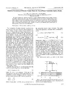
of stuttering that included disfluency types suggested by Williams, Silverman, and Kools (1968). Disfluencies were defined as stuttering (part-word repetitions, disrhythmic phonationsprolongations, tense pauses) and non-stuttering (word repetitions, phrase repetitions, interjections, revisions) types (Johnson & Associates, 1959). Examples of secondary behaviors included eye blinks, facial grimaces, and other bodily movements (Riley, 2009). Procedures Once baseline speech sample data had been obtained (i.e., verification of at least 10% disfluency in conversational speech for the person who stutters and less than 2% disfluency for the one who does not), the participants asked to sit in comfortable chairs and perform several tasks in the following order: (a) sing happy birthday (16 words); (b) count 1-10 (10 words); (c) recite the Pledge of Allegiance (31 words); (d) read a passage aloud (177 words); (e) read a different passage with choral reading with a NFS (177 words); and (f) speak for a minute— monologue (PWS 101 words; NFS 161 words). These tasks were used because many prior imaging studies of stuttering employed similar tasks to elicit CBF differences. During the procedure, participants faced a research assistant who assigned speaking tasks and documented disfluencies and secondaries. A second research assistant digitally recorded and timed all speech. It took approximately 1 hour per participant to complete all speaking tasks. The protocol was approved by the University of Pennsylvania Institutional Review Board, where the trial was carried out. Measurements DCS probes were placed on the head over four different regions of the brain: Broca’s area, left pre-frontal cortex, between the left pre-frontal cortex and Broca’s area, and the frontal region of the right hemisphere (i.e., to act as a control). The probe locations were chosen because (a) it is easier to detect light in the frontal lobe and (b) previous studies that employed other imaging techniques revealed findings in the regions studied during speech tasks. The probes consisted of two sources (200 µm diameter optical fibers) and eight detectors (5 µm diameter single-mode optical fibers)—all held onto the skin with foam pads and Velcro bands. The fibers were custom made with the tips that contact the scalp bent at 90°. The sourcedetector geometry is shown in Figure 1(a). The source-detector distance varied between 1.5 and 2.5 cm. At these distances, DCS can probe a maximum depth of approximately 1 cm below the scalp, enough to reach the most external parts of the cortex. The geometry chosen accounted for eight combinations of source-detector (channels). Figure 1
The fibers were connected one end to portable, custom-built DCS opto-electronics. Briefly, the DCS instrument housed two continuous-wave, long coherence length (>20 m) lasers in the near-infrared region (785nm, CrystaLaser) for the sources. For the detector side, eight photon-counting avalanche photodiodes fed an 8-channel autocorrelator board that computed the temporal intensity autocorrelation functions of the collected light every 2 seconds.
99
DCS data analysis was performed assuming a semi-infinite homogeneous medium and a Brownian diffusion approximation for the mean-square displacement of scatterers in tissue. In every task, the baseline period was taken 10 seconds prior to starting a task, and changes in cerebral blood flow were calculated relative to this baseline period (ΔCBF). For statistical measurements of maximum and average changes, trend estimation was performed with a standard smoothing spline with smoothing parameter of 0.95. Reliability To assess reliability in the analysis of the speech samples, both intra-judge and interjudge tests were performed. For intra-judge reliability assessment, measurements of speech samples (e.g., counting, monologue, etc.) were performed by the same researcher twice. Average number of agreements compared to disagreements for disfluencies and secondary behaviors were calculated with Spearman’s rank order correlation (Fraenkel & Wallen, 1996). For interjudge reliability, two different specialists compared the same speech samples, and their observations were cross-correlated. In this case, the agreement quotient (i.e., number of agreements of specialist 1 divided by [the number of agreements of specialist 2 + disagreements] x 100; Blood, 1993) was used to provide a quantitative measurement of reliability for each speech sample. Intra-judge reliability for the speech samples was 96%, while inter-judge reliability was 92%.
Results Both participants completed all tasks. Table 1 provides details of the analysis during the speech tasks performed by the two participants. The PWS stuttered during the Pledge of Allegiance, reading a passage aloud, choral reading, and the monologue. The NFS had no disfluencies during any tasks.
100
Table 1. Analysis of the speech samples
Singing
Counting
Words per minute
Total number of disfluencies
Percentage of words stuttered
Percentage of syllables stuttered
107
0
0
0
Person who stutters
96
0
0
0
Fluent person
69
0
0
0
Person who stutters
67
0
0
0
186
0
0
0
69
5
16%
10%
177
0
0
0
97
14
7.9%
6%
Fluent person
Pledge recitation
Fluent person
Passage aloud
Fluent person
Choral reading
Fluent person
177
0
0
0
Person who stutters
174
1
0.6%
0.4%
Monologue
Fluent person
161
0
0
0
Person who stutters
101
17
17%
15%
Person who stutters
Person who stutters
Figure 1(b) shows a representative time-course of cerebral blood flow changes in the left pre-frontal lobe (averaged over all the source-detector separations in this region) for the person who stutters and the fluent person during the monologue task. Table 2 summarizes the maximum CBF changes, averaged over the channels at the same location, for three of the six tasks for each participant. For shorter tasks (i.e., duration of less than 30 seconds singing happy birthday, counting 1-10, reciting the Pledge of Allegiance, etc.), we found no significant CBF changes relative to the baseline (no task was completed between 30 seconds and 60 seconds). For longer tasks (i.e., 60-second duration or longer; e.g., monologue), we observed significant CBF increases both in Broca’s area and in the left pre-frontal cortex. In general, CBF changes in the left hemisphere tended to be higher in the fluent person, compared to the person who stutters. The probe placed on the right frontal lobe also showed significant increases in CBF for the person who stutters when the task was long, achieving significant change from baseline in the monologue.
101
Table 2. Comparison of the cerebral blood flow (CBF) changes measured with DCS for the NFS and the PWS. CBF was averaged over all the different source-detector separations in each area of the brain, and the median (standard deviation between the channels) is shown. MedianCBF (Standard Deviation)
Broca’s area
Left pre-frontal
Pledge of Allegiance
Passage Aloud
Monologue
(31 words)
(177 words)
(101/161 words)
8.2 (9.4) %
19.4 (14.2) %
31.6 (14.2) %
Person who stutters
-3.1 (12.1) %
22.7 (15.2) %
17.2 (15.9) %
Fluent person
11.3 (8.6) %
21.9 (8.1) %
24.5 (9.4) %
5.7 (8.3) %
16.1 (11.0) %
4.0 (6.7) %
4.7 (12.4) %
14.5 (9.2) %
27.1 (12.4) %
2.4 (7.1) %
15.1 (12.3) %
17.1 (14.1) %
Fluent person
-2.4 %
3.9 %
5.2 %
Person who stutters
0.6 %
13.1 %
12.1 %
Fluent person
Person who stutters
In between
Fluent person Person who stutters
Right frontal (control)
Among all the tasks, only the monologue exhibited statistically significant differences between the two participants. The fluent subject displayed increases in blood flow in the prefrontal lobe, whereas the subject who stutters showed no significant change in CBF during the one minute monologue in the same location. During this one-minute monologue, the subject who stutters spoke 101 words per minute and stuttered on 15% of the syllables. The other subject spoke 161 words per minute and had no disfluencies.
Discussion The American Speech-Language Hearing Association’s (ASHA) current view is that, as technology advances, our abilities to improve research methodologies and enhance services to patients will improve. Numerous authors agree that technology can improve services and research outcomes (Duffy, Werven, & Aronson, 1997; Howard, Perkins, & Martland, 2001; Tellis, Meloy, Henning, & Jarvie, 2004; Tellis, Cimino, & Alberti, 2010). These preliminary data suggest that optical DCS-measured cerebral blood flow may have potential in stuttering research, and may someday provide analysis of stuttering disfluencies in clinic services. DCS detected significant CBF changes in Broca’s area and in the left pre-frontal cortex when the speech task was sufficiently long (i.e., 60-second duration or longer). In this condition, we consistently observed larger CBF increases in the subject who did not stutter in two cortical areas. This observation is in agreement with previous studies that reported overactivation in motor areas, but minor changes in auditory and cortical speech and language areas in PWS (Blomgren et al., 2003; Brown, Ingham, Ingham, Laird, & Fox, 2005). Our observations also agree with the lack of left advantage in phonemic contrast over the prosodic contrast task in a recent study with NIRS involving PWS (Sato et al., 2011). The probe on the right frontal lobe had only one channel (i.e., it was not possible to calculate the standard deviation over different channels), and it was originally placed as a control for the speech activation tasks. CBF variation in this channel, however, revealed an increasing trend for the person who stutters in long speech tasks, achieving significant change from baseline in tasks such as the monologue. The increased CBF in the right hemisphere for the subject who
102
stutters could be explained by anomalous right-laterality and/or bilateral activation, also previously reported in the literature (Blomgren et al.; Brown et al.; Salmelin et al., 2000). Significant differences between participants were not found on hemodynamic responses to tasks. The only exception was the monologue—where we found a significant increase of CBF in the left frontal lobe in the fluent subject compared to the subject who stutters (i.e., during this task, the person who stutters stuttered on 15% of syllables). We found no significant CBF change from baseline when the tasks were short in duration. Because the hemodynamic change might be expected to be proportional to the duration of the task, short tasks could have caused changes smaller than the signal-to-noise ratio of the experiment. In addition, our temporal resolution of 2 seconds may have been too slow to reliably detect dynamics in the short tasks. Ideally, the combination of better DCS time resolution and tasks longer than 45 seconds should provide better contrast, and, consequently, may improve differentiation between subjects across a broad range of tasks. The present study employed only one diffuse optical technique (DCS). In our next study, we will combine DCS with NIRS; the latter method is another diffuse optical technique that derives changes in oxy- and deoxy-hemoglobin concentration from tissue absorption changes at multiple wavelengths. The combination of DCS and NIRS offers more extensive information about tissue hemodynamics (i.e., both blood flow and blood concentration changes) and provides access to metabolic response. We also plan to test the performance of a real-time algorithm to provide brain hemodynamic response to activation during the task. In summary, this is the first known study in which researchers used DCS to measure CBF during speech tasks for an individual who stutters and one who does not. It suggests that DCS is a feasible technique to assess brain hemodynamic responses in verbal speech tasks. The approach may have significant implications to further understand speech disorders such as stuttering. In future research, we will use a hybrid DCS/NIRS system to assess opticalderived cortical hemodynamic response of 60 participants (PWS and NFS) during several speaking tasks. Based on observations presented in this paper, we will increase the length of the speech samples. We also plan to enroll PWS who are more than 10% disfluent. Both of these changes should increase the differentiation between the two groups. With the strengths of DCS and NIRS, we believe that the combined techniques will provide the field of stuttering research with a novel method to determine differences in CBF and oxygenation levels during speech tasks. If differences are noted, DCS/NIRS may someday provide real-time analysis of stuttering disfluencies.
Acknowledgments The authors would like to thank Meeri N. Kim and Alex Thames for assistance with DCS operation and analysis, David R. Busch for helpful discussions and feedback, and Nicholas Barone and Molly Correll for analyzing the speech samples. This work was supported by the National Institutes of Health through R01-NS060653 and P41-RR002305 (AGY). This work was also supported by the Brassington Research Grant.
References Blomgren, M., Nagarajan, S. S., Lee, J. N., Li, T., & Alvord, L. (2003). Preliminary results of a functional MRI study of brain activation patterns in stuttering and nonstuttering speakers during a lexical access task. Journal of Fluency Disorders, 28(4), 337-55. Blood, G. W. (1993). Treatment efficacy in adults who stutter: Review and recommendations. Journal of Fluency Disorders, 18(2 & 3), 303-318. Boas, D. A., Campbell, L. E., & Yodh, A. G. (1995). Scattering and imaging with diffusing temporal field correlations. Physical Review Letters, 75(9), 1855–1858. Boas, D. A. & Yodh, A. G. (1997). Spatially varying dynamical properties of turbid media probed with diffusing temporal light correlation. Journal of the Optical Society of America A, 14(1), 192–215.
103
Brown, S., Ingham, R. J., Ingham, J. C., Laird, A. R., & Fox, P. T. (2005). Stuttered and fluent speech production: An ALE meta-analysis of functional neuroimaging studies. Human Brain Mapping, 25, 105117. Buckley, E. M., Cook, N. M., Durduran, T., Kim, M. N., Zhou, C., Choe, R.,…Yodh, A. G. (2009). Cerebral hemodynamics in preterm infants during positional intervention measured with diffuse correlation spectroscopy and transcranial Doppler ultrasound. Optics Express, 17, 12571-12581. Carp, S. A., Dai, G. P., Boas, D. A., Franceschini, M. A., & Kim, Y. R. (2010). Validation of diffuse correlation spectroscopy measurements of rodent cerebral blood flow with simultaneous arterial spin labeling MRI; towards MRI-optical continuous cerebral metabolic monitoring. Biomed. Optics Express, 1, 553-565. Chang, S. E., Erickson, K., & Ambrose, N. (2005, November). An MRI study on childhood stuttering persistence and recovery. Paper presented at the annual convention of the American Speech-LanguageHearing Association, San Diego, CA. Chang, S. E., Kenny, M. K., Loucks, T. M. J., & Ludlow, C. L. (2009). Brain activation abnormalities during speech and non-speech in stuttering speakers. NeuroImage, 46, 201-212. De Nil, L. F., Kroll, R. M. Lafaille, S. J. & Houle, S. (2003). A positron emission tomography study of short and long term treatment effects on functional brain activation in adults who stutter. Journal of Fluency Disorders, 28, 357-380. Duffy, J. R., Werven, G. W., & Aronson, A. E. (1997). Telemedicine and the diagnosis of speech and language disorders. Mayo Clinic Proceedings, 72, 1116-1122. Durduran, T., Choe, R., Baker, W. B., Yodh, A.G. (2010). Diffuse optics for tissue monitoring and tomography. Reports on Progress in Physics, 73, 076701-1-076701-43. Durduran, T., Yu, G., Burnett, M. G., Detre, J. A., Greenberg, J. H., Wang, J.,…Yodh, A. G. (2004). Diffuse optical measurements of blood flow, blood oxygenation and metabolism in human brain during sensorimotor cortex activation. Optics Letters, 29, 1766–1768. Durduran, T., Zhou, C., Edlow, B. L., Yu, G., Choe, R., Kim, M. N.,…Detre, J. A. (2009). Transcranial optical monitoring of cerebrovascular hemodynamics in acute stroke patient. Optics Express, 17(5), 38843902. Durduran, T., Zhou, C., Kim, M. N., Buckley, E. M., Yu, G., Choe, R.,…Licht, D. J. (2008). Validation of diffuse correlation spectroscopy for non-invasive, continuous monitoring of CBF in neonates with congenital heart defects. p. Abstract 299 (American Neurological Association, Salt Lake City, UT). Durduran, T., Zhou, C., Yu, G., Choe, R., Silvestre, D., Wang, J. J.,…Licht, D. (2007). Preoperative measurement of CO2 reactivity and cerebral autoregulation in neonates with severe congenital heart defects. San Jose, CA: SPIE Photonics West. Durduran, T., Zhou, C., Yu, G., Edlow, B., Choe, R., Shah, Q.,…Detre, J. A. (2007). Bed-side monitoring of cerebral blood flow (CBF) in acute stroke patients during changes in head of bed position. International Stroke Conference, vol. P37 (American Heart Association). Fraenkel, J. R., & Wallen, N. E. (1996). How to design and evaluate research in education (3rd ed.). New York, NY: McGraw-Hill. Fukui, Y., Ajichi, Y., & Okada, E. (2003). Monte Carlo prediction of near-infra-red light propagation in realistic adult and neonatal head models. Applied Optics, 42, 2881–2887. Hampton, A., & Weber-Fox, C. (2008). Non-linguistic auditory processing in stuttering: evidence from behavior and even-related brain potentials. Journal of Fluency Disorders, 33(4), 253-273. Howard, S., Perkins, M., & Martland, P. (2001). An integrated multi-media package for learning clinical linguistics and phonetics. International Journal of Language & Communication Disorders, 36 (Suppl.), 327332. Ingham, R. J., Fox, P. T., Costello Ingham, J., & Zamarripa, F. (2000). Is overt stuttered speech a prerequisite for the neural activations associated with chronic developmental stuttering? Brain and Language, 75, 163–194. Johnson, W., & Associates. (1959). The onset of stuttering: Research findings and implications. Minneapolis, MN: The University of Minnesota Press.
104
Kim, M. N., Durduran, T., Frangos, S., Buckley, E. M., Zhou, C., Yu, G.,…Yodh, A. G. (2008). Diffuse optical measurements of cerebral blood flow and oxygenation in patients after traumatic brain injury or subarachnoid hemorrhage. OSA Biomedicals Topicals, p. CN237 (St Petersburg, FL). Li, J., Dietsche, G., Iftime, D., Skipetrov, S. E., Maret, G., Elbert, T.,… Gisler, T. (2005). Noninvasive detection of functional brain activity with near-infrared diffusing-wave spectroscopy. Journal of Biomedical Optics, 10(4), 044002-1-044002-12. Maret, G., & Wolf, P. E. (1987). Multiple light scattering from disordered media. The effect of Brownian motion of scatterers. Zeitschrift für Physik B Condensed Matter, 65, 409–413. Mesquita, R. C., & Yodh, A.G. (2011). Diffuse optics: fundamentals and tissue applications. In R. Kaiser, D. S. Weirsma, & L. Fallini (Eds.) Proceedings of the International School of Physics “Enrico Fermi” Course CLXXIII “Nano optics and atomics: transport of light and matter waves (pp. 51-74). Amsterdam, The Netherlands: IOS Press. Ninck, M., Untenberger, M., & Gisler, T. (2010). Diffusing-wave spectroscopy with dynamic contrast variation: disentangling the effects of blood flow and extravascular tissue shearing on signals from deep tissue. Biomedical Optics Express, 1, 1502-1513. Pine, D. J., Weitz, D. A., Chaikin, P. M., & Herbolzheimer, E. (1988). Diffusing-wave spectroscopy. Physical Review Letters, 60, 1134–1137. Riley, G. (2009). Stuttering severity instrument for children and adults (4th ed.). Austin, TX: PRO-ED. Roche-Labarbe, N., Carp, S. A., Surova, A., Patel, M., Boas, D. A., Grant, P. E.,…Franceschini, M. A. (2009). Noninvasive optical measures of CBV, StO2, CBF index, and rCMRO2 in human premature neonates’ brains in the first six weeks of life. Human Brain Mapping, 31, 341-352. Salmelin, R., Schnitzler, A., Schmitz, F., & Freund, H. (2000). Single word reading in developmental stutterers and fluent speakers. Brain, 123(6), 1184-1202. Sato, Y., Mori, K., Koizumi, T., Minagawa-Kawai, Y., Tanaka, A., Ozawa, E.,…Mazuka, R. (2011). Functional lateralization of speech processing in adults and children who stutter. Frontiers in Psychology, 2(70), 1-10. Sommer, M., Koch, M. A., Paulus, W., Weiller, C., & Büchel, C. (2004). Disconnection of speech-relevant brain areas in persistent and developmental stuttering. The Lancet, 360, 380-383. Sunar, U., Quon, H., Durduran, T., Zhang, J., Du, J., Zhou, C.,…Yodh, A. G. (2006). Non-invasive diffuse optical measurement of blood flow and blood oxygenation for monitoring radiation therapy in patients with head and neck tumors: A pilot study. Journal of Biomedical Optics, 11(06), 064021-1-064021-13. Tellis, G. M., Cimino, L., & Alberti, J. (2010). Advanced digital technology for supervising graduate clinicians. Perspectives on Administration and Supervision, 20, 9-13. Tellis, G. M., Meloy, T., Henning, M., & Jarvie, D. (2004). Advanced digital capture technology in identification of stuttering disfluencies. Proceedings from the Fourth World Conference of Fluency Disorders (pp. 56-59). Nijmegen, The Netherlands: Nijmegen University Press. Williams, D. E., Silverman, F. H., & Kools, J. A. (1968) Disfluency behavior of elementary school stutterers and nonstutterers: The adaptation effects. Journal of Speech & Hearing Research, 11, 622-630. Yamamoto, T., Maki, A., Kadoya, T., Tanikawa, Y., Yamad, Y., Okada, E.,…Koizumi, H. (2002). Arranging optical fibers for the spatial resolution improvement of topographical images. Physics in Medicine and Biology, 47, 3429–3440. Yamashita, Y., Maki, A., & Koizumi, H. (1996). Near-infrared topographic measurement system: Imaging of absorbers localized in a scattering medium. Review of Scientific Instruments, 67, 730–732. Yodh, A., & Chance, B. (1995). Spectroscopy and imaging with diffusing light. Physics Today, 48, 34-40. Yu, G., Durduran, T., Zhou, C., Zhu, T. C., Finlay, J. C., Busch, T. M.,…Yodh, A. G. (2006). Real-time in situ monitoring of human prostate photodynamic therapy with diffuse light. Photochemistry and Photobiology, 82, 1279–84. Yu, G., Floyd, T., Durduran, T., Zhou, C., Wang, J. J., Detre, J. A.,…Yodh, A. G. (2007). Validation of diffuse correlation spectroscopy for muscle blood flow with concurrent arterial-spin-labeling perfusion. Optics Express, 15, 1064–75.
105
Zhou, C., Choe, R., Shah, N., Durduran, T., Yu, G., Durkin, A.,…Yodh, A. G. (2007). Diffuse optical monitoring of blood flow and oxygenation in human breast cancer during early stages of neoadjuvant chemotherapy. Journal of Biomedical Optics, 12(5), 051, 90.
106