PROGRESS IN STEM CELL 2016, 3(1):65-72
BioMedPress (BMP) Journal homepage: http://www.cellstemcell.org
Mini-review
DOI: http://dx.doi.org/10.15419/psc.v2i1.82
What are markers for breast cancer stem cells ? Phuc Van Pham*, Nhan Lu-Chinh Phan Laboratory of Stem Cell Research and Application, University of Science, Vietnam National University, Ho Chi Minh city, Viet Nam
ARTICLE INFO Article history: Received 15 Nov 2015 Accepted 20 Feb 2016 Published 20 March 2016
ABSTRACT
Breast cancer stem cells were firstly discovered by Al-Hajj et al. (2003). Many scientists interested in targeting them as an important solution to "remove" the root of breast cancer. However, more and more publications showed that breast cancer stem cells are the heterogenous population that expression of some markers can be different between them. Particularly, some different markers were considered as markers of breast cancer stem cells. Therefore, what are gold standards of these cells? In this publication, we discussed some conflicts about markers of breast cancer stem cells and suggested some gold standards for breast cancer stem cells. Keywords: Stem Cells, Cancer Stem Cells, Breast Cancer Stem Cells, Gold Standards.
Moreover, they also observed that CD44+CD24-/lowESA+
Breast cancer stem cells: From beginning Cancer stem cells (CSCs) in breast tumors were first discovered by Al-Hajj et al. from the University of Michigan in 2003 (Al-Hajj, et al., 2003). They cultured primary breast tumors and analyzed the tumor cells for the expression of CD44, CD24, and ESA. The results showed that
only
cancer
cells
with
+
-/low
CD44 CD24
+
ESA
phenotype were highly tumorigenic. In contrast, cells with CD44-CD24+phenotype did not induce tumor formation.
* Corresponding author: Phuc Van Pham, Email:
[email protected]
cells could maintain their tumorigenic potential when serially passaged for long-term. Based on these results, Al-Hajj et al. concluded that CD44+CD24-/lowcells satisfied the criteria for CSCs, such as self-renewal, differentiation, and high tumorigenicity. These results were further confirmed by several other studies (Cho, et al., 2008, Liang, et al., 2013, Pece, et al., 2010, Pham, et al., 2011, Ponti,
et
al.,
2005,
Saadin
and
White,
2013,
Vassilopoulos, et al., 2008, Walia and Elble, 2010).
© The Author(s) 2016. This article is published with open access by BioMedPress (BMP). This article is distributed under the terms of the Creative Commons Attribution License (CC-BY 4.0) which permits any use, distribution, and reproduction in any medium, provided the original author(s) and the source are credited.
PROGRESS IN STEM CELL 2016, 3(1): 65-72
non-stem cells originated from the heterogeneity of breast
Where are breast CSCs from? Breast mechanisms.
CSCs Some
are in
formed
vitro
through
studies
and
these clinical
investigations suggest that breast CSCs are produced in vivo
through
the
following
2
mechanisms:
(1)
accumulation of mutations in mammary stem cells during development that causes these cells to lose their selfrenewal capacity and (2) development of breast CSCs from non-stem cells
through
different
evolutionary
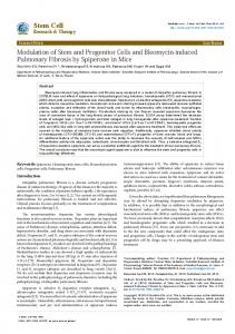
mechanisms. Increased risk of breast cancer in children exposed to radiationindicates that breast cancer may develop directly from long-lived stem or progenitor cells present in the mammary glands (Miller, et al., 1989, Modan, et al., 1989). Importantly, luminal progenitor cells are known to induce the formation of BRCA1-driven tumors (Lim, et al., 2009, Molyneux, et al., 2010, Proia, et al., 2011). Pece et al. (2010) showed that poorly differentiated cancers had higher number of CSCs than well-differentiated cancers (Pece, Tosoni, Confalonieri, Mazzarol, Vecchi, Ronzoni, Bernard, Viale, Pelicci and Di Fiore, 2010) (Fig. 1). In a study involving 61 patients with breast cancer, normal breast tissue and breast tumor tissue from the same patients were analyzed to determine the presence of stem cells in both the tissues. In all, nine patients had triple-negative cancer (TNC) and 52 patients had ER+and Her2+cancer. The results showed that 100% (9/9)
patients
with
TNC
had
CSCs
with
CD44+CD49f+CD133/2+phenotypein both the normal and tumor tissues and that only 13.4% (7/52) patients with ER+ and/or Her2+cancerhad CSCs in the normal breast tissue. Based on this result, Atkinson et al. (2013) proposed that CSCs in breast cancer originated from mutated mammary stem cells present in the normal breast tissue (Atkinson, et al., 2013). The idea that breast CSCs were derived from
cancer cell lines. Almost all breast cancer cell lines contain a subpopulation of cells with breast CSC phenotypes. Interestingly, some studies have shown that different subpopulations of cells in the same breast cancer cell line can interconvert between phenotypes. Analysis of some commercial breast cancer cell lines such as SUM159, SUM149, Ca1a, MDA-MB231,BT474,SKBR3,andMCF7 indicated that these cell lines included a small population of stem cells that underwent self-renewal to form mammospheres in serum-free cultures and differentiation to generate other cells that resulted in heterogeneity(Gupta, et al., 2011, Meyer, et al., 2009, Piggott, et al., 2011). Interconversion of CSCs in breast cancer cell lines was clearly recorded by Gupta et al. (2011). They sorted breast CSCs with stem-like basal and luminal phenotypes from 2 breast cancer cell lines SUM159 and SUM149and observed that these cells exhibited the properties of their parental cell lines after 11 days in culture(Gupta, Fillmore, Jiang, Shapira, Tao, Kuperwasser and Lander, 2011). In another study, Piggott et al. (2011) depleted CSCs from BT474 breast cancer cell line and observed that this depleted cell line re-established progenitor-like cells after4weeksinculture (Piggott, Omidvar, Marti Perez, Eberl and Clarkson, 2011). In some cell lines such asCa1a, MCF7, SUM159, and MDA-MB-231, breast cells without a CSC phenotype (CD44+CD24+) produced breast CSCs with CD44+CD24- phenotype in vitro (Meyer, Fleming, Ali, Pesesky, Ginsburg and Vonderhaar, 2009). In addition, non-CSCs
(CD44+CD24+
phenotype)
isolated
from
Ca1a,ZR75-1,and MCF7cell lines successfully produced heterogeneous tumors showing local invasion in immuno compromisedmice (Meyer, Fleming, Ali, Pesesky, Ginsburg and Vonderhaar, 2009). These evidences confirmed that breast non-CSCs could be automatically converted into CSCs both in vitro and in vivo (Fig. 1).
66
PROGRESS IN STEM CELL 2016, 3(1): 65-72
Some recent studies have shown that actively
and cells from 2 breast cancer cell lines MDA-MB-231
transdifferentiated breast non-CSCs can be converted into
(MDA) and MA11 show increased metastatic capacity.
CSCs after treatment with some defined factors. CSC-like
Because MSCs can migrate and localize to breast
cells were produced after the transfection of MCF10Acells
cancers, formation of MSC–breast cancer cell hybrids
with SRC oncogene (Iliopoulos, et al., 2011). Moreover, the
maybe a potential mechanism for generating breast CSCs
CSC-conditioned medium can convert non-CSCs into
(Rappa, et al., 2012). Fusion of M2 macrophages with
CSCs (Iliopoulos, Hirsch, Wang and Struhl, 2011). Shaffer
cells from breast cancer cell lines MCF-7 and MDA-MB-
et al. also produced CSCs by transfecting non-CSCs
231 in the presence of polyethylene glycol produces
withSV40andH-ras (Chaffer, et al., 2011).
hybrids that show increased migration, invasion, and
Some studies have shown that breast CSCs can be produced in vitro by fusing breast cancer cells with
tumorigenicity and haveCD44+CD24-/low phenotype (Ding, et al., 2012).
mesenchymal stromal cells or macrophages. Hybrids of bone marrow-derived mesenchymal stem cells (MSCs)
Figure 1. Relations of breast cancer stem cells and normal mammary stem cells.
What the markers of breast cancer stem cells?
previously published reports have described the isolation of
BCSCs
from
malignant
tumors
using
specific
markers(Ponti, Costa et al. 2005, Pham, Phan et al. 2011,
Specific markers Studies have shown that BCSCs are presentin tumors and are defined by a unique set of markers. Many
Lin, Hutzen et al. 2013, Wang, Lv et al. 2014). However, other studies
have
shown that
BCSCs
exists a
heterogeneous population in tumors (Lorico and Rappa
67
PROGRESS IN STEM CELL 2016, 3(1): 65-72
2011, Hwang-Verslues, Lee et al. 2012, Wong, Fuller et
was shown that only subpopulations of basal A and
al. 2012). This indicated that BCSCs could express
luminal B contain small populations of cells with
different markers with different levels of expression at
CD44highand CD24low. This suggested that in the normal
various times. This was consistent with other theories
breast tissue there are at least two subpopulations of cells
about cancer stem cells (CSCs).
with markers CD44highandCD24low. cell
Similarly, there are three populations of cells in
heterogeneity. The first is related to the existence of
breast cancer tissue, including, basal A, luminal B, and
different CSCs in different tumors. The second is related
luminal C. However, there are differences in their ratios. In
to the theory of clonal evolution, which describes how
fact, the number of basal A cells significantly decreases as
different tumors can originate from a single stem cell. A
the number of luminal C cells increases. In contrast to
recent report showed that maybe there is a dynamic
normal tissue, in cancer tissue both luminal B and luminal
balance between cancer progenitor cells and CSCs(Li and
C
Laterra 2012). A CSC can differentiate into a progenitor
However, CD44highandCD24low cells of luminal B can form
cancer cell, and a progenitor cancer cell can de-
mammospheres more easily than luminal C cells. In
differentiate into a CSC. This suggested that CSCs and
comparison to subpopulations, MUC-1-, ALDH+, and
their progenitor cancer cells co-exist in a dynamic form (Li
CD10+, CD44highCD24low cells of luminal B have the
and Laterra 2012). The relationship between CSCs and
greatest mammosphere-forming capacity. In summary,
cancer progenitor cells creates the necessity to specify a
the CD44highCD24lowcell population from a breast tumor
combination of markers for the identification of CSCs.
can contain subpopulations that differ in the expression of
There
are
two
theories
about
stem
Heterogeneity in the BCSC population: which is the strongest sub-population? Several recent studies have shown that BCSCs contain
sub-populations
tumorigenicity.
To
date,
that
exhibit
only
some
different of
these
subpopulations have been identified. Ghebeh et al. determined that there are 3 main cell populations in the normal breast tissue, including, “basal A” progenitor cells with the markers Ep-CAM-/lowandCD49f+, “luminal B” progenitor cells with the markers Ep-CAMhighandCD49f+, and “luminal C” differentiated cells with the markers EpCAMhighandCD49f- (Ghebeh, Sleiman et al. 2013). These populations have been culturedin order to identify their mammosphere forming potential. The results show that
cells
contain
the
markers
CD44highandCD24low.
Ep-CAM and CD49f. Hence, the results of this analysis indicate that CD44highCD24lowEp-CAM+CD49f+could be a marker profile for BCSCs. Molyneux et al. (2010) showed that breast cancer cells originate from luminal epithelial progenitors and not from basal stem cells(Molyneux, Geyer et al. 2010). Uchoa Dde et al. (2014) determined that breast cancer cells express the BCSC phenotype (Uchoa Dde, Graudenz
et
al.
2014).
In
a
previous
study,
immunohistochemistry was used to show that there were BCSCs in hormone-receptor-positive breast cancer, but there was no correlation between markers of CSCs and the response to endocrine therapy and overall clinical outcome (Hashimoto, Shimizu et al. 2012).
only subpopulations basal A and luminal B could form a
The heterogeneity of the BCSC population based
mammosphere whereas luminal C could not form a
on cell markers CD44highCD24lowhas resulted in conflicting
mammosphere. Moreover, the mammosphere-forming
conclusions. For example, Lin et al. (2012) showed that
capacity of basal A is stronger than that of luminal B. It
there
is
a correlation
between
of
prevalence of
68
PROGRESS IN STEM CELL 2016, 3(1): 65-72
CD44+CD24- tumor cells and invasive ductal carcinoma,
genes. Second, ALDH1A can metabolize and detoxify
high recurrence, and shorter DFS and OS. Therefore, this
chemotherapeutics (Magni, Shammah et al. 1996). Due to
marker is important for breast cancer diagnosis and
this property, ALDH1A is known as a contributing factor to
treatment. Conversely, Zhong et al. (2014) suggested that
chemotherapy resistance of hematopoietic stem cells.
+
ALDH is a better clinical indicator for relapse and invasive ductal carcinoma than CD44+CD24- (Table 1).
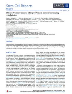
In the recent years, ALDH1A has been thought to be expressed in a sub-population of breast cancer cells (Fig. 2). These ALDH+ breast cancer cells exhibit stem cell properties (Ginestier, Hur et al. 2007). Clinically, ALDH+ is
Table 1: Makers of breast cancer stem cells Markers
Tumorigenicity
suggested to be a prognostic marker to predict metastasis
References
and poor patient outcome (Balicki 2007, Ginestier, Hur et
(cells) CD44(+)CD24(-
al. 2007, Charafe-Jauffret, Ginestier et al. 2010, Marcato,
100
(Al-Hajj, Wicha et al. 2003)
/low)Lineage(-) CD44 /CD24 +
-
expression 100
(Fillmore and Kuperwasser
/low/ESA+ CD44+/CD24-
2008) 1000
breast
tumors
is
associated
with
chemotherapy resistance (Tanei, Morimoto et al. 2009,
(Ponti, Costa et al. 2005, 2008, Pham, Phan et al. 2011, Yan, Chen et al.
What is the difference between the three populations considered to be BCSCs? There is a population with the phenotype of CD44+CD24-, another with ALDH, and another with CD133. There are also
2013) 50-100
in
Alamgeer, Ganju et al. 2014).
Honeth, Bendahl et al.
CD133
Dean et al. 2011, Liu, Lv et al. 2014). In fact, ALDH1A
(Wright, Calcagno et al. 2008)
combinations of phenotypes such as CD44+CD24-, CD133+, and ALDH+. Some populations of BCSCs also include +
phenotypesCD44+CD133+,
CD44+ALDH+,
or
+
CD133 ALDH . These differences seem to be related to distinct levels of differentiation status in BCSCs (Ricardo,
ALDH Aldehyde dehydrogenases (ALDHs) are a group
Vieira et al. 2011).
of enzymes that catalyze the oxidation (dehydrogenation) of aldehydes (Marchitti, Brocker et al. 2008). There are 19 kinds of ALDH identified in humans. One of these is ALDH1A, a well-known enzyme responsible for oxidizing aldehydes to carboxylic acids (Marchitti, Brocker et al. 2008). ALDH1A is highly expressed in the epithelium of testis, brain, eye, liver, kidney, and some stem cells. To date,
ALDH1A
exhibits
two
main
functions
in
hematopoietic stem cells and neural stem cells. The first is the oxidation of retinal to retinoic acid (Collins 2008). The retinoic acid activates nuclear retinoic acid receptors (RARs) which in turn regulate the expression of related
Figure 2. Breast cancer stem cells with different markers.
69
PROGRESS IN STEM CELL 2016, 3(1): 65-72
Zhong et al. (2014) demonstrated that ALDH1+ +
-
defined as a minor cell population that exhibits low-
and CD44 /CD24 breast cancer cells play significant roles
intensity staining with dyes such as Hoechst 33342
in metastasis. However, the ALDH1 marker is a better
(H33342) or rhodamine 123 (R123). Both H33342 and
predictive marker for breast cancer metastasis than the
R123 easily stain cells because they can cross the cell
+
-
CD44 /CD24 phenotype (Zhong, Shen et al. 2014). In a
membrane. However, SP cells absorb less of the dye.
previous study, Tanei et al. (2009) found that ALDH1-
Some investigations have suggested that side population
+
-
positive but not CD44 /CD24 BCSCs played a significant
cells over-expressed trans-membrane transporters like
role in resistance to chemotherapy(Tanei, Morimoto et al.
ATP-binding cassette (ABC) molecule ABCG2/BCRP
2009). This marker was also highly expressed in TNBC
(ATP-binding
compared to non-TNBC (Li, Ma et al. 2013).
resistance protein-1). Due to the high expression of this
cassette,
sub-family
G/breast
cancer
ABC transporter, SP cells can extrude some dyes and
Side population A side population (SP) is defined as a subpopulation of cells that expresses distinct properties compared to the main population in flow cytometry analysis. The SP cell phenotype was first discovered in 1996 in mouse bone marrow. SP cells in bone marrow are related to hematopoietic stem cells (Goodell, Brose et al. 1996). Using this established technique, some authors have also detected SP cells in other normal tissues
drugs (Hadnagy, Gaboury et al. 2006, Wu and Alman 2008). Therefore, this population is clearly visible in flow cytometry analysis. SPs are present in both normal breast tissue and breast cancer tissue but are limited in number. These cells are characterized as having some of the same properties as stem cells. However, it has been shown that SPs exist in almost all breast cancer cell lines (Patrawala, Calhoun et al. 2005).
including the mammary gland (Alvi, Clayton et al. 2003)and tumors including breast tumor (Britton, Kirby et al. 2011). In the stem cell analysis, a side population is
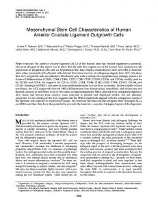
Figure 3. Strategies for breast cancer stem cell characterization and isolation
70
PROGRESS IN STEM CELL 2016, 3(1): 65-72
What is the relationship between SP cells and the cancer stem cell phenotype? To date, whether the SP cell population is enrichedby cancer stem cells remains a controversial topic. Some studies have showed that SPs contain cells with higher clonogenic and tumorigenic potential than non-SPs (Patrawala, Calhoun et al. 2005, Hiraga, Ito et al. 2011). However, other studies have
Open Access This article is distributed under the terms of the Creative Commons Attribution License (CC-BY 4.0) which permits any use, distribution, and reproduction in any medium, provided the original author(s) and the source are credited.
claimed that SP cells do not relate to cancer stem cells and, more importantly, that the cancer stem cell population is outside of the SP cells. It is thought that cancer
stem
cells
highly
express
ABCG2,
which
contributes to anti-tumor drug resistance and that cancer stem cells are SP cells. However, Patrawala et al. (2005) demonstrated that ABCG2 cancer cells also exhibit high clonogenic
and
tumorigenic
potential,
while
also
expressing several genes for “stemness” (Patrawala, Calhoun et al. 2005) (Fig. 3).
Conclusion
Acknowledgment This work was funded by Vietnam National University, Ho Chi Minh city.
Competing interests The authors declare that they have no competing interests.
References Al-Hajj M, Wicha MS, Benito-Hernandez A, Morrison SJ, Clarke MF (2003) Prospective identification of tumorigenic breast cancer cells. Proceedings of the National Academy of Sciences of the United States of America 100:3983-3988 Atkinson RL, Yang WT, Rosen DG, Landis MD, Wong H, Lewis MT, Creighton CJ, Sexton KR, Hilsenbeck SG, Sahin AA, Brewster AM, Woodward WA, Chang JC (2013) Cancer stem cell markers are enriched in normal tissue adjacent to triple negative breast cancer and inversely correlated with DNA repair deficiency. Breast cancer research : BCR 15:R77 Chaffer CL, Brueckmann I, Scheel C, Kaestli AJ, Wiggins PA, Rodrigues LO, Brooks M, Reinhardt F, Su Y, Polyak K, Arendt LM, Kuperwasser C, Bierie B, Weinberg RA (2011) Normal and neoplastic nonstem cells can spontaneously convert to a stemlike state. Proceedings of the National Academy of Sciences of the United States of America 108:7950-7955 Cho RW, Wang X, Diehn M, Shedden K, Chen GY, Sherlock G, Gurney A, Lewicki J, Clarke MF (2008) Isolation and molecular characterization of cancer stem cells in MMTV-Wnt-1 murine breast tumors. Stem cells (Dayton, Ohio) 26:364-371 Ding J, Jin W, Chen C, Shao Z, Wu J (2012) Tumor associated macrophage x cancer cell hybrids may acquire cancer stem cell properties in breast cancer. PloS one 7:e41942 Gupta PB, Fillmore CM, Jiang G, Shapira SD, Tao K, Kuperwasser C, Lander ES (2011) Stochastic state transitions give rise to phenotypic equilibrium in populations of cancer cells. Cell 146:633-644 Iliopoulos D, Hirsch HA, Wang G, Struhl K (2011) Inducible formation of breast cancer stem cells and their dynamic equilibrium with non-stem cancer cells via IL6 secretion. Proceedings of the National Academy of Sciences of the United States of America 108:1397-1402 Liang S, Furuhashi M, Nakane R, Nakazawa S, Goudarzi H, Hamada J, Iizasa H (2013) Isolation and characterization of human breast cancer cells with SOX2 promoter activity. Biochemical and biophysical research communications 437:205-211 Lim E, Vaillant F, Wu D, Forrest NC, Pal B, Hart AH, Asselin-Labat ML, Gyorki DE, Ward T, Partanen A, Feleppa F, Huschtscha LI, Thorne HJ, Fox SB, Yan M, French JD, Brown MA, Smyth GK, Visvader JE, Lindeman GJ (2009) Aberrant luminal progenitors as the candidate target population for basal tumor development in BRCA1 mutation carriers. Nature medicine 15:907-913 Meyer MJ, Fleming JM, Ali MA, Pesesky MW, Ginsburg E,
71
PROGRESS IN STEM CELL 2016, 3(1): 65-72
Vonderhaar BK (2009) Dynamic regulation of CD24 and the invasive, CD44posCD24neg phenotype in breast cancer cell lines. Breast cancer research : BCR 11:R82 Miller AB, Howe GR, Sherman GJ, Lindsay JP, Yaffe MJ, Dinner PJ, Risch HA, Preston DL (1989) Mortality from breast cancer after irradiation during fluoroscopic examinations in patients being treated for tuberculosis. The New England journal of medicine 321:1285-1289 Modan B, Chetrit A, Alfandary E, Katz L (1989) Increased risk of breast cancer after low-dose irradiation. Lancet 1:629-631 Molyneux G, Geyer FC, Magnay FA, McCarthy A, Kendrick H, Natrajan R, Mackay A, Grigoriadis A, Tutt A, Ashworth A, ReisFilho JS, Smalley MJ (2010) BRCA1 basal-like breast cancers originate from luminal epithelial progenitors and not from basal stem cells. Cell stem cell 7:403-417 Pece S, Tosoni D, Confalonieri S, Mazzarol G, Vecchi M, Ronzoni S, Bernard L, Viale G, Pelicci PG, Di Fiore PP (2010) Biological and molecular heterogeneity of breast cancers correlates with their cancer stem cell content. Cell 140:62-73
8:149-163 Rappa G, Mercapide J, Lorico A (2012) Spontaneous formation of tumorigenic hybrids between breast cancer and multipotent stromal cells is a source of tumor heterogeneity. The American journal of pathology 180:2504-2515 Saadin K, White IM (2013) Breast cancer stem cell enrichment and isolation by mammosphere culture and its potential diagnostic applications. Expert review of molecular diagnostics 13:49-60 Vassilopoulos A, Wang RH, Petrovas C, Ambrozak D, Koup R, Deng CX (2008) Identification and characterization of cancer initiating cells from BRCA1 related mammary tumors using markers for normal mammary stem cells. International journal of biological sciences 4:133-142 Walia V, Elble RC (2010) Enrichment for breast cancer cells with stem/progenitor properties by differential adhesion. Stem cells and development 19:1175-1182
Pham PV, Phan NL, Nguyen NT, Truong NH, Duong TT, Le DV, Truong KD, Phan NK (2011) Differentiation of breast cancer stem cells by knockdown of CD44: promising differentiation therapy. Journal of translational medicine 9:209 Piggott L, Omidvar N, Marti Perez S, Eberl M, Clarkson RW (2011) Suppression of apoptosis inhibitor c-FLIP selectively eliminates breast cancer stem cell activity in response to the anti-cancer agent, TRAIL. Breast cancer research : BCR 13:R88 Ponti D, Costa A, Zaffaroni N, Pratesi G, Petrangolini G, Coradini D, Pilotti S, Pierotti MA, Daidone MG (2005) Isolation and in vitro propagation of tumorigenic breast cancer cells with stem/progenitor cell properties. Cancer research 65:5506-5511 Proia TA, Keller PJ, Gupta PB, Klebba I, Jones AD, Sedic M, Gilmore H, Tung N, Naber SP, Schnitt S, Lander ES, Kuperwasser C (2011) Genetic predisposition directs breast cancer phenotype by dictating progenitor cell fate. Cell stem cell
Cite this article as:
Pham, P., & Phan, N. (2016). What are markers for breast cancer stem cells ?. Progress In Stem Cell, 3(1), 65-72.
72