Commentary
921
Protease-activated receptor signalling, endocytic sorting and dysregulation in cancer Puneeta Arora, Tiffany K. Ricks and JoAnn Trejo* Department of Pharmacology, School of Medicine, University of North Carolina at Chapel Hill, Chapel Hill, NC 27599-7365, USA *Author for correspondence (e-mail:
[email protected])
Journal of Cell Science
Accepted 15 January 2007 Journal of Cell Science 120, 921-928 Published by The Company of Biologists 2007 doi:10.1242/jcs.03409
Summary Protease-activated receptors (PARs) are G-protein-coupled receptors (GPCRs) that are activated by a unique proteolytic mechanism. PARs play crucial roles in hemostasis and thrombosis, as well as in inflammation and vascular development. Coagulant proteases, which are generated at sites of vascular injury, act mainly through PARs to elicit signalling in a variety of cell types. Since PARs are irreversibly activated signalling must be tightly regulated. Desensitization and trafficking of proteolytically activated PARs control the magnitude, duration and spatial aspects of receptor signalling. Recent studies have revealed novel endocytic sorting mechanisms that regulate PAR signalling. PARs have also been implicated in tumor progression. PARs are overexpressed in several types of Introduction Protease-activated receptors (PARs) are G-protein-coupled receptors (GPCRs) that signal in response to extracellular proteases. There are four PARs encoded in the mammalian genome. PAR1, the prototype for this family, transmits cellular responses to thrombin, the main effector protease of the coagulation cascade. PAR3 and PAR4 also respond to thrombin, whereas PAR2 is activated by trypsin-like serine proteases but not by thrombin. PARs have important functions in hemostasis and thrombosis, as well as in inflammatory and proliferative responses triggered by vascular injury, a topic recently reviewed (Coughlin, 2005). The proteolytic nature of PAR activation, which results in irreversible activation, is distinct from most GPCRs. Thus, PAR signalling is tightly regulated by rapid desensitization† at the plasma membrane and by receptor trafficking. PARs are overexpressed in several types of malignant cancer. Coagulant proteases and PARs can promote tumor growth, invasion and metastasis but precisely how PARs contribute to cancer progression is not known. The zincdependent matrix metalloprotease 1 (MMP-1), also known as interstitial collagenase, has recently been reported to promote tumor growth and invasion through activation of PAR1 (Boire et al., 2005), providing an important link between tumorgenerated metalloproteases and PAR signalling. In addition to PAR overexpression, breast carcinoma cells display aberrant PAR1 trafficking, which causes persistent signalling and cellular invasion (Booden et al., 2004). These studies provide the first example of how aberrant trafficking may cause †
Desensitization is defined as a loss in the responsiveness of a signalling system
malignant cancer, transmit signals in response to tumorgenerated proteases and promote tumor growth, invasion and metastasis. Recent work also indicates that matrix metalloprotease 1 (MMP-1) signals through PAR1 to promote tumor growth and invasion. In addition to PAR overexpression, tumor cells display aberrant PAR1 trafficking, which causes persistent signalling and cellular invasion. Thus, a novel type of gain-of-function in GPCR signalling in cancer can be acquired through dysregulation of receptor trafficking. Key words: Arrestin, GPCR, Thrombin, Trafficking, Coagulant protease
constitutive GPCR activation to promote tumor cell invasion. Here, we discuss activation of PARs, signal regulation and their dysregulation in cancer. The PAR family There are four PAR members, encoded by distinct genes in the GPCR superfamily, the largest family of signalling receptors in the mammalian genome. PAR1 was the first proteaseactivated receptor discovered: it was found in a search for a receptor that confers thrombin signalling on human platelets and other cell types (Rasmussen et al., 1991; Vu et al., 1991a). Hence, PAR1 was originally dubbed the thrombin receptor. PAR2 was then identified in a mouse genomic library screen using probes homologous to the transmembrane regions of the substance K receptor (Nystedt et al., 1994). The search for other PARs was prompted by studies of PAR1-knockout mice. Fibroblasts derived from PAR1-knockout mice show complete loss of thrombin signalling, whereas PAR1-null platelets respond normally to thrombin (Connolly et al., 1996). These unexpected findings led to the search for and identification of PAR3 and PAR4 and the discovery of species-specific differences in PAR expression in platelets (Ishihara et al., 1997; Kahn et al., 1998; Xu et al., 1998). PAR3 and PAR4 mediate thrombin signalling in mouse platelets, whereas PAR1 and PAR4 are the functional thrombin receptors in human platelets. PARs have different but overlapping expression patterns. PAR1, PAR3 and PAR4 are expressed primarily by cells in the vasculature and are the major physiological effectors of thrombin signalling in vivo (Coughlin, 2000). However, other proteases can cleave and activate these receptors. PAR2 is
922
Journal of Cell Science 120 (6)
Journal of Cell Science
expressed by vascular, intestinal and airway cells and mediates inflammatory and proliferative responses associated with tissue injury. Multiple serine proteases can cleave and activate PAR2, including trypsin (Nystedt et al., 1994), mast cell tryptase (Molino et al., 1997) and coagulation factor (F) VIIa and FXa (Camerer et al., 2000; Riewald and Ruf, 2001) but not thrombin. In many cases, the particular protease that functions as the physiological regulator of PAR activation in a given cellular setting has not been clearly defined. PAR activation Proteases activate PARs by cleavage of an N-terminal peptide bond, which results in the formation of a new N-terminus that acts as a tethered ligand and binds intramolecularly to the receptor to trigger transmembrane signalling (Fig. 1) (Coughlin, 1999). Although typically proteolytic cleavage leads to the activation of the same receptor, there is evidence of crosstalk between different PARs. PAR3 binds to and localizes thrombin for activation of PAR4, a receptor that has a low affinity for thrombin (Nakanishi-Matsui et al., 2000). PAR3 is efficiently cleaved by thrombin but does not appear to signal on its own. PAR3 is expressed together with PAR1, but not PAR4, in human endothelial cells, and might modulate the activity of other PARs in certain cell types. Another type of crosstalk has been reported in human endothelial cells: the tethered ligand domain of signalling-defective cleaved PAR1 can transactivate PAR2 (O’Brien et al., 2000), although this mechanism is less efficient than intramolecular activation of the same receptor. The activation mechanisms of PAR1 and PAR2 have been most extensively studied. PAR1 is activated mainly by thrombin and FXa, which are coagulant proteases formed during vascular injury. PAR3 and PAR4 are also cleaved by thrombin. Thrombin binds to and cleaves PARs with exquisite specificity, whereas other proteases interact with cell surface cofactors to facilitate PAR recognition and cleavage. The PAR1 N-terminal LDPR41-S42 sequence, which has a basic residue in the P1 position, is essential for thrombin recognition and cleavage (Fig. 2) (Vu et al., 1991b). A second interaction between thrombin’s anion-binding exosite‡ and an acidic region C-terminal to the PAR1 cleavage site increases its affinity for and remarkable potency towards PAR1. This highly acidic region is similar to a sequence present in the leech anticoagulant peptide hirudin (Rydel et al., 1990). Thrombin cleaves PAR1 at the R41-S42 peptide bond, exposing the tethered ligand domain. Synthetic peptides that mimic the tethered ligand sequence can activate the receptor independently of proteolytic cleavage. In addition to thrombin, FXa can cleave and activate PAR1 as a monomer or in a complex with tissue factor (TF) and FVIIa (Camerer et al., 2000; Riewald and Ruf, 2001). TF, a single-span membrane protein, is the principal initiator of blood clotting, binds to FVII and supports both its activation and activation of FXa. FXa, together with its cofactor FVa, generates thrombin to initiate coagulation and PAR signalling in response to vascular injury and in thrombotic disease. PAR1 is also cleaved and activated by the anticoagulantproteases activated protein C (APC) and plasmin. Protein C, ‡ An exosite is an additional substrate-binding site on a protease distinct from the catalytic core
the precursor of APC, circulates in the blood as a two-chain plasma glycoprotein and is cleaved and activated by thrombin bound to thrombomodulin on the endothelial cell surface. APC bound to its cofactor endothelial protein C receptor (EPCR), an integral membrane protein, can activate PAR1 (Riewald et al., 2002). Although APC and thrombin are both thought to proteolytically activate PAR1 in endothelial cells, they promote anti-inflammatory and pro-inflammatory responses, respectively (Feistritzer and Riewald, 2005). How activation of the same receptor by two different proteases elicits distinct cellular responses is not known. Plasmin cleaves PAR1 at multiple sites, which either activates or incapacitates the receptor, depending on the position of the cleavage site. At high concentrations, plasmin cleaves PAR1 at the R41-S42 peptide bond, generating the N-terminal tethered ligand and consequently cellular signalling (Kuliopulos et al., 1999). Recently, MMP-1 was reported to proteolytically activate PAR1. Boire et al. showed that the addition of purified MMP1 to cells ectopically expressing PAR1 results in cleavage of the N-terminus and mobilization of intracellular Ca2+ (Boire et al., 2005). MMPs generally require hydrophobic residues at the P1⬘ position and prefer hydrophobic or basic residues at the P2⬘ position, which are present in the known PAR1 cleavage site (Fig. 2). However, a comprehensive analysis of MMP-1 substrates using peptide libraries failed to identify the PAR1 cleavage site as a compatible substrate (Turk et al., 2001). The ability of MMP-1 to cleave PAR1 may thus be facilitated by interaction with a cell surface cofactor, which could localize the protease to the cell surface and allosterically modulate its activity toward PAR1. Clearly, further research is needed to establish how MMP-1 acts on PAR1 to generate a functional ligand and/or signalling. PAR2, the only PAR not activated by thrombin, is cleaved and activated by physiological concentrations of trypsin and mast cell tryptase. Cleavage of PAR2 at the N-terminal R34-S35 peptide bond is responsible for proteolytic activation and initiation of receptor signalling (Fig. 2) (Nystedt et al., 1994). Synthetic peptides that mimic the tethered ligand domain can bypass proteolytic cleavage to activate PAR2. An N-linked glycosylation site is present in the N-terminus of PAR2 and appears to regulate signalling by tryptase but not trypsin or agonist peptide (Compton et al., 2002), which indicates that posttranslational modifications may confer protease specificity. In addition, cell surface cofactors may orient the protease and allosterically modulate its activity to favor PAR2 cleavage. FVIIa can activate PAR2 either directly in a complex with TF or indirectly through generation of FXa, which may signal more efficiently as a ternary TF-VIIa-Xa complex rather than as a monomer (Riewald and Ruf, 2001). Moreover, a recent study showed that a distinct cryptic form of TF stabilized by cleavage of a disulfide bond interacts with FVIIa but not FXa to elicit signalling through PAR2 (Ahamed et al., 2006). Several other proteases are also capable of cleaving and activating PAR2, as well as other PARs, but whether these proteases function as physiological regulators in vivo awaits validation. In addition, some proteases, including certain MMPs, disable PARs by cleaving downstream of the activation site, resulting in loss of the tethered ligand domain (Ludeman et al., 2004), and thus revealing a potentially important mechanism for regulation of PAR signalling in various cell types.
923
PAR signalling and trafficking
Journal of Cell Science
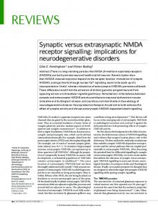
Fig. 1. PAR1 activation and signalling. PAR1 is a seven-transmembrane-span G-protein-coupled receptor that is activated by a unique proteolytic mechanism. Thrombin (␣-Th), a serine protease, binds to and cleaves the N-terminal exodomain of PAR1. The newly unmasked N-terminus of PAR1 then acts as a tethered ligand and binds intramolecularly to the receptor to trigger transmembrane signalling. Synthetic peptides that mimic the tethered ligand domain can activate PAR1 independently of proteolysis. PAR1 couples to G␣q, G␣12/13, G␣i and G␥ to activate a variety of signalling cascades and cellular responses. The G␣12/13 subunits bind RhoGEFs, which activate small G-proteins such as Rho. G␣q activates phospholipase C, triggering phosphoinositide hydrolysis, resulting in inositol (1,4,5)-trisphosphate [Ins(1,4,5)P3] and diacylglycerol (DAG) production and Ca2+ mobilization, protein kinase C (PKC) and MAP kinase activation. G␥ can activate PI-3-kinase, G-protein-coupled receptor kinases (GRKs) and other effectors. PAR1 is uncoupled from G-protein signalling by rapid phosphorylation and arrestin binding.
N
N α-Th
α-Th
Arrestin2
C
C
Gα12/13
Gαq
RhoGEFs Phospholipase Cβ Rho
Gα i Adenylyl cyclase
PtdIns(1,4,5)P3, DAG
C
P
P
Gβγ PI-3-kinase, GRKs, K+ channels, phospholipase Cβ
Ca2+, PKC, MAP kinases (ERK1,2)
Cell shape, adhesion, secretion, growth and motility
PAR signalling responses but promotes ERK1/2 signalling comparable to that Once activated, PARs are likely to undergo conformational elicited by thrombin (Ludeman et al., 2005; Riewald et al., changes to facilitate coupling to heterotrimeric G-proteins. 2002). Clearly, the molecular mechanisms that dictate PAR–GPAR1 couples to the G␣q, G␣i and G␣12/13 subtypes and protein selectivity deserve more research. induces activation of MAP kinases, mobilization of intracellular Ca2+, RhoGEF-mediated Rho and Rac signalling PAR signal regulation and regulation of other effectors to promote diverse cellular The proteolytic activation of PARs is irreversible; thus, responses (Fig. 1) (Coughlin, 2005). PAR4 also appears to signalling must be tightly regulated. Desensitization of couple to G␣q and G␣12/13, but not G␣i, at least in fibroblasts activated GPCRs occurs by rapid phosphorylation and arrestin (Faruqi et al., 2000), whereas PAR3 has not been shown to binding, which promotes uncoupling of the receptor from Gsignal through G-proteins. Although there is no direct evidence protein signalling within seconds. GPCR internalization ensues linking PAR2 to G-protein activation, numerous studies show within minutes and removes activated receptor from G-proteins that PAR2-selective agonists increase second messenger and signalling effectors at the plasma membrane. Like most responses suggestive of G␣q, G␣i and perhaps G␣12/13 GPCRs, activated PAR1 is rapidly desensitized by signalling. In addition, activated PAR2 binds to and internalizes with arrestin, a multifunctional adaptor protein (DeFea et al., 2000; Stalheim et al., 2005). The interaction of internalized PAR2 with arrestin is thought to sustain ERK1/2 signalling in the cytoplasm independently of Gprotein activation. Activation of distinct G-protein subtypes and non-G-protein effectors by PARs is crucial for eliciting cell type-specific responses; however, the mechanisms responsible are not known. PAR1 appears to display differential G-protein coupling when activated proteolytically by its tethered ligand versus free peptide agonists (McLaughlin et al., 2005). Several GPCRs Fig. 2. Human PAR N-terminal and C-terminal sequences. Residues flanking the display such functional selectivity, and different cleavage site specify protease recognition. These are defined as unprimed (P) and conformational states of the activated receptor primed sites (P⬘), respectively (i.e. P3-P2-P1/P1⬘-P2⬘-P3⬘, where / indicates the could couple selectively to distinct G-protein cleavage site). Only transmembrane domain (TM) 1 and TM7 are shown. The subtypes. The compartmentalization of PARs number of N-terminal residues omitted before the cleavage site is shown in and G-proteins in plasma membrane lipid raft parentheses and the sequences of the tethered ligand domains are shown in yellow. microdomains such as caveolae might also The hirudin-like sequence in PAR1 and PAR3 responsible for high-affinity thrombin confer PAR–G-protein selectivity. This could binding is underlined. The potential sites of PAR phosphorylation are shown in red explain why APC bound to EPCR is markedly and the PAR1 distal tyrosine-based motif important for 2 binding is underlined. Asterisks indicate the end of the PAR sequences. less efficient at stimulating G␣q-mediated
924
Journal of Cell Science 120 (6)
Constitutive internalization
PAR1
Trypsin TF-VIIa-Xa
Thrombin N
PAR4
Thrombin N
N
N
PAR3
PAR2
Agonist-induced internalization
Thrombin N
N
P Adaptor
AP2 C
Ub
?
C
P
Arrestin
C
C
Ub
Adaptor
C
? ?
Recycling endosome
Early endosome
Early endosome
Early endosome
N
SNX1
P P C
C
t1/2=1h
Journal of Cell Science
Lysosome
Arrestin C
ERK1/2
C
Ub
?
t1/2=3h Lysosome
Lysosome
Fig. 3. PAR endocytic sorting. PAR1 displays both constitutive and agonist-induced internalization. Uncleaved PAR1 constitutively cycles between the plasma membrane and an intracellular compartment. The clathrin adaptor AP2, which binds to a tyrosine-based motif in the PAR1 cytoplasmic tail, mediates constitutive internalization. PAR1 is basally ubiquitylated. Agonist-induced internalization of PAR1 requires phosphorylation and ubiquitylation specifies internalization through clathrin-coated pits. The clathrin adaptor that mediates agonist-induced internalization of ubiquitylated PAR1 has not been identified. Internalized PAR1 is sorted from endosomes to lysosomes through a sorting nexin-1 (SNX1)-dependent pathway, which is independent of ubiquitylation and the ESCRT-1 machinery. Arrestins mediate PAR2 internalization. Internalized PAR2 bound to arrestins appear to signal to ERK1/2 from an endocytic compartment. Sorting of PAR2 from endosomes to lysosomes requires ubiquitylation. The sorting mechanism(s) and adaptor proteins that mediate PAR4 internalization and lysosomal sorting are not known, and whether PAR3 redistributes from the cell surface remains to be determined.
phosphorylation and arrestin binding (Fig. 1) (Ishii et al., 1994; Paing et al., 2002). However, unlike most GPCRs, which are internalized and then recycled, activated PAR1 is internalized, sorted directly to lysosomes and rapidly degraded independently of arrestins (Fig. 3) (Hoxie et al., 1993; Trejo and Coughlin, 1999). Internalization of activated PAR1 removes it from signalling effectors, whereas lysosomal sorting prevents it from returning to the cell surface and continuing to signal, terminating signalling (Trejo et al., 1998). Internalization of activated PAR4 also appears to be a mechanism for signal termination, but phosphorylation of the activated receptor has not been detected (Shapiro et al., 2000). A slower internalization rate and/or lack of phosphorylation of activated PAR4 are thought to promote sustained signalling, which is important for thrombin-stimulated platelet aggregation (Covic et al., 2000; Shapiro et al., 2000). The mechanisms that control PAR2 signalling are not clearly defined. On the basis of pharmacological inhibitor studies, a role for phosphorylation in shutting off PAR2 signalling has been suggested but not directly demonstrated, despite the abundance of multiple serine and threonine residues in the cytoplasmic tail (Fig. 2) (Bohm et al., 1996). However, arrestins do mediate desensitization and internalization of activated PAR2 (Fig. 3) (Stalheim et al., 2005). In addition to uncoupling PAR2 from G-proteins, arrestins appear to promote sustained ERK1/2 signalling by activated PAR2 in the cytoplasm (DeFea et al., 2000; Stalheim et al., 2005). The mechanism(s) that regulate signalling by internalized PAR2 are not known.
PAR1 displays two modes of trafficking important for regulation of signalling (Fig. 3). Uncleaved PAR1 cycles constitutively between the cell surface and an intracellular compartment, generating a protected receptor pool that replenishes the cell surface after protease exposure and leads to rapid resensitization independently of de novo receptor synthesis (Hein et al., 1994; Paing et al., 2006). Activated PAR1, by contrast, is internalized, sorted to lysosomes and degraded (Hoxie et al., 1993; Trejo and Coughlin, 1999). Constitutive internalization and agonist-induced internalization of PAR1 are clathrin- and dynamin-dependent (Trejo et al., 2000). However, in contrast to most GPCRs, neither constitutive nor activated PAR1 internalization requires arrestins (Paing et al., 2002). Arrestins interact with clathrin and clathrin adaptor protein complex 2 (AP2) to facilitate internalization of activated GPCRs through clathrin-coated pits (Goodman, et al., 1996; Laporte et al., 1999). Rather than arrestins, AP2 is crucial for PAR1 constitutive internalization and is essential for cellular resensitization to thrombin signalling (Paing et al., 2006). The 2 subunit of AP2 binds directly to a tyrosine-based motif in the cytoplasmic tail of PAR1 (Figs 2 and 3). This motif and AP2 function are each crucial for maintaining an intracellular pool of receptors that replenish the cell surface with uncleaved PAR1 after protease exposure. Interestingly, internalization of activated PAR1 through clathrin-coated pits is not dependent on arrestins or AP2, which suggests that the two internalization routes are specified by distinct endocytic machineries. The internalization mechanism of activated PAR1 remains to be defined.
Journal of Cell Science
PAR signalling and trafficking
925
Arrestins are required for internalization of activated PAR2 in fibroblasts (Stalheim et al., 2005), a process that is dynamin-dependent (Roosterman et al., 2003). Dynamin, a large GTPase, promotes endocytosis by facilitating release of clathrin-coated pits or caveolae from the plasma membrane. Whether PAR2 is sequestered in clathrin-coated pits or caveolae or both remains to be determined. Interestingly, the recovery of uncleaved PAR2, unlike PAR1, to the cell surface after protease exposure does not involve constitutive cycling but is instead because of movement of naïve receptor from a pre-existing Golgi pool as well as de novo receptor synthesis (Dery et al., 1999). The mechanisms that regulate PAR4 internalization are not known, and whether cleaved PAR3 leaves the cell surface has not been Fig. 4. PARs function in the vasculature and in tumor invasion and metastasis. determined. PAR function is shown in the context of a blood vessel. Thrombin, the main The best-characterized route for lysosomal effector protease of the coagulation cascade, activates PAR1, PAR3 and PAR4 degradation of integral membrane proteins involves to elicit signalling in a variety of cell types. Thrombin activates PAR1 and PAR4 in human platelets and generates fibrin, which is important for thrombus ubiquitin-dependent sorting by the ESCRT formation and tumor cell survival and metastasis. FXa can also cleave and machinery (Katzmann et al., 2002). Ubiquitylation is activate PAR1. Activated endothelial cells express tissue factor (TF) and are involved in lysosomal sorting of the 2-adrenergic represented by an oval shape. PAR2 is activated by TF-bound FVIIa and FXa and chemokine CXCR4 GPCRs (Marchese and but not by thrombin. PAR1 and PAR2 are also cleaved and activated by tumorBenovic, 2001; Shenoy et al., 2001). Recent work generated proteases, which contribute to tumor cell growth, invasion and shows that PAR1 is basally ubiquitylated (B. L. metastasis. Wolfe, A. Marchese and J.T., unpublished). However, in contrast to the 2-adrenergic receptor and CXCR4, metastatic colony formation as well as for angiogenesis. In ubiquitylation of PAR1 is not required for lysosomal addition, thrombin can promote tumor progression by acting degradation since a non-ubiquitylatable PAR1 mutant is directly on tumor cell-expressed PARs. PAR1 is overexpressed degraded comparably to the wild-type receptor in fibroblasts. in aggressive melanoma (Tellez and Bar-Eli, 2003), colon Moreover, agonist-induced PAR1 lysosomal degradation is cancer (Darmoul et al., 2003), prostate cancer (Chay et al., independent of Hrs and Tsg101 (Gullapalli et al., 2006), which 2002) and invasive breast cancer (Even-Ram et al., 1998). The promote assembly of a multiprotein ESCRT-I complex that expression of PAR1 and PAR2 is also increased in stromal sorts ubiquitylated cargo into the involuting membrane of fibroblasts of malignant tissues but not those in normal or multivesicular endosomes (Raiborg et al., 2003). The novel benign breast tissue specimens (D’Andrea et al., 2001). lysosomal sorting pathway for activated PAR1 does, however, Overexpression of PAR1 transforms NIH 3T3 fibroblasts involve sorting nexin 1, a phox homology (PX)-domain(Martin et al., 2001; Whitehead et al., 1995) and induces containing protein that binds phosphoinositides and functions hyperplasia of mammary gland epithelial cells, an oncogenic in membrane trafficking (Fig. 3) (Gullapalli et al., 2006). phenotype (Yin et al., 2003a). Moreover, targeted Sorting nexin 1 is not responsible for lysosomal sorting of all overexpression of human PAR1 in mouse mammary glands PARs since ubiquitylation of PAR2 mediates lysosomal sorting activates the Wnt and -catenin pathway, which is crucial for and degradation (Jacob et al., 2005) similarly to other GPCRs. tumor progression in certain types of malignant cancer (Yin et al., 2006). PARs and cancer PARs also have the capacity to transduce signals in response PARs elicit cellular responses to coagulant proteases triggered to multiple tumor-generated proteases. Tumors are replete with by vascular injury (Coughlin, 2005). In addition, coagulant proteases, including urokinase-plasminogen activator (uPA) proteases and PARs have been implicated in several types of and MMPs. Tumor cells upregulate uPA expression, which is malignant cancer. A link between hyperactivation of associated with poor prognosis (Nicolai and Blasi, 2003). UPA coagulation and tumor progression is well documented. In fact, binds to its cell surface receptor uPAR especially at the invasive tumors have been referred to as ‘wounds that do not heal’ since front (Bissell and Radisky, 2001) and cleaves plasminogen to similar signalling pathways operate in wound healing and generate plasmin. Plasmin can proteolytically activate PAR1 tumor progression (Bissell and Radisky, 2001). In contrast to (Kuliopulos et al., 1999). Plasmin also degrades extracellular wound healing, however, in cancer these signalling pathways matrix proteins and cleaves and activates MMPs. Reactive contribute to tumor growth and metastasis. stromal cells produce several types of MMPs, including MMPActivation of coagulation can support tumor progression at 1, which can activate PAR1 (Boire et al., 2005). TF is also multiple levels. Thrombin activates platelets and endothelial upregulated in many tumor cell types and supports FVIIa and cells and cleaves fibrinogen to generate fibrin (Fig. 4). Tumor FXa activity (Riewald and Ruf, 2002). TF bound to FVIIa can cells lodge in vascular sites with platelet and fibrin thrombi, activate PAR2, whereas FXa can signal through either PAR1 which promotes tumor cell survival and metastasis (Camerer or PAR2 (Fig. 4). In addition, FXa is responsible for proteolytic et al., 2004; Gasic et al., 1968; Palumbo et al., 2000; Rickles conversion of prothrombin to thrombin, the main effector and Edwards, 1983). Fibrin also serves as a matrix for
Journal of Cell Science
926
Journal of Cell Science 120 (6)
protease of PAR1. A recent study shows that tumor cells are able to synthesize and secrete FVII ectopically, indicating that coagulant proteases can be generated in tumors independently of their production in liver (Koizume et al., 2006). PAR1 can promote tumor progression through a variety of mechanisms. Thrombin stimulates proliferation of colon cancer and melanoma cells (Darmoul et al., 2003; Tellez and Bar-Eli, 2003). The mitogenic activity of thrombin is mediated by PAR1 and is associated with prolonged ERK1/2 activation (Kahan et al., 1992; Trejo et al., 1996). Sustained ERK1/2 signalling not only promotes cell cycle progression but also contributes to cellular transformation, migration and survival. PAR1 promotes cell motility through a mechanism involving activation of the EGFR in renal carcinoma (Bergmann et al., 2006). In addition, PAR1 activates ␣v5 integrin in melanoma to promote migration and invasion (Even-Ram et al., 2001). PAR1 may also contribute to tumor cell survival since it can prevent apoptosis of certain cell types (Guo et al., 2004; Zania et al., 2006). However, the role of PAR1 in tumor cell apoptosis has not been firmly established. Other studies also suggest an important role for PAR1 in tumor growth and invasion. The expression of PAR1 in noninvasive MCF7 breast carcinoma is sufficient to promote growth and invasion in a xenograft nude mouse model (Boire et al., 2005). In addition, antisense- or RNAi-mediated reduction of PAR1 levels diminishes invasiveness of several breast carcinoma cell lines in vitro (Booden et al., 2004; EvenRam et al., 1998; Morris et al., 2006). The ability of PAR1 to promote tumor growth and invasion could involve its regulation of angiogenesis. Approximately 50% of Par1-null mice die at midgestation owing to abnormal vascular development, whereas restoration of PAR1 expression in endothelial cells prevents death (Connolly et al., 1996; Griffin et al., 2001). Thus, PAR1 signalling in endothelial cells is important for blood vessel formation. Moreover, induction of PAR1 expression and signalling in melanoma or prostate carcinoma increases tumor growth, angiogenesis and vascular endothelial growth factor (VEGF) production (Yin et al., 2003b). PAR1 signalling is also dysregulated in tumor cells. Activated PAR1 fails to be downregulated in highly invasive breast carcinoma, and consequently PAR1 persistently activates ERK1/2 and induces cellular invasion (Booden et al., 2004). One phenomenon contributing to persistent signalling is slowed receptor internalization and/or recycling and a lack of lysosomal degradation. These defects appear to be specific to breast cancer cells, because PAR1 ectopically expressed in normal human mammary epithelial cells displays proper trafficking and signal termination (Booden et al., 2004). Increased PAR1 levels in invasive carcinoma cells may thus be due, at least partially, to defective trafficking. Several other crucial mediators of tumor progression, including EGFR, HER2/ErbB2 and CXCR4, also display defective trafficking in breast carcinoma (Harari and Yarden, 2000; Li et al., 2004). Other mechanisms contributing to increased PAR1 mRNA levels and protein involve changes in gene transcription regulated in part by the transcription factor AP2 (Tellez et al., 2003). Although Par1 mutations acquired during tumorigenesis could also lead to increased stability of the receptor or constitutive activity, we found no missense mutations in the Par1 coding sequence in several breast carcinoma cell lines. The mechanisms responsible for defective
PAR1 trafficking in invasive breast carcinoma remain to be defined. There is increasing evidence that PAR2 is an important mediator of tumor progression. The levels of trypsin, a potent and physiological activator of PAR2, are elevated in gastric, colon, ovarian and lung tumors (Ducroc et al., 2002). TF is also linked to cancer progression (Belting et al., 2005). TF-FVIIa and FXa stimulate breast carcinoma cell migration and invasion through activation of PAR2 (Hjortoe et al., 2004; Morris et al., 2006). Induction of breast carcinoma cell migration by activated PAR2 involves arrestin-dependent ERK1/2 activation (Ge et al., 2004). In colon and gastric carcinoma, activated PAR2 stimulates EGFR activity, ERK1/2 signalling and cellular proliferation (Caruso et al., 2006; Darmoul et al., 2004). Interestingly, a TF mutant lacking its cytoplasmic tail domain enhances angiogenesis through a PAR2-dependent mechanism, which suggests that the TF cytoplasmic tail negatively regulates the proangiogenic actions of PAR2 (Belting et al., 2004). Activated PAR2 stimulates protein-kinase-C-mediated phosphorylation of the TF cytoplasmic tail, alleviating this inhibition, and thereby enhances angiogenesis and cell migration (Ahamed and Ruf, 2004; Belting et al., 2004). Thus, one can envision a scenario in which TF-VIIa-mediated activation of PAR2 causes dysregulated signalling and sustained TF cytoplasmic tail phosphorylation, resulting in aberrant tumor cell migration and angiogenesis. Conclusions and perspectives PARs are uniquely activated by proteolytic cleavage, which results in irreversible activation, unlike normal ligand-activated GPCRs. Thus, rapid desensitization and receptor trafficking tightly regulate PAR signalling. The temporal and spatial regulation of PAR signalling is crucial for a variety of biological responses, including proper growth and cell migration. Recent studies have advanced our understanding of the molecular mechanisms that control constitutive endocytosis of PAR1 and its importance in control of cellular resensitization. We will now need to explore the mechanism by which activated PAR1 is internalized, which is essential for signal termination. The mechanisms that control signalling and trafficking of other PARs have yet to be fully elucidated and are also vital to our understanding of coagulant protease signalling. Fundamental questions regarding the mechanisms that control PAR–G-protein selectivity also remain. PARs are implicated in tumor progression but precisely how these receptors contribute to tumor cell growth, invasion and metastasis is not known. Thus, establishing this will be crucial to our understanding of the molecular basis of metastatic disease and for identifying new drug targets for cancer treatment. Proteases are viewed as essential for tumor progression and metastasis, but the development of protease inhibitors has been hampered by lack of specificity and associated toxic effects. Thus, a major challenge in the field is to identify specific protease effectors important for cancer progression. PARs are one class of protease effector implicated in tumor progression that might serve as ideal drug targets. We thank members of the J. Trejo laboratory for comments and advice. This work is supported by NIH HL073328, an American Heart Association Established Investigator Award and a Susan G. Komen
PAR signalling and trafficking Breast Cancer Foundation Award (to J.T.). T.K.R. is supported by a UNCF-Merck predoctoral fellowship. We also acknowledge that owing to space limitations several important studies in the field could not be cited.
Journal of Cell Science
References Ahamed, J. and Ruf, W. (2004). Protease-activated receptor 2-dependent phosphorylation of the tissue factor cytoplasmic domain. J. Biol. Chem. 279, 2303823044. Ahamed, J., Versteeg, H. H., Kerver, M., Chen, V. M., Mueller, B. M., Hogg, P. J. and Ruf, W. (2006). Disulfide isomerization switches tissue factor from coagulation to cell signaling. Proc. Natl. Acad. Sci. USA 45, 12020-12028. Belting, M., Dorrell, M., Sandgren, S., Aguilar, E., Ahamed, J., Dorfleutner, A., Carmeliet, P., Mueller, B. M., Friedlander, M. and Ruf, W. (2004). Regulation of angiogenesis by tissue factor cytoplasmic domain signaling. Nat. Med. 10, 502-509. Belting, M., Ahamed, J. and Ruf, W. (2005). Signaling of the tissue factor coagulation in angiogenesis and cancer. Arterioscler. Thromb. Vasc. Biol. 25, 1545-1550. Bergmann, S., Junker, K., Henklein, P., Hollenberg, M. D., Settmacher, U. and Kaufmann, R. (2006). PAR-type thrombin receptors in renal carcinoma cells: PAR1mediated EGFR activation promotes cell migration. Oncol. Rep. 15, 889-893. Bissell, M. J. and Radisky, D. (2001). Putting tumors in context. Nat. Rev. Cancer 1, 4654. Bohm, S. K., Khitin, L. M., Grady, E. F., Aponte, G., Payan, D. G. and Bunnett, N. W. (1996). Mechanisms of desensitization and resensitization of proteinase-activated receptor-2. J. Biol. Chem. 271, 22003-22016. Boire, A., Covic, L., Agarwal, A., Jacques, S., Sherifi, S. and Kuliopulos, A. (2005). PAR1 is a matrix metalloprotease-1 receptor that promotes invasion and tumorigenesis of breast cancer cells. Cell 120, 303-313. Booden, M. A., Ekert, L., Der, C. J. and Trejo, J. (2004). Persistent signaling by dysregulated thrombin receptor trafficking promotes breast carcinoma cell invasion. Mol. Cell. Biol. 24, 1990-1999. Camerer, E., Huang, W. and Coughlin, S. R. (2000). Tissue factor- and factor Xdependent activation of protease-activated receptor 2 by factor VIIa. Proc. Natl. Acad. Sci. USA 97, 5255-5260. Camerer, E., Qazi, A. A., Duong, D., Cornelissen, I., Advincula, R. and Coughlin, S. R. (2004). Platelets, protease-activated receptors, and fibrinogen in hematogenous metastasis. Blood 104, 397-401. Caruso, R., Pallone, F., Fina, D., Gioia, V., Peluso, I., Caprioli, F., Stolfi, C., Perfetti, A., Spagnoli, L. G., Palmieri, G. et al. (2006). Protease-activated receptor-2 activation in gastric cancer cells promotes epidermal growth factor receptor trans-activation and proliferation. Am. J. Pathol. 169, 268-278. Chay, C. H., Cooper, C. R., Gendernalik, J. D., Dhanasekaran, S. M., Chinnaiyan, A. M., Rubin, M. A., Schmaier, A. H. and Pienta, K. J. (2002). A functional thrombin receptor (PAR1) is expressed on bone-derived prostate cancer cell lines. Urology 60, 760-765. Compton, S. J., Sandhu, S., Wijesuriya, S. J. and Hollenberg, M. D. (2002). Glycosylation of human protease-activated receptor-2 (hPAR2): role in cell surface expression and signalling. Biochem. J. 368, 495-505. Connolly, A. J., Ishihara, H., Kahn, M. L., Farese, R. V. and Coughlin, S. R. (1996). Role of the thrombin receptor in development and evidence for a second receptor. Nature 381, 516-519. Coughlin, S. R. (1999). How the protease thrombin talks to cells. Proc. Natl. Acad. Sci. USA 96, 11023-11027. Coughlin, S. R. (2000). Thrombin signalling and protease-activated receptors. Nature 407, 258-264. Coughlin, S. R. (2005). Protease-activated receptors in hemostasis, thrombosis and vascular biology. J. Thromb. Haemost. 3, 1800-1814. Covic, L., Gresser, A. L. and Kuliopulos, A. (2000). Biphasic kinetics of activation and signaling for PAR1 and PAR4 thrombin receptors in platelets. Biochemistry 39, 54585467. D’Andrea, M. R., Derian, C. K., Santulli, R. J. and Andrade-Gordon, P. (2001). Differential expression of protease-activated receptors-1 and -2 in stromal fibroblasts of normal, benign, and malignant human tissues. Am. J. Pathol. 158, 2031-2041. Darmoul, D., Gratio, V., Devaud, H., Lehy, T. and Laburthe, M. (2003). Aberrant expression and activation of the thrombin receptor protease-activated receptor-1 induce cell proliferation and motility in human colon cancer cells. Am. J. Pathol. 162, 15031513. Darmoul, D., Gratio, V., Devaud, H. and Labrurthe, M. (2004). Protease-activated receptor-2 in colon cancer: trypsin-induced MAPK phosphorylation and cell proliferation are mediated by epidermal growth factor transactivation. J. Biol. Chem. 279, 20927-20934. DeFea, K. A., Zalevski, J., Thoma, M. S., Dery, O., Mullins, R. D. and Bunnett, N. W. (2000). -arrestin-dependent endocytosis of proteinase-activated receptor-2 is required for intracellular targeting of activated ERK1/2. J. Cell Biol. 148, 1267-1281. Dery, O., Thoma, M. S., Wong, H., Grady, E. F. and Bunnett, N. W. (1999). Trafficking of proteinase-activated receptor-2 and -arrestin-1 tagged with green fluorescent protein. J. Biol. Chem. 274, 18524-18535. Ducroc, R., Bontemps, C., Marazova, K., Devaud, H., Darmoul, D. and Laburthe, M. (2002). Trypsin is produced by and activates protease-activated receptor-2 in human cancer colon cells: evidence for new autocrine loop. Life Sci. 70, 1359-1367. Even-Ram, S., Uziely, B., Cohen, P., Grisaru-Granovsky, S., Maoz, M., Ginzburg, Y., Reich, R., Vlodavsky, I. and Bar-Shavit, R. (1998). Thrombin receptor
927
overexpression in malignant and physiological invasion processes. Nat. Med. 4, 909914. Even-Ram, S., Maoz, M., Pokroy, E., Reich, R., Katz, B. Z., Gutwein, P., Altevogt, P. and Bar-Shavit, R. (2001). Tumor cell invasion is promoted by protease-activated receptor-1 in cooperation with the alpha vbeta 5 integrin. J. Biol. Chem. 276, 1095210962. Faruqi, T. R., Weiss, E. J., Shapiro, M. J., Huang, W. and Coughlin, S. R. (2000). Structure-function analysis of protease-activated receptor-4 tethered ligand peptides. Determinants of specificity and utility in assays of receptor function. J. Biol. Chem. 275, 19728-19734. Feistritzer, C. and Riewald, M. (2005). Endothelial barrier protection by activated protein C through PAR1-dependent sphingosine-1-phosphate receptor-1 crossactivation. Blood 105, 3178-3184. Gasic, G., Gasic, T. and Stewart, C. (1968). Antimetastatic effects associated with platelet reduction. Proc. Natl. Acad. Sci. USA 61, 46-52. Ge, L., Shenoy, S. K., Lefkowitz, R. J. and DeFea, K. (2004). Constitutive proteaseactivated-receptor-2 mediated migration of MDA-MB-231 breast cancer cells requires both -arrestin-1 and 2. J. Biol. Chem. 279, 55419-55424. Goodman, O. B., Jr, Krupnick, J. G., Santini, F., Gurevich, V. V., Penn, R. B., Gagnon, A. W., Keen, J. H. and Benovic, J. L. (1996). Beta-arrestin acts as a clathrin adaptor in endocytosis of the Beta2-adrenergic receptor. Nature 383, 447-450. Griffin, C. T., Srinivasan, Y., Zheng, Y.-W., Huang, W. and Coughlin, S. R. (2001). A role for thrombin receptor signaling in endothelial cells during embryonic development. Science 293, 1666-1670. Gullapalli, A., Wolfe, B. L., Griffin, C. T., Magnuson, T. and Trejo, J. (2006). An essential role for SNX1 in lysosomal sorting protease-activated receptor-1: evidence for retromer, Hrs and Tsg101 independent functions of sorting nexins. Mol. Biol. Cell 17, 1228-1238. Guo, H., Liu, D., Gelbard, H., Cheng, T., Insalasco, R., Fernandez, J. A., Griffin, J. H. and Zlokovic, B. V. (2004). Activated protein C prevents neuronal apoptosis via protease activated receptors 1 and 3. Neuron 41, 563-572. Harari, D. and Yarden, Y. (2000). Molecular mechanisms underlying ErbB2/HER2 action in breast cancer. Oncogene 19, 6102-6114. Hein, L., Ishii, K., Coughlin, S. R. and Kobilka, B. K. (1994). Intracellular targeting and trafficking of thrombin receptors: a novel mechanism for resensitization of a G protein-coupled receptor. J. Biol. Chem. 269, 27719-27726. Hjortoe, G. M., Peterson, L. C., Albrektsen, T., Sorensen, B. B., Norby, P. L., Mandal, S. K., Pendurthi, U. R. and Rao, L. V. M. (2004). Tissue factor-factor VIIa-specific up-regulation of IL-8 expression in MDA-MB-231 cells is mediated by PAR-2 and results in increased cell migration. Blood 103, 3029-3037. Hoxie, J. A., Ahuja, M., Belmonte, E., Pizarro, S., Parton, R. and Brass, L. F. (1993). Internalization and recycling of activated thrombin receptors. J. Biol. Chem. 268, 13756-13763. Ishihara, H., Connolly, A. J., Zeng, D., Kahn, M. L., Zheng, Y. W., Timmons, C., Tram, T. and Coughlin, S. R. (1997). Protease-activated receptor 3 is a second thrombin receptor in humans. Nature 386, 502-506. Ishii, K., Chen, J., Ishii, M., Koch, W. J., Freedman, N. J., Lefkowitz, R. J. and Coughlin, S. R. (1994). Inhibition of thrombin receptor signaling by a G proteincoupled receptor kinase. Functional specificity among G protein-coupled receptor kinases. J. Biol. Chem. 269, 1125-1130. Jacob, C., Cottrell, G. S., Gehringer, D., Schmidlin, F., Grady, E. F. and Bunnett, N. W. (2005). c-Cbl mediates ubiquitination, degradation, and down-regulation of human protease-activated receptor 2. J. Biol. Chem. 280, 16076-16087. Kahan, C., Seuwen, K., Meloche, S. and Pouyssegur, J. (1992). Coordinate, biphasic activation of p44 mitogen-activated protein kinase and S6 kinase by growth factors in hamster fibroblasts. J. Biol. Chem. 267, 13369-13375. Kahn, M. L., Zheng, Y. W., Huang, W., Bigornia, V., Zeng, D., Moff, S., Farese, R. V., Jr, Tam, C. and Coughlin, S. R. (1998). A dual thrombin receptor system for platelet activation. Nature 394, 690-694. Katzmann, D. J., Odorizzi, G. and Emr, S. D. (2002). Receptor downregulation and multivesicular-body sorting. Nat. Rev. Mol. Cell Biol. 3, 893-905. Koizume, S., Jin, M. S., Miyagi, E., Hirahara, F., Nakamura, Y., Piao, J. H., Asai, A., Yoshida, A., Tsuchiya, E., Ruf, W. et al. (2006). Activation of cancer cell migration and invasion by ectopic synthesis of coagulation factor VII. Cancer Res. 66, 9453-9460. Kuliopulos, A., Covic, L., Seely, S. K., Sheridan, P. J., Helin, J. and Costello, C. E. (1999). Plasmin desensitization of the PAR1 thrombin receptor: kinetics, sites of truncation, and implications for thrombolytic therapy. Biochemistry 38, 4572-4585. Laporte, S., Oakley, R. H., Zhang, J., Holt, J. A., Ferguson, S. S. G., Caron, M. G. and Barak, L. S. (1999). The 2-adrenergic receptor/arrestin complex recruits the clathrin adaptor AP-2 during endocytosis. Proc. Natl. Acad. Sci. USA 96, 3712-3717. Li, Y. M., Pan, Y., Wei, Y., Cheng, X., Zhou, B. P., Tan, M., Zhou, X., Xia, W., Hortobagyi, G. N., Yu, D. et al. (2004). Upregulation of CXCR4 is essential for HER2-mediated tumor metastasis. Cancer Cell 6, 459-469. Ludeman, M. J., Zheng, Y. W., Ishii, K. and Coughlin, S. R. (2004). Regulated shedding of PAR1 N-terminal exodomain from endothelial cells. J. Biol. Chem. 279, 18592-18599. Ludeman, M. J., Kataoka, H., Srinivasas, Y., Esmon, N. L., Esmon, C. T. and Coughlin, S. R. (2005). PAR1 cleavage and signaling in response to activated protein C and thrombin. J. Biol. Chem. 280, 13122-13128. Marchese, A. and Benovic, J. L. (2001). Agonist-promoted ubiquitination of the G protein-coupled receptor CXCR4 mediates lysosomal sorting. J. Biol. Chem. 276, 45509-45512.
Journal of Cell Science
928
Journal of Cell Science 120 (6)
Martin, C. B., Mahon, G. M., Klinger, M. B., Kay, R. J., Symons, M., Der, C. J. and Whitehead, I. P. (2001). The thrombin receptor, PAR-1, causes transformation by activation of Rho-mediated signaling pathways. Oncogene 20, 1953-1963. McLaughlin, J. N., Shen, L., Holinstat, M., Brooks, J. D., DiBenedetto, E. and Hamm, H. E. (2005). Functional selectivity of G protein signaling by agonist peptides and thrombin for the protease-activated receptor-1. J. Biol. Chem. 280, 25048-25059. Molino, M., Barnathan, E. S., Numerof, R., Clark, J., Dreyer, M., Cumashi, A., Hoxie, J. A., Schechter, N., Woolkalis, M. and Brass, L. F. (1997). Interactions of mast cell tryptase with thrombin receptors and PAR-2. J. Biol. Chem. 272, 4043-4049. Morris, D. R., Ding, Y., Ricks, T. K., Gullapalli, A., Wolfe, B. L. and Trejo, J. (2006). Protease-activated receptor-2 is essential for factor VIIa and Xa-induced signaling, migration and invasion of breast cancer cells. Cancer Res. 66, 307-314. Nakanishi-Matsui, M., Zheng, Y.-W., Weiss, E. J., Sulciner, D. and Coughlin, S. R. (2000). PAR3 is a cofactor for PAR4 activation by thrombin. Nature 404, 609-613. Nicolai, S. and Blasi, F. (2003). The urokinase plasminogen activator system in cancer: recent advances and implication for prognosis and therapy. Cancer Met. Rev. 22, 205222. Nystedt, S., Emilsson, K., Wahlestedt, C. and Sundelin, J. (1994). Molecular cloning of a potential novel proteinase activated receptor. Proc. Natl. Acad. Sci. USA 91, 92089212. O’Brien, P. J., Prevost, N., Molino, M., Hollinger, M. K., Woolkalis, M. J., Woulfe, D. S. and Brass, L. F. (2000). Thrombin responses in human endothelial cells. Contributions from receptors other than PAR1 include transactivation of PAR2 by thrombin-cleaved PAR1. J. Biol. Chem. 275, 13502-13509. Paing, M. M., Stutts, A. B., Kohout, T. A., Lefkowitz, R. J. and Trejo, J. (2002). arrestins regulate protease-activated receptor-1 desensitization but not internalization or down-regulation. J. Biol. Chem. 277, 1292-1300. Paing, M. M., Johnston, C. A., Siderovski, D. P. and Trejo, J. (2006). Clathrin adaptor AP2 regulates thrombin receptor constitutive internalization and endothelial cell resensitization. Mol. Cell. Biol. 28, 3221-3242. Palumbo, J. S., Kombrinck, K. W., Drew, A. F., Grimes, T. S., Kiser, J. H., Degen, J. L. and Bugge, T. H. (2000). Fibrinogen is an important determinant of the metastatic potential of circulating tumor cells. Blood 96, 3302-3309. Raiborg, C., Rusten, T. E. and Stenmark, H. (2003). Protein sorting into multivesicular endsomes. Curr. Opin. Cell Biol. 15, 446-455. Rasmussen, U. B., Vouret-Craviari, V., Jallat, S., Schlesinger, Y., Pagers, G., Pavirani, A., Lecocq, J. P., Pouyssegur, J. and Van Obberghen-Schilling, E. (1991). cDNA cloning and expression of a hamster alpha-thrombin receptor coupled to Ca2+ mobilization. FEBS Lett. 288, 123-128. Rickles, F. R. and Edwards, R. L. (1983). Activation of blood coagulation in cancer: Trousseau’s syndrome revisited. Blood 62, 14-31. Riewald, M. and Ruf, W. (2001). Mechanistic coupling of protease signaling and initiation of coagulation by tissue factor. Proc. Natl. Acad. Sci. USA 98, 7742-7747. Riewald, M. and Ruf, M. (2002). Orchestration of coagulation protease signaling by tissue factor. Trends Cardiovasc. Med. 12, 149-154. Riewald, M., Petrovan, R. J., Donner, A., Mueller, B. M. and Ruf, W. (2002). Activation of endothelial cell protease activated receptor-1 by the protein C pathway. Science 296, 1880-1882. Roosterman, D., Schmidlin, F. and Bunnett, N. W. (2003). Rab5a and rab11a mediate agonist-induced trafficking of protease-activated receptor-2. Am. J. Physiol. Cell Physiol. 284, C1319-C1329. Rydel, T. J., Ravichandran, K. G., Tulinsky, A., Bode, W., Huber, R., Roitsch, C. and Fenton, J. W., 2nd (1990). The structure of a complex of recombinant hirudin and human alpha-thrombin. Science 249, 277-280. Shapiro, M. J., Weiss, E. J., Faruqi, T. R. and Coughlin, S. R. (2000). Protease-
activated receptors 1 and 4 are shut off with distinct kinetics after activation by thrombin. J. Biol. Chem. 275, 25216-25221. Shenoy, S. K., McDonald, P. H., Kohout, T. A. and Lefkowitz, R. J. (2001). Regulation of receptor fate by ubiquitination of activated 2-adrenergic receptor and -arrestin. Science 294, 1307-1313. Stalheim, L., Ding, Y., Gullapalli, A., Paing, M. M., Wolfe, B. L., Morris, D. R. and Trejo, J. (2005). Multiple independent functions of arrestins in regulation of proteaseactivated receptor-2 signaling and trafficking. Mol. Pharmacol. 67, 1-10. Tellez, C. and Bar-Eli, M. (2003). Role and regulation of the thrombin receptor (PAR1) in human melanoma. Oncogene 22, 3130-3137. Tellez, C., McCarty, M., Ruiz, M. and Bar-Eli, M. (2003). Loss of activator protein-2a results in overexpression of protease-activated receptor-1 and correlates with the malignant phenotype of human melanoma. J. Biol. Chem. 278, 46632-46642. Trejo, J. and Coughlin, S. R. (1999). The cytoplasmic tails of protease-activated receptor-1 and substance P receptor specify sorting to lysosomes versus recycling. J. Biol. Chem. 274, 2216-2224. Trejo, J., Connolly, A. J. and Coughlin, S. R. (1996). The cloned thrombin receptor is necessary and sufficient for activation of mitogen-activated protein kinase and mitogenesis in mouse lung fibroblasts. Loss of responses in fibroblasts from receptor knockout mice. J. Biol. Chem. 271, 21536-21541. Trejo, J., Hammes, S. R. and Coughlin, S. R. (1998). Termination of signaling by protease-activated receptor-1 is linked to lysosomal sorting. Proc. Natl. Acad. Sci. USA 95, 13698-13702. Trejo, J., Altschuler, Y., Fu, H.-W., Mostov, K. E. and Coughlin, S. R. (2000). Proteaseactivated receptor downregulation: a mutant HeLa cell line suggests novel requirements for PAR1 phosphorylation and recruitment to clathrin-coated pits. J. Biol. Chem. 275, 31255-31265. Turk, B. E., Huang, L. L., Piro, E. T. and Cantley, L. C. (2001). Determination of protease cleavage site motifs using mixture-based oriented peptide libraries. Nat. Biotechnol. 19, 661-667. Vu, T.-K. H., Hung, D. T., Wheaton, V. I. and Coughlin, S. R. (1991a). Molecular cloning of a functional thrombin receptor reveals a novel proteolytic mechanism of receptor activation. Cell 64, 1057-1068. Vu, T.-K. H., Wheaton, V. I., Hung, D. T. and Coughlin, S. R. (1991b). Domains specifying thrombin-receptor interaction. Nature 353, 674-677. Whitehead, I., Kirk, H. and Kay, R. (1995). Expression cloning of oncogenes by retroviral transfer of cDNA libraries. Mol. Cell. Biol. 15, 704-710. Xu, W. F., Andersen, H., Whitmore, T. E., Presnell, S. R., Yee, D. P., Ching, A., Gilbert, T., Davie, E. W. and Foster, D. C. (1998). Cloning and characterization of human protease-activated receptor 4. Proc. Natl. Acad. Sci. USA 95, 6642-6646. Yin, Y.-J., Salah, Z., Grisaru-Granovsky, S., Cohen, I., Even-Ram, S., Maoz, M., Uziely, B., Peretz, T. and Bar-Shavit, R. (2003a). Human protease-activated receptor1 expression in malignant epithelia: a role in invasivness. Aterioscler. Thromb. Vasc. Biol. 23, 940-944. Yin, Y.-J., Salah, Z., Grisaru-Granovsky, S., Cohen, I., Even-Ram, S., Maoz, M., Uziely, B., Peretz, T. and Bar-Shavit, R. (2003b). Oncogenic transformation induces tumor angiogenesis: a role for PAR1 activation. FASEB J. 17, 163-174. Yin, Y.-J., Katz, V., Salah, Z., Maoz, M., Cohen, I., Uziely, B., Turm, H., GrisaruGranovsky, S., Suzuki, H. and Bar-Shavit, R. (2006). Mammary gland tissue targeted overexpression of human protease-activated receptor 1 reveals a novel link to -catenin stabilization. Cancer Res. 66, 5224-5233. Zania, P., Kritikou, S., Flordellis, C. S., Maragoudakis, M. E. and Tsopanoglou, N. E. (2006). Blockade of angiogenesis by small molecule antagonists to proteaseactivated receptor-1: association with endothelial cell growth suppression and induction of apoptosis. J. Pharmacol. Exp. Ther. 318, 246-254.