W396–W401 Nucleic Acids Research, 2009, Vol. 37, Web Server issue doi:10.1093/nar/gkp449
Published online 29 May 2009
ProteDNA: a sequence-based predictor of sequence-specific DNA-binding residues in transcription factors Wen-Yi Chu1, Yu-Feng Huang1, Chun-Chin Huang2, Yi-Sheng Cheng3, Chien-Kang Huang2,* and Yen-Jen Oyang1,4,5,* 1
Department of Computer Science and Information Engineering, 2Department of Engineering Science and Ocean Engineering, 3Department of Life Science, 4Graduate Institute of Biomedical Electronics and Bioinformatics, and 5Center for Systems Biology and Bioinformatics, National Taiwan University, Taipei, Taiwan, ROC
Received March 4, 2009; Revised May 11, 2009; Accepted May 12, 2009
ABSTRACT This article presents the design of a sequence-based predictor named ProteDNA for identifying the sequence-specific binding residues in a transcription factor (TF). Concerning protein–DNA interactions, there are two types of binding mechanisms involved, namely sequence-specific binding and nonspecific binding. Sequence-specific bindings occur between protein sidechains and nucleotide bases and correspond to sequence-specific recognition of genes. Therefore, sequence-specific bindings are essential for correct gene regulation. In this respect, ProteDNA is distinctive since it has been designed to identify sequence-specific binding residues. In order to accommodate users with different application needs, ProteDNA has been designed to operate under two modes, namely, the high-precision mode and the balanced mode. According to the experiments reported in this article, under the high-precision mode, ProteDNA has been able to deliver precision of 82.3%, specificity of 99.3%, sensitivity of 49.8% and accuracy of 96.5%. Meanwhile, under the balanced mode, ProteDNA has been able to deliver precision of 60.8%, specificity of 97.6%, sensitivity of 60.7% and accuracy of 95.4%. ProteDNA is available at the following websites: http://protedna.csbb.ntu.edu.tw/ http://protedna.csie.ntu.edu.tw/ http://bio222.esoe.ntu.edu.tw/ProteDNA/. INTRODUCTION In recent years, prediction of residues in a protein chain that may be involved in interaction with the DNA has
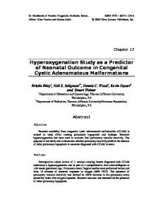
been a research topic that attracts a high level of interest. Some of the studies were purely based on analysis of the polypeptide sequence (1–5), while the others took the structural information into account (3,6). In this respect, as it has been reported in a recent article that the tertiary structures of a large number of transcription factors (TFs) are mostly disordered (7), sequence-based analysis aimed at identifying the residues in a highly disordered TF that play key roles in interaction with the DNA is essential for obtaining a comprehensive picture of how the TF functions. Concerning protein–DNA interactions, there are two types of binding mechanisms involved, namely sequence-specific binding and nonspecific binding (8). Sequence-specific bindings occur between protein sidechains and nucleotide bases, while nonspecific bindings occur between protein sidechains and the DNA sugar/ phosphate backbone. In molecular biology, sequencespecific bindings correspond to sequence-specific recognition of genes and therefore are essential for correct gene regulation. This article presents the design of a sequence based predictor named ProteDNA for identifying the residues in a TF that are involved in sequence-specific binding with the DNA. In this article, a residue is regarded as involved in sequence-specific binding with the DNA, if one or more heavy atoms in its sidechain fall within 4.5 A˚ from the nucleobases of the DNA. Figure 1 illustrates the function carried out by ProteDNA. Figure 1(a) shows the prediction output of ProteDNA for the polypeptide sequence of Yeast TF GCN4 in the complex with Protein Data Bank (PDB) (9) ID 1YSA. Figure 1(b) depicts the output of ProteDNA in the tertiary structure of PDB complex 1YSA. In Figure 1(b), the residues colored by red are those sequence-specific binding residues correctly identified by ProteDNA, while the residue colored by blue is a false negative. In this case, there is no
*To whom correspondence should be addressed. Tel: +886 2 3366 5736; Fax: +886 2 2392 9885; Email:
[email protected] Correspondence may also be addressed to Dr Yen-Jen Oyang. Tel: +886 2 3366 4888; Email:
[email protected] ß 2009 The Author(s) This is an Open Access article distributed under the terms of the Creative Commons Attribution Non-Commercial License (http://creativecommons.org/licenses/ by-nc/2.0/uk/) which permits unrestricted non-commercial use, distribution, and reproduction in any medium, provided the original work is properly cited.
Nucleic Acids Research, 2009, Vol. 37, Web Server issue W397
(a)
(b)
Figure 1. Illustration of the function of ProteDNA. (a) The partial prediction output of ProteDNA with the polypeptide sequence of Yeast TF GCN4 in PDB complex 1YSA. (b) The tertiary structure of the complex with PDB ID 1YSA. The residues colored by red are those sequence-specific binding residues correctly identified by ProteDNA, while the residues colored by blue are the false negatives. In this case, there is no false positive.
false positive. However, this case contains a residue for which ProteDNA makes no prediction. The reason that causes ProteDNA providing no prediction in some cases is that some TF–DNA complexes deposited in the PDB contain disordered regions and therefore ProteDNA cannot learn any clues in order to make predictions for residues located in a similar polypeptide segment. In this article, the performance of ProteDNA is reported based on the following metrics: TP TP , sensitivity ¼ , TP þ FP TP þ FN TN TP þ TN specificity ¼ , accuracy ¼ : TN þ FP TP þ TN þ FP þ FN precision ¼
where TP, TN, FP and FN stand for the number of true positive samples, the number of true negative samples, the number of false positive samples and the number of false negative samples, respectively. In order to accommodate users with different application needs, ProteDNA has been designed to operate under two modes, namely, the high-precision mode and the balanced mode. In this respect, the user can select either mode when submitting a query to the web server. The experiments reported in this article show that under the high-precision mode, ProteDNA delivers precision of 82.3%, specificity of 99.3%, sensitivity of 49.8% and accuracy of 96.5%. Meanwhile, under the balanced mode, ProteDNA delivers precision of 60.8%, specificity of 97.6%, sensitivity of 60.7% and accuracy of 95.4%.
Figure 2. Overview of the architecture of ProteDNA.
METHODS Overview Figure 2 presents an overview of the architecture of ProteDNA. The entire hybrid predictor consists of the primary predictor and the auxiliary predictor. The primary predictor is a support vector machine (SVM) with its parameter settings optimized for delivering high precision. As a result, one can expect that sensitivity of the SVM-based primary predictor has been traded, since tuning the parameters of a predictor aimed at raising precision typically means that sensitivity is traded and vice versa. Accordingly, as shown in Figure 2, in the design of ProteDNA, we have incorporated a mechanism derived from the secondary structure element alignment (SSEA)
W398 Nucleic Acids Research, 2009, Vol. 37, Web Server issue
approach first proposed by Gewehr and Zimmer (10) to complement the prediction power of the SVM. With the primary and auxiliary predictors, ProteDNA can operate under the high-precision mode as well as the balanced mode in order to accommodate users with different application needs. Under the high-precision mode, only the SVM-based primary predictor is enabled. On the other hand, under the balanced mode, both predictors are enabled and a residue is predicted to be involved in specific binding with the DNA if either the primary or the secondary predictor makes such a prediction. For evaluating the performance of ProteDNA, we have created a data set containing 253 TF–DNA complexes, among which 227 complexes were extracted from the 691 protein–DNA complexes that Ofran et al. (11) collected from the PDB and the remaining 26 TF–DNA complexes are those that were deposited into the PDB during September 2007 and November 2008. During the process to extract the 227 complexes from the Ofran collection, we excluded those complexes that do not contain a TF and then queried the PFAM server (12) to exclude those complexes in which no polypeptide segment is within the DNA-binding domain predicted by the PFAM server. In this respect, we submitted the full sequences of the proteins in the complex to the PFAM server and adopted only those predicted binding domains with the P-value computed by the PFAM server