lipopeptide is not completely soluble in the Bradford Reagent. 3 Methods. 3.1 Semi- quantitative Methods. 3.1.1 Surface Tension. The reduction in surface ...
Protocols for Measuring Biosurfactant Production in Microbial Cultures Roger Marchant and Ibrahim M. Banat Abstract Microbial biosurfactants have wide structural and functional diversity which consequently requires the adoption of a range of techniques to investigate these amphiphilic molecules. Literature on the production and analytical detection of biosurfactants is overwhelmed with assertions of high yields for such products which are mostly over exaggerated estimates due to the use of flawed or inaccurate analytical techniques. In this chapter we focus on quantitative methods available to allow accurate estimates of production and yield data to be generated. Keywords Colorimetric interfacial tension, UPLC
1
methods,
HPLC/MS,
Rhamnolipid,
Sophorolipid,
Surface/
Introduction Biosurfactants are amphiphilic molecules mainly produced by bacteria, yeasts and fungi. They possess both hydrophilic and hydrophobic moieties and are able to display a variety of surface activities that, among other roles, help solubilising hydrophobic substrates and are the subject of intense investigation [1, 2]. One of the major potential applications is their use as replacements for synthetic surfactants in many existing and future industrial and environmental applications [3]. Diverse functional properties, viz. emulsification, wetting, foaming, cleansing, phase separation, surface activity and reduction in viscosity of crude oil, makes it feasible to utilise them for numerous environmental, food, pharmaceutical, medical, cleaning and other industrial application purposes [4–9]. Accurate measurement of biosurfactant production and determination of final yields from microbial fermentations are things which have been rarely or never achieved by researchers up to the present time. The reasons for this are complex but are due to factors such as the diversity of the congeners produced and their chemical
T.J. McGenity et al. (eds.), Hydrocarbon and Lipid Microbiology Protocols, Springer Protocols Handbooks, DOI 10.1007/8623_2014_10, © Springer-Verlag Berlin Heidelberg 2014
Roger Marchant and Ibrahim M. Banat
similarity plus the need for sophisticated methodology for analysis. There have been many publications describing several qualitative methods for the screening and detection of biosurfactant producing cultures and product in cell culture broths. Such methods include haemolysis of erythrocytes, axisymmetric drop shape analysis, cell surface hydrophobicity, drop collapse, oil spread on surfaces, tilted glass slide, blue agar plate method, emulsification activity, agar plate method and direct colony or broth chromatographic technique. All these methods have been adequately reviewed by Satpute et al. [10]. In this chapter we will not deal with these varied methods that have traditionally been employed for screening for biosurfactant production as they are generally indirect methods using the physicochemical properties of surfactants and emulsifiers as indicators of production rather than direct quantitative measures. There are also a number of methods that have been used quantitatively to asses production, but which in reality are only semi-quantitative at best, these methods will be described and evaluated. Finally we will focus on the critical quantitative methods that are now being developed to allow accurate production and yield data to be generated. The field of microbial biosurfactant research is beset with claims for efficient and large product yields which on closer examination can be shown to be massive overestimates based on the use of flawed methodology.
2
Materials Bradford Reagent: Dissolve 100 mg Coomassie Brilliant Blue G250 in 50 ml 95 % (v/v) ethanol, and add 100 ml 85 % (w/v) phosphoric acid. Dilute to 1 l when the dye has completely dissolved and then filter through Whatman Number 1 filter paper just before use. 1 M NaOH can be used in the assay mixture if the lipopeptide is not completely soluble in the Bradford Reagent.
3
Methods
3.1 Semiquantitative Methods 3.1.1 Surface Tension
The reduction in surface tension of a culture broth as the organism grows is indicative that the organism may be producing a specific surface active compound, although many molecules show the same characteristic and may only be subsidiary metabolites. It is a relatively simple procedure to measure the surface tension (ST) of a culture broth during the growth cycle using the the Du Nouy method using a digital tensiometer equipped with a platinumiridium ring. Distilled water is used to calibrate the instrument and mean values of replicates are necessary for reproducible results.
Protocols for Measuring Biosurfactant Production in Microbial Cultures
The ST value for pure water is 72.8 mN/m at 20� C and microbial biosurfactants may reduce the value to around 27 mN/m. Although ST measurements do give numerical values, the data are of limited use in quantifying the biosurfactant production for the following reasons: 1. The ST value falls to a minimum at the Critical Micelle Concentration (CMC) for a specific biosurfactant molecule and any further increases in biosurfactant concentration produce no further decline in ST value. For the most active biosurfactants, the CMC value may be very low, e.g. rhamnolipid CMC values have been variously determined as in the range 24–65 mg/L. Since biosurfactant production may be in the range of tens to hundreds of grams per litre, the initial decline in ST in a culture covers only a small portion of the production cycle. The value of the method can be extended, however, by using a dilution series and calculating approximate yields on the basis of titre. 2. Microbial biosurfactants are not produced as a single molecular species but as a mixture of different congeners. Very often this mixture is exceedingly complex and the variations in structure lead to each congener having different surface-active properties. A further complication is that different culture conditions may alter the proportion of the individual congeners. It is clear therefore that an overall measurement like ST can only give an indication of biosurfactant production and no specific quantitative information concerning individual congeners. 3. Measuring the ST of a culture broth also fails to take account of the contribution to the ST value by constituents of the growth medium and the changes that take place during microbial growth. For example, the use of oleic acid as a carbon source may lead to its emulsification and reduction of surface tension measurements in the broth culture. 4. Theoretically isolating and purifying the individual congeners from the mixture should allow true CMC values to be determined, however, separation of a complex mixture of different but similar congeners is seldom feasible [11]. 3.1.2 Interfacial Tension
Interfacial tension (IFT) is a measure of the cohesive energy present at an interface, e.g. liquid/liquid or gas/liquid phases. The surface activity of biosurfactants reduces the IFT in a similar fashion to the ST effect. The IFT can be measured using specific pieces of equipment, or through examination of the axisymmetric shape of a hanging or supine drop [12], the units of measurement for IFT are the same as ST, i.e. mN/m. The same comments made about the limitations of ST measurement apply to IFT.
Roger Marchant and Ibrahim M. Banat
3.2 Colorimetric Methods 3.2.1 Orcinol Method
Colorimetric methods to determine the production of biosurfactants in microbial cultures have many immediate attractions. They offer the prospect of a simple, rapid, reproducible and accurate methodology; however, in the case of biosurfactants, this prospect is not one that is fulfilled. The orcinol method for the determination of rhamnolipids [13] is one that has been used extensively in the literature to report production and yields of rhamnolipids; unfortunately in most applications, it is fatally flawed as we will discuss later. The method is carried out as follows: cell-free supernatant from a microbial culture is diluted 10 times and 100 μl of this sample added to 900 μl of fresh orcinol reagent (0.19% orcinol in 53% sulphuric acid). The sample is then incubated in a water bath at 80� C for 30 min, cooled to room temperature for 15 min and finally the absorbance measured at 421 nm (Abs421nm). The rhamnose content is calculated by comparison with a standard curve prepared with L-rhamnose at concentrations of 0–50 μg/ml. Rhamnolipid concentration is obtained by multiplying the rhamnose content by 3.0, which represents the rhamnolipid/rhamnose correlation [14]. The measurements should be carried out in duplicate and may be expected to consistently show less than 5% variation. The underlying problems with the orcinol method for the determination of glycolipid biosurfactants is that the reagent reacts with any pentose sugar and is not specific for rhamnose; indeed orcinol has been used for the quantification of both DNA and RNA. The second point is that it is in any case an indirect method for rhamnolipid determination, and the final calculation of rhamnolipid concentration depends on an estimated proportion of rhamnose to total glycolipid quantity which will vary from culture to culture and between different fermentation runs in addition to variations of sugar content and proportions between the different biosurfactant congeners such as mono- and di-rhamnose sugar containing rhamnolipids. The effect of these problems is that the orcinol method always overestimates the quantity of rhamnolipid present, sometimes to an alarming degree. Carrying out the determination on the culture broth will compound the error, but even using an extraction and purification protocol as described by Smyth et al. [11, 15] will not remove the errors. Some workers have attempted to improve the accuracy of the method by using a standard of rhamnolipid rather than pure rhamnose; however, this does not prevent the interference of contaminating pentoses in the sample. In conclusion we can say that the orcinol method is really only of value in a comparative quantitative role within a single experiment and should not be used where absolute values are required to make comparisons between different studies.
Protocols for Measuring Biosurfactant Production in Microbial Cultures 3.2.2 Bradford Method
Just as orcinol can be used as an indirect method to measure the pentose portion of a glycolipid biosurfactant, the Bradford method [16] to estimate protein can used as an indirect method for lipopeptide biosurfactant quantification. The assay is carried out as follows: Samples should contain between 5 and 100 μg protein in a volume of 100 μl. If the lipopeptide to be assayed is not soluble in the Bradford reagent, add an equal volume of 1 M NaOH to each sample and vortex (if this option is used, the NaOH should also be added to the standards). Standards containing 5–100 μg of a suitable protein, e.g. albumin, should be prepared for calibration of the method. Add 5 ml of the Bradford Reagent to each sample and incubate for 5 min and then measure the absorbance at 595 nm. The Bradford Reagent reacts primarily with arginine residues and less so with histidine, lysine tyrosine, tryptophan and phenylalanine residues. This means that the overall reaction is very dependent on the composition of the peptide being assayed. It is therefore obvious that this method, like the orcinol method, has severe limitations for the quantification of biosurfactant production and should only be used to obtain relative yields under one set of conditions rather than as an absolute quantification method.
3.3 Gravimetric Methods
The most obvious and perhaps the simplest quantification method is gravimetric analysis, i.e. isolating/separating and then directly weighing the amount of product. While this approach has a great deal of merit, there are significant problems in applying it to biosurfactant production. Methods for the isolation and purification of a range of low molecular weight and high molecular weight biosurfactants have been detailed by Smyth et al. [11, 15]. One major complication for the isolation of the ‘pure’ biosurfactant is the type of substrate used for the growth of the microbial producer. Very often this takes the form of an excess amount of a pure fatty acid or fatty acid mixture such as rapeseed oil, which has to be removed before the further purification of the biosurfactant can take place. Solvent extraction, e.g. hexane, can be used for this removal; however, it can be difficult to determine whether excess fatty acid removal has been effectively achieved. The purity of the finally isolated biosurfactant can be checked before gravimetric analysis using analytical methods such as direct injection mass spectrometry; however, not all contaminating molecules will give a signal and this methodology can only really be used in a negative mode; i.e. if a contaminant is seen then the surfactant preparation is not pure, but if no contaminants are seen this does not necessarily mean the preparation is pure. Despite the drawbacks of the gravimetric method, it does provide a reasonably good comparative measure providing care is taken with the isolation and purification steps.
Roger Marchant and Ibrahim M. Banat
a
14.92;761.54
Rha-Rha-C14-C14
100
%
Rha-Rha-C12-C14/ Rha-Rha-C14-C12
Rha-Rha-C12-C12
11.79 733.52
Rha-C12-C14/ Rha-C14-C12
Rha-C14-C14
14.42 587.42
0.03 190.91
17.74 615.48
12.21 535.36
2.37 546.23
18.06 789.58
8.86 705.50
0
Time 2.00
4.00
6.00
8.00
10.00
b
12.00
14.00
16.00
Rha-Rha-C14-C14
100
18.00 761.5
Rha-Rha-C12-C14/ Rha-Rha-C14-C12
%
Rha-C14-C14 Rha-Rha-C12-C12
733.5 762.6
Rha-C12-C14/ Rha-C14-C12 615.5 734.5 763.6
587.5
616.5
588.5 543.0 559.4 563.4 585.1
705.5
617.5 596.6
0 540 550 560 570 580 590 600 610 620
640.5
647.5
682.4 689.4 657.5
789.6
735.5 790.6
765.5
717.4
736.5 721.4
745.4
789.4
791.6 m/z
630 640 650 660 670 680 690 700 710 720 730 740 750 760 770 780 790
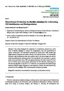
Fig. 1 Separation of rhamnolipid congeners produced by B. thailandensis E264. (a) LC-QToF-MS of rhamnolipid crude extract after SPE purification. Static phase, Agilent poroshell SB-C3, 2.1 � 100 mm, particle size 2.7 μm. Mobile phase 1, H2O (4 mM ammonium acetate), and mobile phase 2, MeCN, were used for chromatographic separation as follows: 0–17 min 50–70% mobile phase 2, 17.0–17.5 min 70% mobile
Protocols for Measuring Biosurfactant Production in Microbial Cultures
3.4 Rigorous Quantitative Methods
The very nature of most biosurfactants produced by microorganisms makes absolute quantification a very difficult task. The mixtures of different congeners differing only slightly in structure, but with possibly widely variant functionalities, provide a major obstacle to accurate differential quantification. As we have seen with the indirect methods or the measurement of a fractional component of the molecules, these techniques can produce grossly misleading results which are usually overestimates of quantity. An effective approach therefore must include separation and characterisation technologies which allow individual components of the mixtures to be identified and quantified. These requirements effectively limit the possible methods to combinations of HPLC and mass spectrometry; see Fig. 1 which shows an example of such separation for rhamnolipid congeners produced by B. thailandensis. HPLC is a practical and efficient method for separating individual components of biosurfactant mixtures, particularly glycolipids. The precise conditions which need to be used for complete separation of a specific set of biosurfactant congeners have to be determined experimentally each time, but we can give the conditions for separation of some of the systems that have already been developed.
3.4.1 Rhamnolipids and Sophorolipids
The first step in the process of analysing a culture broth involves the removal of the cells by centrifuging followed by some measure of purifying the biosurfactant through acidification to precipitate the glycolipids combined with solvent extraction [11]. The purified extract (5 μl of a 1 mg/mL solution) of the glycolipid is then diluted in methanol (95 μl) and 10 μl used for HPLC analyses coupled to a tandem quadrupole mass spectrometer. Analysis of mixtures containing predominantly C10C10 rhamnolipids can be performed using a Luna C18 250 � 4.6 mm 5 μm column (Phenomenex, Cheshire, UK) with a mobile phase consisting of water + 4 mM ammonium acetate (phase A) and acetonitrile (phase B) and the operating conditions as follows: 70:30 (A:B) to 30:70 (A:B) over 50 min and back to 70:30 (A:B) over 5 min with hold for 5 min. Analysis of sophorolipids can be carried out using a similar C18 column with the same mobile phases and chromatographic conditions as above. Flow rates of 0.5 ml/min are appropriate. The mass
ä Fig. 1 (continued) phase 2, 17.5–18.0 min 70%–50% mobile phase 2 and 18–20 min 50% mobile phase 2. (b) QToF-MS profile of rhamnolipid crude extract after SPE purification. SPE purification was carried out using Strata SI-1 silica (55 μm, 70 A) 2 g/12 ml Giga Tubes. CHCl3 was used to clean the column before the sample was added. The sample was then dissolved in CHCl3 and added to the column; pure CHCl3 was ran through the column to clean and remove any unwanted products from the sample. Finally, the purified rhamnolipid was eluted using a 1:1, v/v solution of CHCl3:MeOH
Roger Marchant and Ibrahim M. Banat
spectrometer should be operated in ionisation negative mode with a scanning range of 50–1,200 Da. Using these conditions, good separation of congeners should be achieved and relative abundance of the individual congeners will be observable from the HPLC peak areas. The MS data will also allow each peak to be directly identified. Ideally for fully accurate quantification, a glycolipid standard should be run for each congener to calibrate the peak areas from the HPLC trace. This approach, however, is not practicable since it is probably only realistic to obtain pure samples of the major components of any mixture in useable quantities. An alternative approach is to use an internal standard with each glycolipid sample run. Costa et al. [17] used this technique for the analysis of rhamnolipids from Burkholderia glumae, adding 16-hydroxyhexadecanoic acid or 5,6,7,8-tetradeutero-4-hydroxy-2-heptylquinoline at a final concentration of 10 mg/L. Burkholderia glumae produces mono and di-rhamnolipids with alkyl chains ranging in length between C12C12 and C16C16 with the most abundant congener Rha-RhaC14C14; effective separation of these congeners was achieved using a 4.6 � 50 mm 300SB-C3 Zorbax 5 μm reverse-phase column using a water/acetonitrile gradient with a constant 2 mM concentration of ammonium acetate. The choice of internal standard to use in this system is governed by the ability to obtain a good signal in the MS system. The examples of HPLC separation conditions given above cannot be applied directly to separation and quantification of other glycolipid biosurfactants without selecting an appropriate column and suitable elution phases and gradients. Each new type of surfactant mixture will require careful optimisation of the conditions. Although the system outlined above for quantification of biosurfactant produced in a microbial culture is accurate and reproducible, it does have disadvantages, notably the length of time required to run each sample means that monitoring of the progress of a fermentation run can only be carried out off-line and at relatively infrequent intervals. More recently an improved technique has been developed (Rudden, Smyth, Marchant, & Banat) which uses UPLC technology with much improved separation of congeners achieved in 3 min with a further 2 min conditioning of the column between runs; this compares to a normal run time of 55 min using HPLC. This method potentially allows accurate quantification to be carried out at short time intervals during a fermentation run. The UPLC system operates at ultrahigh pressure (600 bar) through a narrow bore column (21 x 100 mm) and for separation of C10C10 rhamnolipids uses water plus 4 mM ammonium acetate as mobile phase A and acetonitrile as phase B with a gradient from 50% A to 90% B over 2.2 min. The peaks produced are analysed as with the HPLC system using a tandem quadrupole
Protocols for Measuring Biosurfactant Production in Microbial Cultures
mass spectrometer. Rhamnolipid standards used for calibration and validation of the system were prepared from a mixture of up to 14 different congeners by separating the congeners into two fractions: one containing mono-rhamnolipids and the other di-rhamnolipids. Since separation of the fractions into individual congeners was impracticable (some congeners are present in very low amounts), the relative proportion of each congener was established from the % peak area from the mixed sample, and then 1 mg/mL of the most abundant rhamnolipid congener present in each of the purified fractions was analytically weighed to obtain standard solutions; i.e. in fraction R1, Rha-C10-C10 (503 m/z [M-H] ) was the most abundant congener comprising 83.8% of the total sample, and in fraction R2, Rha-Rha-C10-C10 (649 m/z [M-H] ) was the most abundant congener comprising 85.1% of the total sample. Standard curves could then be constructed to allow accurate quantification of all the congeners present in the initial sample. The major advantages of the UPLC method are the approximately 12fold reduction in run time for the separation of the rhamnolipid congeners compared to the HPLC method, and with the use of multiple reaction monitoring (MRM), which eliminates other interfering compounds in the sample, preliminary preparation of the sample can be reduced to the removal of particulate material, i.e. microbial cells.
4
Conclusion The overall conclusion at present is that published data on the yields of biosurfactant from different fermentation systems cannot be directly compared and that great care should be exercised in evaluating claims for high production yields. The fact that microorganisms produce a mixture of very similar congeners also means that absolute quantification of yield is always going to be difficult to achieve. The use of appropriate standards in conjunction with techniques that allow accurate quantification, e.g. HPLC or UPLC, is most likely to give the accurate and reproducible yield data that are required to evaluate the economic feasibility of any process.
Acknowledgement We would like to thank Dr Thomas J.P. Smyth and Mr Scot Funston for valuable comments and results from analytical methods.
Roger Marchant and Ibrahim M. Banat
References 1. Marchant R, Banat IM (2012) Biosurfactants: a sustainable replacement for chemical surfactants? Biotechnol Lett 34:1597–1605 2. Marchant R, Banat IM (2012) Microbial biosurfactants: challenges and opportunities for future exploitation. Trends in Biotechnol 30:558–565 3. Banat IM, Franzetti A, Gandolfi I, Bestetti G, Martinotti MG, Fracchia L, Smyth TJP, Marchant R (2010) Microbial biosurfactants production, applications and future potential. Appl Microbiol Biotechnol 87:427–444 4. Perfumo A, Smyth TJP, Marchant R, Banat, IM (2010) Production and roles of biosurfactants and bioemulsifiers in accessing hydrophobic substrates. In: Timmis KN (ed) Handbook of hydrocarbon and lipid microbiology, Chap 47, vol 2, part 7. Springer-Verlag, Berlin Heidelberg, pp 1501–1512 5. Fracchia L, Cavallo M, Martinotti MG and Banat IM (2012) Biosurfactants and bioemulsifiers biomedical and related applications – present status and future potentials. In: Ghista DN (ed) Biomedical science, engineering and technology, Chap 14. InTech Open Access Publisher, pp 325–370. ISBN: 978-953-307471-9 6. Franzetti A, Gandolfi I, Raimondi C, Bestetti G, Banat IM, Smyth TJP, Papacchini M, Cavallo M, Fracchia L (2012) Environmental fate, toxicity, characteristics and potential applications of novel bioemulsifiers produced by Variovorax paradoxus 7bCT5. Bioresource Technol 108:245–51 7. Mueller MM, Kugler JH, Henkel M, Gerlitzki M, Hormann B, Pohnlein M, Syldatk C, Hausmann R (2012) Rhamnolipids-next generation surfactants? J Biotechnol 162:366–380 8. Campos JM, Montenegro Stamford TL, Sarubbo LA, de Luna JM, Rufino RD, Banat IM (2013) Microbial biosurfactants as additives for food industries; a review. Biotechnol Prog 29:1097–1108 9. Quinn GA, Maloy AP, Banat MM, Banat IM (2013) A comparison of effects of broad-
spectrum antibiotics and biosurfactants on established bacterial biofilms. Curr Microbiol 67:614–623 10. Satpute SK, Banpurkar AG, Dhakephalkar PK, Banat IM, Chopade BA (2010) Methods for investigating biosurfactants and bioemulsifiers: a review. Crit Rev Biotechnol 30:127–144 11. Smyth TJP, Perfumo A, Marchant R, Banat IM (2010) Isolation and analysis of low molecular weight microbial glycolipids. In: Timmis KN (ed) Handbook of hydrocarbon and lipid microbiology, vol 5, part 2, Chap 28. Springer-Verlag, Berlin Heidelberg, pp 3705–3723 12. Van der Vegt W, van der Mei HC, Noordmans J, Busscher HJ (1991) Assessment of bacterial biosurfactant production through axisymmetric drop shape analysis by profile. Appl Microbiol Biotechnol 35:766–770 13. Chandrasekaran EV, Bemiller JN (1980) Constituent analysis of glycosaminoglycans. In: Whistler RL (ed) Methods in carbohydrate chemistry. Academic, New Jersey, pp 89–96 14. Abalos A, Pinazo A, Infante MR, Casals M, Garcia F, Manresa A (2001) Physicochemical and antimicrobial properties of new rhamnolipids produced by Pseudomonas aeruginosa AT10 from soybean oil refinery wastes. Langmuir 17:1367–1371 15. Smyth TJP, Perfumo A, McClean S, Marchant R, Banat IM (2010) Isolation and analysis of lipopeptides and high molecular weight biosurfactants. In: Timmis KN (ed) Handbook of hydrocarbon and lipid microbiology, vol 5, part 2, Chap 27. Springer-Verlag, Berlin Heidelberg, pp 3688–3704 16. Bradford MM (1976) A rapid and sensitive method for the quantitation of microgram quantities of protein utilizing the principle of protein-dye binding. Anal Biochem 72:248–254 17. Costa SGVAO, De´ziel E, Le´pine F (2011) Characterization of rhamnolipid production by Burkholderia glumae. Lett Appl Microbiol 53:620–627