Psychological Medicine http://journals.cambridge.org/PSM Additional services for Psychological Medicine: Email alerts: Click here Subscriptions: Click here Commercial reprints: Click here Terms of use : Click here
Neural correlates of visuospatial working memory in the ‘atrisk mental state’ M. R. Broome, P. FusarPoli, P. Matthiasson, J. B. Woolley, L. Valmaggia, L. C. Johns, P. Tabraham, E. Bramon, S. C. R. Williams, M. J. Brammer, X. Chitnis, F. Zelaya and P. K. McGuire Psychological Medicine / Volume 40 / Issue 12 / December 2010, pp 1987 1999 DOI: 10.1017/S0033291710000280, Published online: 10 March 2010
Link to this article: http://journals.cambridge.org/abstract_S0033291710000280 How to cite this article: M. R. Broome, P. FusarPoli, P. Matthiasson, J. B. Woolley, L. Valmaggia, L. C. Johns, P. Tabraham, E. Bramon, S. C. R. Williams, M. J. Brammer, X. Chitnis, F. Zelaya and P. K. McGuire (2010). Neural correlates of visuospatial working memory in the ‘atrisk mental state’. Psychological Medicine, 40, pp 19871999 doi:10.1017/S0033291710000280 Request Permissions : Click here
Downloaded from http://journals.cambridge.org/PSM, IP address: 86.20.35.14 on 15 Jan 2013
Psychological Medicine (2010), 40, 1987–1999. f Cambridge University Press 2010 doi:10.1017/S0033291710000280
O R I G I N A L AR T I C LE
Neural correlates of visuospatial working memory in the ‘at-risk mental state’ M. R. Broome1,2*, P. Fusar-Poli1,3, P. Matthiasson1, J. B. Woolley1, L. Valmaggia1,4, L. C. Johns1, P. Tabraham1, E. Bramon1, S. C. R. Williams5, M. J. Brammer6, X. Chitnis6, F. Zelaya5 and P. K. McGuire1 1
Psychosis Clinical Academic Group, Institute of Psychiatry, King’s College London, UK Health Sciences Research Institute, Warwick Medical School, University of Warwick, Coventry, UK 3 Department of Applied and Psychobehavioural Health Sciences, University of Pavia, Italy 4 Department of Psychiatry and Neuropsychology, Maastricht University, The Netherlands 5 Neuroimaging Research Group, Department of Neurology, Institute of Psychiatry, King’s College London, UK 6 Brain Image Analysis Unit, Department of Biostatistics and Computing, Institute of Psychiatry, King’s College London, UK 2
Background. Impaired spatial working memory (SWM) is a robust feature of schizophrenia and has been linked to the risk of developing psychosis in people with an at-risk mental state (ARMS). We used functional magnetic resonance imaging (fMRI) to examine the neural substrate of SWM in the ARMS and in patients who had just developed schizophrenia. Method. fMRI was used to study 17 patients with an ARMS, 10 patients with a first episode of psychosis and 15 agematched healthy comparison subjects. The blood oxygen level-dependent (BOLD) response was measured while subjects performed an object–location paired-associate memory task, with experimental manipulation of mnemonic load. Results. In all groups, increasing mnemonic load was associated with activation in the medial frontal and medial posterior parietal cortex. Significant between-group differences in activation were evident in a cluster spanning the medial frontal cortex and right precuneus, with the ARMS groups showing less activation than controls but greater activation than first-episode psychosis (FEP) patients. These group differences were more evident at the most demanding levels of the task than at the easy level. In all groups, task performance improved with repetition of the conditions. However, there was a significant group difference in the response of the right precuneus across repeated trials, with an attenuation of activation in controls but increased activation in FEP and little change in the ARMS. Conclusions. Abnormal neural activity in the medial frontal cortex and posterior parietal cortex during an SWM task may be a neural correlate of increased vulnerability to psychosis. Received 19 January 2009 ; Revised 26 January 2010 ; Accepted 26 January 2010 ; First published online 10 March 2010 Key words : ARMS, imaging, memory, prodrome, psychosis, visuospatial.
Introduction Although it is known that schizophrenia is associated with neurocognitive dysfunction, the extent to which this is related to the disorder, as opposed to vulnerability to schizophrenia, is unclear. There is also increasing evidence that neuroimaging abnormalities may change over the course of psychotic disorders (Lieberman, 1999 ; Rapoport et al. 1999 ; Lieberman et al. 2001 ; Pantelis et al. 2003) and can be affected by treatment (Chakos et al. 2005 ; Dazzan et al. 2005). Determining variables that are linked to vulnerability
* Address for correspondence : Dr M. R. Broome, Warwick Medical School, University of Warwick, Gibbet Hill, Coventry CV4 7AL, UK. (Email :
[email protected])
to schizophrenia, rather than to the disorder itself, is important for identifying those who may benefit from interventions that may prevent the onset of the disorder, and also allows understanding of how the disorder develops and progresses. One way of clarifying the relative contribution of these factors is to compare individuals who are at very high risk of psychosis, patients who have just developed schizophrenia and have had minimal treatment, and healthy volunteers. People with ‘ prodromal ’ symptoms of psychosis have a 25–40 % risk of developing a psychotic disorder in the next 12 months (Yung et al. 2003) and thus have an ‘ at-risk mental state ’ (ARMS). However, this rate of transition has not remained the same in other centres or, indeed, over time. Other groups have reported higher rates of transition (Miller et al. 2002), and the
1988
M. R. Broome et al.
Melbourne Personal Assessment and Crisis Evaluation (PACE) service has recently reported a transition rate of less than 10 % (Yung et al. 2007). Within our own service, OASIS, current transition rates are at approximately 21 % (Valmaggia et al. 2009). Knowledge of neurocognitive function in this group is growing rapidly. Neuropsychological studies point to an impairment of executive and memory functions (Brewer et al. 2005) with some deficits only evident when the task demands are relatively high (Broome et al. 2007). In general, neuropsychological performance in ARMS subjects has been found to be at an intermediate level relative to patients with schizophrenia and controls (Wood et al. 2003 ; Brewer et al. 2005 ; Lencz et al. 2006 ; Wagner et al. 2006 ; Pukrop et al. 2007), with evidence suggesting that spatial working memory (SWM) is impaired. Structural magnetic resonance imaging (MRI) studies suggest that the ARMS is associated with reduced grey matter volumes in the prefrontal, cingulate and temporal cortex (Pantelis et al. 2003) whereas functional MRI (fMRI) studies have reported differential prefrontal activation in ARMS subjects relative to controls and patients with schizophrenia during a visual oddball paradigm (Morey et al. 2005) and during verbal fluency and the N-back verbal working memory tasks (Broome et al. 2009). In both these studies, the clinical high-risk group demonstrated activations intermediate between those with schizophrenia and healthy controls, with the control subjects typically showing greatest activation and those with psychosis, the least. Working memory refers to the retention of information in conscious awareness when it is not present in the environment. Working memory has been implicated as an important contributor to language processing, learning, planning, reasoning and general fluid intelligence (Postle, 2006). It can be subdivided into a memory component (holding information ‘ online ’) and a manipulation component (working on the information being held). It has been further subdivided according to the form of the information involved (verbal versus non-verbal ; spatial versus nonspatial ; verbal versus object memory) (Pollmann & von Cramon, 2000). In our previous imaging work with the ARMS groups we studied verbal working memory (Broome et al. 2009). In the present study our focus is on SWM. SWM impairments have been well documented in schizophrenia (Park & Holzman, 1992 ; Fleming et al. 1997) and have been highlighted as a neuropsychological dysfunction that is core to the disorder (Silver et al. 2003 ; Joyce & Huddy, 2004). Impairments in visuospatial working memory are evident early in the course of schizophrenia (Wood et al. 2002, 2003 ; Smith et al. 2006 ; Vance et al. 2006), but it is unclear whether impairments in SWM predate
the onset of psychosis. Studies of monozygotic and dizygotic twins pairs discordant for schizophrenia (Cannon et al. 2000 ; Glahn et al. 2005) indicate that SWM deficits are associated with increased genetic risk for schizophrenia, and it has been suggested that a higher genetic loading for disease-related traits is linked to greater cognitive impairment (Saperstein et al. 2006). Impaired spatial memory performance has also been reported in subjects with high levels of schizotypy (Park et al. 1995) or schizotypal personality disorder (Farmer et al. 2000), and in those with a history of very preterm birth (Narberhaus et al. 2009). Several studies have reported impaired memory performance in the ARMS (Wood et al. 2003 ; Brewer et al. 2005 ; Francey et al. 2005 ; Lencz et al. 2006 ; Pukrop et al. 2007). Brewer et al. (2005) found that ARMS subjects showed impairments on measures of visual reproduction and verbal memory, and that this deficit was specific to the subgroup that went on to develop psychosis. Brewer and colleagues performed a pairedassociate task, but one that assessed verbal, rather than spatial, memory. To date, functional neuroimaging studies of working memory in the ARMS have been limited to the verbal domain (Broome et al. 2009). However, SWM has been studied in the offspring of people with schizophrenia using a memory-guided saccade task ; this genetically high-risk group showed decreased activation in the dorsolateral prefrontal and inferior parietal cortex while performing the task relative to controls (Keshavan et al. 2002). In the present study, we used fMRI to assess cortical activation during an object–location paired-associate memory task. This task is complex, comprising elements of encoding, recognition, learning and discrimination (Narberhaus et al. 2009). Interpretation of data from non-verbal associative learning tests can be compromised if the stimuli are easy to verbalize (Goldstein et al. 1988). Paired-associate learning (PAL) tasks attempt to overcome this problem by pairing abstract designs with spatial locations (Brewer et al. 2005). The paradigm we used also incorporated different levels of mnemonic load, which allowed us to examine whether functional deficits were related to the demands on working memory. In addition, the repetition of trials over the course of the study enables us to examine whether abnormalities were related to the ability to learn the relationship between the pairs of stimuli and their spatial location. We studied three groups : (1) patients with a first episode of schizophrenia, (2) subjects with an ARMS, and (3) healthy controls. We hypothesized that, relative to controls, individuals with an ARMS would show qualitatively similar functional abnormalities to patients with firstepisode psychosis (FEP) but that the magnitude of these abnormalities would be less severe. More
Neural correlates of visuospatial working memory specifically, we predicted that group differences in activation would be evident in the frontal and parietal cortex (Curtis, 2006), with the superior frontal cortex implicated in the maintenance of spatial information and the dorsolateral cortex implicated in its manipulation (Postle et al. 2000), and that these differences would become more apparent as the mnemonic demands of the task were increased (Gould et al. 2003). A further prediction was that differential frontal and parietal activation would be evident in association with differential learning across repeated trials of the task (Brewer et al. 2005 ; Lencz et al. 2006). Method Subjects ARMS group Individuals meeting PACE criteria for the ARMS were recruited from Outreach and Support in South London (OASIS ; Broome et al. 2005a). The diagnosis was based on assessment by two experienced clinicians using the Comprehensive Assessment for the ARMS (CAARMS ; Yung et al. 1998, 2003) and a consensus meeting with the clinical team. None of the subjects had ever received antipsychotic medication. An individual can meet criteria for the ARMS in one or more of three ways : first, a recent decline in function coupled with either schizotypal personality disorder or a firstdegree relative with psychosis ; second, ‘ attenuated ’ positive symptoms ; and third, a brief psychotic episode of less than 1 week’s duration that resolves without antipsychotic medication. FEP group Patients who had recently presented with a first episode of psychosis (n=10) were recruited from Lambeth Early Onset (LEO) Services (www.slam.nhs. uk/services/). All met ICD-10 criteria for schizophreniform psychosis at the time of scanning and met OPCRIT criteria (McGuffin et al. 1991) for schizophrenia when subsequently reassessed 12 months after first presentation. Three of these patients were unmedicated. The other seven had been treated with either oral risperidone or quetiapine for a mean of 10 days [95 % confidence interval (CI) 4.7–16.3] at mean doses of 1.7 and 63.75 mg respectively. Controls Healthy volunteers (n=15) were recruited through advertisements in the local media. All subjects lived in the borough of Lambeth (London), were native speakers of English and were right-handed. The groups were matched on sociodemographic variables
1989
Table 1. Age, IQ, gender and psychopathology ratings
Age (years) NART IQ Gender (M : F) PANSS Total PANSS Positive PANSS Negative PANSS General
Controls (n=15)
ARMS (n=17)
FEP (n=10)
25.4 (4.9) 111.2 (7.2) 11 : 4 N.A. N.A. N.A. N.A.
24.2 (4.1) 102.9 (11.9) 12 : 5 51. 9 (12.7) 11.7 (3.4) 10.6 (4.1) 20.9 (9.2)
25.5 (5.9) 103.6 (9.2) 7:3 58.1 (9.5) 18.5 (4.6) 10.0 (2.3) 29.6 (5.9)
NART, National Adult Reading Test ; M, male ; F, female ; PANSS, Positive and Negative Syndrome Scale ; ARMS, at-risk mental state ; FEP, first-episode psychosis ; N.A., not applicable. Values given as mean (standard deviation).
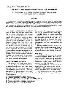
(Table 1), including age (F=0.35, p=0.71) and handedness. Subjects were excluded if there was a history of neurological disorder or they met DSM-IV criteria for a substance misuse disorder. General intellectual function was estimated in all subjects using the National Adult Reading Test (NART). The severity of symptoms in the clinical groups was assessed with the Positive and Negative Syndrome Scale (PANSS ; Kay, 1990) on the day of scanning by a psychiatrist (M.R.B. or P.M.) trained in its use. Experimental task Stimuli were presented in 22.5 s epochs, alternating with 34.5 s epochs of cross-hair fixation ; this cycle was repeated 12 times (for a total of 24 epochs) so the total duration of the experiment was 686 s or 343 images [repetition time (TR)=2 s]. Cognitive load was manipulated by presenting trials at one of three levels of difficulty (easy, intermediate, and hard) in a block design, with four blocks of each level of difficulty. Thus, there were a total of 12 blocks of trials alternating with 12 blocks of cross-hair fixation. The blocks of trials were always presented in the same sequence with respect to level of difficulty : easy, intermediate, and then hard. Each block comprised seven trials. In an easy trial, two stimuli (highly discriminable coloured shapes) were shown either side (left and right) of a central cross-hair, followed by the central cross-hair alone, then the central presentation of one of the two original stimuli. Subjects had been trained to move a joystick with their right hand in the direction of the location originally occupied by the central stimulus. Intermediate and hard trials were the same except that four and eight stimuli were presented around the central cross-hair respectively. The speed
1990
M. R. Broome et al.
+
Easy
Display array
+ Fixation Test stimulus
Baseline
22.5 s
Intermediate
+
34.5 s
Display array
+ Fixation
Test stimulus
Baseline
Hard
+ Display array
+ Fixation
Test stimulus
Fig. 1. The paired-associate learning task.
and accuracy of the joystick movements were recorded during scanning. To avoid habituation of the subject, every stimulus had a randomly varied time of presentation, either between stimuli or before the presentation of the probe. As the stimuli were jittered randomly in every block, we did not need to take account of this in the block design analysis (Fig. 1). Behavioural data All behavioural data, response accuracy and response latency, were recorded on a personal computer using Visual Basic (Microsoft Corp., USA) and analysed in SPSS version 11.0 (SPSS Inc., USA). Image acquisition Images were acquired on a 1.5-T Signa (GE) system at the Maudsley Hospital, London. T2*-weighted images were acquired in 38r3 mm slices, with a 0.3 mm gap in 14 axial planes, and a TR of 2 s, echo time (TE) 40 ms, and flip angle 90x. To facilitate anatomical localization of activation, a high-resolution inversion recovery image dataset was also acquired, with 3 mm contiguous slices and an in-plane resolution of 3 mm [TR 1600 ms, inversion time (TI) 180 ms, TE 80 ms]. Image analysis Individual brain activation maps The data were analysed with software developed at the Institute of Psychiatry, using a non-parametric
approach. Data were realigned (Bullmore et al. 1999b) and then smoothed using a Gaussian filter [full-width at half-maximum (FWHM) 7.2 mm]. Responses to the experimental paradigms were detected by convolving each component of the design with each of two gamma variate functions (peak responses at 4 and 8 s respectively). The best fit between the weighted sum of these convolutions and the time series at each voxel was computed using the constrained blood oxygen level-dependent (BOLD) effect model (Friman et al. 2003). A goodness-of-fit statistic comprising the ratio of the sum of squares of deviations from the mean image intensity (over the whole time series) divided by the sum of squares of deviations due to the residuals (SSQratio) was then computed at each voxel. The data were then permuted by a wavelet-based method (Bullmore et al. 2001) to calculate the null distribution of SSQratios under the assumption of no experimentally determined response. This was used to calculate the critical value of SSQratio needed to threshold the maps at a type I error rate of