ORIGINAL RESEARCH ARTICLE Tanaffos (2005) 4(13), 33- 37 ©2005 NRITLD, National Research Institute of Tuberculosis and Lung Disease, Iran
New Chest Tube to Facilitate Intrapleural Injections Azizollah Abbasi Dezfouli, Farid Aghayee Meybodi, Abolghasem Daneshvar Kakhki, Sanaz Sasani, Roya Farzanegan, Mohammad Reza Lashgari
D
Department of Thoracic Surgery, NRITLD, Shaheed Beheshti University of Medical Sciences and Health Services,TEHRAN-IRAN. ABSTRACT
SI
Background: In some cases, apart from the evacuation of the pleural space, it is required to inject a medicine into it. This procedure through the ordinary chest tubes is not an easy task since it might cause some unwanted problems for the patient. A new chest tube designed and produced by a few researchers contains a catheter in its thickness, which enables it to be used for both pleural drainage and drug entrance simultaneously.
of
Materials and Methods: Instead of the ordinary chest tube, the new chest tube(with catheter)was inserted in 50 patients suffering from various thoracic disorders which needed intrapleural drainage and injection due to different reasons .Amongst all,26 cases had traumatic hemothorax and pneumothorax. Following pleural drainage, bupivacaine was injected into the pleural space through the catheter of the new chest tube in order to reduce pain. In 10 patients suffering from malignant
ive
pleural effusion, bleomycin was introduced into the pleural cavity as sclerosing substance to create pleurodesis. Finally, irrigation of pleural space was performed by means of the catheter for the remaining 14 patients with empyema. Results: In all cases, intrapleural injections were done easily without the need to detach the chest tube connections and the consequences were satisfactory to all the physicians. No serious complication was observed and merely mild and avoidable
ch
side effects took place in 4 patients.
Conclusion: The new introductory tube provides a sterile and appropriate pathway to release different kinds of medications into the pleural space, without consuming any extra time or budget.(Tanaffos 2005; 4(13): 33-37)
Ar
Key words: Chest tube, Pleural cavity evacuation, Intrapleural injection
INTRODUCTION
Chest tube was presented to the medical world 150 years ago (1). Since then, this instrument has undergone numerous reforms to reach its current shape. Nowadays, while being strong enough, chest tubes produced are flexible as well. They also cause minimal tissue reactions (2). Today, chest tube is Correspondence to: Aghayee Meybodi F Tel: +98-912-1976772 Email address:
[email protected]
utilized to cure empyema, malignant effusions, chylothorax, pneumothorax, and hemothorax. Also, from the 1992 onwards, the necessity of chest tube insertion after thoracotomy has been admitted and made it an indispensable part of thoracic operations (2). More than 100,000 cardiac and thoracic surgeries are performed annually in the U.S which all need chest tube insertion for re-expansion of the lungs and pleural cavity drainage (3).
www.SID.ir
34 New Chest Tube
Occasionally, in some cases of thoracic diseases,
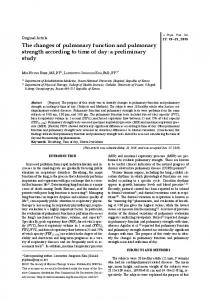
Central part made from latex
Catheter orifice for drug injection
there is a need to introduce medicines into the pleural space such as anesthetic drugs for pain reduction (4,5,6,7), sclerosing substances for pleurodesis (8), chemotherapeutic agents for primary or metastatic malignancies of pleural structures(9,10) , lysogenic
Chest tube
Main tubal orifice for drainage
substances to dissolute fibrinous clots(11,12) , and antibiotic-containing or irrigation fluid into the
medications into the pleural cavity. Some researchers have been using chest tubes for drug injections. Another method, which is especially used in providing pleural anesthesia, is the usage of a separate catheter. This catheter is entered into the procedure (when the thorax is not opened).(14)
At a proper distance, the two catheters are separated so that the smaller catheter does not cause inconvenience while fixing the tube to the chest wall. At the distal end of the thin catheter, a small cap is situated. Its central part is made of latex allowed for the injections. Therefore, there is no need to remove the cap and the risk of pneumothorax will be reduced (Fig 2).
of
pleural space during open thoracotomy or a closed
Figure 1. Schematic view of chest tube.
D
Different methods are applied to introduce
SI
pleural space.(13)
The application of a catheter similar to those used in epidural anesthesia inside the chest tube has also
been reported. This catheter is led into the pleural
ive
cavity through the chest tube and used to introduce
the anesthetic drug.(15) All the methods mentioned to some extent are accompanied by problems. They
ch
are time consuming and may interfere with the
sterility of the injection passage as well. That is what inspired us to devise a new chest tube to ease the Figure 2. Chest tube with catheter.
Ar
process.
MATERIALS AND METHODS
The authors of the article have designed a chest tube that holds a thin catheter in its thickness, while the main duct is identical to typical chest tubes. The narrow catheter has several holes within equal intervals and especial arrangement, that is the diameter
of
the
holes
decreases
gradually
approaching the distal end of the catheter. This characteristic makes it possible for the drug to be distributed cavity.(Fig 1)
monotonously
inside
the
pleural
The new instrument was used in 50 patients who needed chest tube insertion due to different indications. The major reasons included injection of anesthetic drugs in 26 cases, sclerosing substance in 10 patients, and irrigation of the pleural cavity in 14 cases suffering from complicated empyema. For all, intrapleural injections were performed smoothly without disconnecting the tube junctions. No obstacle was observed while releasing the medication into the pleural space or in the subsequent phases. On account of the fact that the new chest tube is not basically different from the previous samples,
Tanaffos 2005; 4(13): 33-37
www.SID.ir
Abbasi Dezfouli A, et al. 35
SI
D
produced in domestic markets at much lower prices. In our study, it was simply used by the surgeons and everybody was satisfied with its comfort and simplicity. Authors believe that there is no similar product in both local and external markets .Hence; the new introduced chest tube can be used as an effective, safe, simple, and cost-effective device by the surgeons and be a substitute for the commonplace methods. In conclusion, it is suggested that more extensive researches and studies with greater sample size must be conducted to assess either the advantages or the disadvantages of the new instrument regarding all the mentioned aspects.
REFERENCES 1.
Hippocrates:Writing .In Hutchins RA Editor .Great Books
of
we did not expect to encounter any significant diversity considering the post treatment complications. The patients were justified comprehensively and the ethical standards were observed. Simple complications occurred in 4 patients, but all were considered to be preventable. In 2 patients, mild pneumothorax happened which appeared spontaneously. In one case of prolonged empyema, clogging of the holes was reported due to thick and dense secretions and absence of regular saline irrigations through the catheter. The last complication was detected as catheter occlusion occurring as a result of fastening the supportive silk thread on the chest tube incorrectly. It was amended over the patient's bedside and then the injection was carried out.
of the Western World.Vol 29 Chicaco,Encyclopedia
DISCUSSION
Britanica;1952.p. 142.
Ar
ch
ive
Today, intrapleural drug injection has several applications. Intrapleural anesthesia, chemotherapy, and streptokinase injection in empyema are known as standard therapeutic methods which are being taken advantages of enormously. Different ways are available to achieve the aforementioned purposes. The simplest method is usage of chest tube itself, but it needs separating the tube connections, increases the probability of pneumothorax, and also provides proper setting for returning of the bacterial florae of the chest tube to the pleural space. Based on a study, patients' chest tubes are colonized with MRSA and resistant gram-negative bacteria by 46.9% after 3-4 days. Also, in 37.5% of cases, anaerobic bacterial florae have been observed (16). Intrapleural catheters or special chemotherapy catheters may be used for intrapleural injections. However, it involves extra measurements and expenses to supply them. Besides, the majority of these catheters should be imported from abroad. In contrast, the new chest tube with catheter can be
2.
Gregoire J,Deslauriers J . Surgical techniques in the pleura.
In:Pearson
F.G
editors.
Thoracic
surgery
USA:
CHURCHILL LIVIGESTONE 2002.P.1281-99. 3.
Puntillo KA. Effects of interpleural bupivacaine on pleural chest tube removal pain: a randomized controlled trial. Am J Crit Care 1996; 5 (2): 102- 8.
4.
Engdahl O, Boe J, Sandstedt S. Interpleural bupivacaine for analgesia during chest drainage treatment for pneumothorax. A randomized double-blind study. Acta Anaesthesiol Scand 1993; 37 (2): 149- 53.
5.
Wallach HW. Intrapleural therapy with tetracycline and lidocaine for malignant pleural effusions. Chest 1978; 73 (2): 246.
6.
Knottenbelt JD, James MF, Bloomfield M. Intrapleural bupivacaine analgesia in chest trauma: a randomized double-blind controlled trial. Injury 1991; 22 (2): 114- 6.
7.
Aykac B, Erolcay H, Dikmen Y, Oz H, Yillar O. Comparison of intrapleural versus intravenous morphine for postthoracotomy pain management. J Cardiothorac Vasc Anesth 1995; 9 (5): 538- 40.
8.
Maddaus M A ,Luketich JD. Chest wall, lung ,mediastinum
Tanaffos 2005; 4(13): 33-37
www.SID.ir
36 New Chest Tube
9.
and pleura.In :Brunicardi F C editors .Schwartz’s principles
complicated parapneumonic effusions. Am J Respir Crit
of surgery USA. MC Graw Hill; 2005.P. 545- 611.
Care Med 2004; 170 (1): 49- 53.
Shoji T, Tanaka F, Yanagihara K, Inui K, Wada H. Phase II study
of
repeated
intrapleural
chemotherapy
using
13. Rosenfeldt
FL,Mc Gibney
D,Braiimbridge MV,Watson
DA .Comparison between irrigation and conventional
implantable access system for management of malignant
treatment
for
empyema
and
pneumonectomy
pleural effusion. Chest 2002; 121 (3): 821- 4.
infection.Thorax 1981;36(4):272-7.
space
10. Matsuzaki Y, Edagawa M, Shimizu T, Hara M, Tomita M,
14. Management of postoperative pain . In: Benumof JL ed.
Ayabe T, et al. Intrapleural hyperthermic perfusion with
Anesthesia for thoracic surgery. philadelphia, USA: WB
chemotherapy increases apoptosis in malignant pleuritis.
saunders, 1995.P. 757-77
Ann Thorac Surg 2004; 78 (5): 1769- 72.
15. Baker JW, Tribble CG. Pleural anesthetics given through an
empyemas
with
intrapleural
streptokinase.
Eur
J
D
Fibrinolytic treatment of complicated pediatric thoracic
epidural catheter secured inside a chest tube. Ann Thorac Surg 1991; 51 (1): 138- 9. 16. Korona-Glowniak I, Rybojad P, Malm A, Furmanik F.
SI
11. Ekingen G, Guvenc BH, Sozubir S, Tuzlaci A, Senel U.
Microflora colonizing pleural drains after thoracic surgery.
Cardiothorac Surg 2004; 26 (3): 503- 7. 12. Diacon AH, Theron J, Schuurmans MM, Van de Wal BW,
Ar
ch
ive
of
Bolliger CT. Intrapleural streptokinase for empyema and
Pneumonol Alergol Pol 2003; 71(1-2): 5-11.
Tanaffos 2005; 4(13): 33-37
www.SID.ir