May 5, 1983 - Determination of the molecular weight of enzyme B by gelfiltration. ... 111), cytochrome oxidase (bovine heart), catalase (bovine liver), egg.
Journal of General Microbwlogy (1983), 129, 3549-3564.
Printed in Great Britain
3549
Purification and Some Properties of Two Principal Enzymes of the Thiosulphate-oxidizing Multi-enzyme System from ThiobaciZZus A2 By W E I - P I N G LU A N D D. P. K E L L Y * Department of Environmental Sciences, University of Warwick, Coventry CV4 7AL, U.K. (Received 5 May 1983; revised 9 August 1983)
A soluble thiosulphate-oxidizing multi-enzyme system, precipitated from a crude cell extract of Thiobacillus A2 with ammonium sulphate, has been resolved into four essential components by DEAE-Sepharose chromatography, gel filtration of Sephadex G-100 and G-200, hydrophobic
interaction chromatography on phenyl-Sepharose and preparative isoelectric focusing. Oxidation of thiosulphate to sulphate coupled to the reduction of horse-heart cytochrome c as electron acceptor was catalysed by two colourless proteins (enzyme A: M,, 16000; and enzyme B :M , ,64000), cytochrome c55 2.5 ( M , , 32OOO) and ‘cytochrome c5 ’ (M,, 300000). Enzymes A and B were purified 110- and 280-fold, respectively. Sulphite :cytochrome c oxidoreductase was also purified 660-fold. The mechanism of action of the system is discussed.
INTRODUCTION
The preparation of a cell extract from Thiobacillus A2, capable of the complete oxidation of thiosulphate to sulphate with the consumption of two mol oxygen for each thiosulphate oxidized, was described previously (Lu & Kelly, 1983a). Complete oxidation of thiosulphate required both a membrane system and a soluble fraction. The soluble fraction coupling thiosulphate oxidation to cytochrome c reduction was separated into three major components (Lu & Kelly, 1983b). The earlier work indicated the thiosulphate :cytochrome c oxidoreduction process to be effected by a soluble multi-enzyme system. Subsequently, the rhodanese present in the system was shown not be required for thiosulphate oxidation (Lu & Kelly, 1983~).Further analysis of this multi-enzyme system was undertaken with an initial objective of establishing the nature of the thiosulphate-cleaving enzyme, believed from earlier work to be a primary step in thiosulphate oxidation, (Suzuki, 1974; Kelly, 1982) and to seek the presence of a ‘sulphanesulphur oxidase’ in addition to the sulphite oxidase already demonstrated (Lu & Kelly, 1983b). The present paper describes the purification and some properties of two principal enzymes from the enzyme system and the reconstitution of the thiosulphate-oxidizing activity with the two enzymes, two partially purified c-type cytochromes, mammalian cytochrome c and cytochrome oxidase. The involvement of thiosulphate cleavage and of sulphite oxidase in thiosulphate oxidation is discussed. METHODS
Organisms and chemostat culrures. Thiobacillus A2, which has recently been designated as a new species, Thiobacillus versutus (Harrison, 1983), was grown in continuous culture as described previously (Lu & Kelly, 1983a, b). Cell suspensions (80-100 mg dry wt m1-I) collected and concentrated from the culture were stored at - 70 “C. This storage had no effect on the thiosulphate-oxidizing activity of crude extracts prepared from the frozen cells. Preparation of cell extract and A65% fraction, and resolution of the A65% fraction into three main fractions involved in thiosulphateoxidation. These procedures were as described previously (Lu & Kelly, 19836) and are summarized in Fig. 1. The c-type cytochromes in the 0-35 M-NaCl(1) and (11) fractions were found to have a bands at 551 and 552-5nm, respectively, rather than at 552 nm, as was previously thought (Lu & Kelly, 1983b). The previous observation was probably due to incomplete separation of the two cytochromes from each other. 0022-1287/83/0001-1230$02.00 0 1983 SGM
0 M NaCl Cytochrome c550
enzyme B
pkl
Preparative isoelectric focusing on Sephadex IEF
I
0.35 M NaCl(I1) Cytochrome c552.5 fraction
oxidoreductase
L
Cytochrome c55, fraction
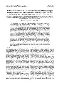
Fig. 1. Fractionation of the A65 % ammonium sulphate fraction into constituent enzymes and cytochromes.
Purified
I
Hydrophobic interaction chromatography on phenyl-Sepharose CL 4B
I
Preparative isoelectric focusing on Sephadex TEF
Gel filtration on Sephadex G-200
0.35M NaCI(1) Sulphite :cytochrome c oxidoreductase Cytochrome cj5, Enzyme B
Sulphite :cytochrome c oxidoreductase
0.2 M NaCl Cytochrome
Gel filtration on Sephadex G-100
I
Hydrophobic interaction chromatography on phenyl-Sepharose CL-4B
I
Ammonium sulphate fraction A90 %
I
0.12 M NaCl Enzyme A
0.1 M NaCI
Ion-exchange chromatography on DEAE-Sepharose CL-6B with stepwise elution with NaCl
I
A65 % FRACTION
0
cn
Thiosulphate oxidation by Thiobacillus A2
3551
1"
5 - 4 W
Y
v
.c
e
3
Y
a
2 I 0
Elution volume (ml) Fig. 2. Elution pattern of the A90% fraction on phenyl-Sepharose CL-4B. For details see Methods. 0 , Protein (Azso); 0 , enzyme A activity; --- ammonium sulphate and ethylene glycol gradient.
Resolution of the A65% fiactwn. Enzymes A and B, sulphite :cytochrome c oxidoreductase and cytochrome were resolved as summarized in Fig. 1. Enzyme assays. Thiosulphate :cytochrome c oxidoreductase activity could only be observed using mixtures of the main fractions or of the further purified enzymes (Fig. 1;Lu & Kelly, 19836, c). The 0.1 M-NaCI and 0-35MNaCl(1) fractions contained activities we shall refer to, respectively, as 'enzyme A' and 'enzyme B'. Both were required for thiosulphate-oxidizing activity. The 0.35 M-NaCl(1) fraction was resolved into two major components, one being enzyme B, the other being cytochrome cS51, both of which were required for full activity. Enzyme A activity was measured as described by Lu & Kelly (1983c), except that a smaller amount of enzyme A solution (0.0141 mg) was used at the later stages of the purification. Enzyme B was assayed by essentially the same procedure as for enzyme A. The reaction mixture (1 ml in a 1 cm light-path cuvette) contained (pmol): Na2St03,2; Tris buffer, pH 7-3,45; cytochromec (horse-heart type 11), 0-07;A65% fraction (0.3 mg protein, as enzyme A); 0.35 M-NaCl(I1) fraction (0.6 mg protein); G-200-1 fraction (0.4 mg protein, containing cytochrome C s S l ; see Results) and enzyme B solution (0.014.08 mg protein). Reaction at 30 "C was initiated by adding the enzyme B solution. Activity was expressed as cytochromec reduction in terms of the protein added as enzyme B. The activities of enzyme A and enzyme B measured and calculated in this way are relative, as the amounts of enzyme B (for assay of enzyme A), enzyme A (for assay of enzyme B), cytochromecS5 and cytochrome c 5 5 2 . 5 used in the assay will affect the activity. In order to obtain a linear relationship between amount of enzyme A assayed and the activity, a ten times or greater excess of enzyme B (in terms of protein) was used. Similarly, enzyme A was used in excess to assay enzyme B. Sulphite :cytochrome c oxidoreductase activity was measured as described before {Lu & Kelly, 1983a), except that less protein (4-50 pg) was used. Pur8cution of Enzyme A. Protein precipitated from the combined 0.1 M- and 0.12 M-NaC1 fractions by precipitation between 60-90% saturation with ammonium sulphate was recovered as described previously and referred to as the A90% fraction (Lu &Kelly, 1983~). This was stored at - 20 "C. The A90% fraction (420 mg) was thawed and applied to the top of a 2-6 cm x 6.5 cm column of phenyl-Sepharose CL-4B equilibrated with 18 mMphosphate buffer, pH 6.5 containing 2 m ~ - N a ~ and S ~ (NH4)2S04 o~ at 17% saturation. After sample addition, elution was continued with one bed volume of equilibrating buffer, followed by a linear gradient of decreasing ammonium sulphate concentration and increasing ethylene glycol concentration, which was produced by constant-head mixing of 250 ml each of (NH,)$04 (17% saturation) and 50% (v/v) ethylene glycol, both in 18 mwphosphate buffer, pH 6.5 containing 2 mM-Na2Sz03at 4 "C and a flow rate of 30 ml h- l . The typical elution pattern is shown in Fig. 2. The active fractions were combined and then concentrated at 4 "C in a 50 ml Amicon ultra-filtrationcell over an Amicon PMlO membrane under nitrogen at 0.7 bar (7 x lo4 N m-*). In the presence of ethylene glycol about 30% of the enzyme passed through the membrane in the filtrate. The ultrafiltration had to be repeated three times to obtain 95% of the enzyme. Gelfiltration on Sephudex G-100. The concentrated enzyme A solution (120 mg) was loaded on the bottom of a 2.6cm x 83cm column of Sephadex G-100 equilibrated with 50mM-Tris buffer, pH 7-3, containing 2mMNa2S20,,and eluted upwards with the same buffer at 4 "C and a flow rate of 16 ml h-l. The elution pattern was as c551
3552
W - P . LU A N D D. P. K E L L Y
-
1.0 -
h 0 N
.-Eu
5
a
0.80.6-
0.40.2 -
0-
1
1
200 240 280 320 Elution volume (ml)
Fig. 3. Elution pattern on Sephadex G-100 of the active fraction from phenyl-Sepharose CL-4B chromatography.For details see Methods. @, Protein ( A z s o ) ;0 ,enzyme A activity. The void volume was 160ml. seen in Fig. 3. The active fractions were pooled and concentrated by (NH4)2S04as described before (Lu & Kelly, 1983~). Preparative isoelectricfocusing. The concentrated enzyme A solution (10-15 mg protein) was dialysed against 1% (w/v) glycine for 3 h at 4 "C and then loaded on a flat bed Sephadex IEF gel containing 1/15 (v/v) Pharmalyte, pH 2.5-5. Preparation of gel and gel bed (115 x 240 x 2 mm), application of sample, focusing condition and recovery of separated protein's were essentially the same as described in the instruction manual from Pharmacia. An LKB Multiphor 21 17 and constant power supply 2197 were used. Cold water (0 "C) was circulated through the cooling plate during focusing. A quick paper print technique for detection of focused proteins as described in the LKB manual was employed. A typical paper print is shown in Fig. 4(a). Recovered enzyme A was desalted by dialysisor gel filtrationon Sephadex G-25, followed by dialysis against solid polyethylene glycol to concentrate the sample. Possible modifications of the purificationprocedure. Thiosulphate was included in the elution buffers as an enzyme stabilizing agent, because purified enzyme A was subsequently found to be unstable in the absence of thiosulphate. The ultrafiltration steps could use membranes with lower molecular weight cut-off levels (e.g. YM5 instead of PMlO) in order to avoid passage of the enzyme in the presence of ethylene glycol. Purijcation of enzyme B. Resolution of 0.35 M-NaCI(I)fractwn into two major fractions containing enzyme B and cytochrome c551. The 0-35M-NaCl(1) fraction (500 mg protein) was applied to the bottom of a column (3.2 x 85.5 cm) of Sephadex G-200 equilibrated with 50 mM-Tris buffer, pH 7.3 and eluted upwards with same buffer at 4 "C and a flow rate of 35 ml h- l . Active fractions were pooled and then concentrated by ultrafiltration under nitrogen pressure (0-7bar) through an Amicon PMlO membrane at 4 "C. Hydrophobic interaction chromatography. Concentrated enzyme B solution (60mg) was brought to about 15% saturation with (NHJ2S04 loaded on to the top of a 2-6 x 6-5cm column of phenyl-Sepharose CL4B equilibrated with 50 m - T r i s buffer, pH 6.5, containing 10%saturation with (NH,),SO,. After sample application, the column was eluted downwards with one bed volume of the same buffer at 4 "C and a flow rate of 30 ml h- l , followed by a linear gradient of decreasing ammonium sulphate concentration, which was produced from two 150 ml volumes of 50 mM-Tris buffer, pH 7.3, one of which contained (NH4)2S04(10% saturation). The active fractions were combined and concentrated by ultrafiltration as stated above. Preparative isoelectricfocusing. The procedure was the same as described for purification of enzyme A (Fig. 4b). Assay of stoicheiometry of thwsulphate oxidation by the reconstituted system. This was done in a Clark oxygen electrode cell essentially as described by Lu & Kelly (1983a). Reaction mixture (final volume 1 ml) contained: purified enzyme A (0.15 mg), pure enzyme B (0.2 mg), cytochrome c551 fraction (G-200-1 fraction, 0.5 mg), cytochrome c 5 5 2 . 5 (0-35M-NaCl(I1) fraction, 0.5 mg), horse-heart cytochrome c (2 mg), bovine-heart cytochrome oxidase (5 units), Tris buffer, pH 7-3(40 pmol) and Na2S203(50 or 100 nmol precisely). Oxygen concentration in the experimental conditions was calibrated using the method described by Robinson & Cooper (1970). PAGE. SDS-PAGE was carried out to monitor the protein purificationand to determine molecular weight using the method of Laemmli ((1970). Acrylamide (12%, w/v) was used in the resolving gel. Protein samples were incubated at 60 "C for 10 min in Tris buffer, pH 6-5, containing 3% (v/v) 2-mercaptoethanol and 1% (w/v) SDS before loading and were electrophoresed at a constant 40 mA for 5 to 6 h at 4 "C.The gels were stained overnight in a mixture of 30% (v/v) methanol, 5% (v/v) acetic acid in distilled water containing 0.2% (w/v) Coomassie brilliant blue G and destained in the same solution without the dye. Standard marker proteins ( M , in parentheses)
Thwsulphate oxidation by Thwbacillus A2
3553
Fig. 4. Paper prints of flat-bed isoelectric focusing gels of purified enzymes A and B. (a) Isoelectric focusing of active material (enzyme A) from Sephadex G-100 chromatography (see Methods). Sample (1 2 mg protein) was loaded and focused for 6 h at 12 W,constant power, 1500 V maximum. The gel was pre-run for 1 h. (b)Isoelectric focusingof the enzyme B fraction (HIC-11) from phenyl-Sepharose CL-4B chromatography (see Methods and Results). Sample (15 mg protein) was loaded and focused for 6 h at 30 W, constant power, 2000 V maximum. The gel was pre-run for 1 h.
used for calibration were bovine albumin (66000), egg albumin (45OOO), glyceraldehyde-3-phosphatedehydrogenase (36000), carbonic anhydrase (29000), trypsinogen (24000), trypsin inhibitor (20 100) and a-lactalbumin (14000). Purity of the samples was also examined by discontinuous PAGE under nondenaturing conditions essentially as described by Davis (1964). Acrylamide (8%, w/v) was used in the resolving gel. The electrophoresis was performed at a constant 30 mA for 4 h at 4 "C. The staining procedure was the same as for the SDS-gel. Determination of the molecular weight of enzyme B by gelfiltration. A method based on Andrews (1965) was used with bovine serum albumin (mol. wt 67000), egg albumin (mol. wt 43000), bovine pancreas chymotrypsinogen A (mol. wt 25000) and bovine ribonuclease A (mol. wt 13700) as marker proteins. Enzyme B (4 mg protein) and marker proteins (5 mg protein each) were run separately on a 2.6 cm x 84 cm column of Sephadex G-100 with 50 mM-Tris buffer, pH 7.3 at 4 "C. The positions of the marker proteins and enzyme B were determined by measuring absorbance at 280 nm and enzyme activity.
3554
W-P. L U AND D . P. KELLY
Determination of isoelectric point. The isoelectric points of the purified proteins were measured by flat bed electrefocusing in polyacrylamide gels using Pharmalyte, pH 2.5-5 and an LKB Multiphor 21 17 and constant power supply 2197. Preparation of gel (100 x 50 x 1 mm), application of samples, running conditions and staining of gel were based on the instruction for PI calibration kits from Pharmacia. The pH gradient profile across the IEF gel was calibrated by using a low PI calibration kit. Reaction of pur@ed enzymes with 35S 20$-Reaction . mixtures (0.5 ml) in Tris/HCl, pH7-3, contained either Na235S-S03or Na2S-35S03(2 pmol at 2-10 x lo6 d.p.m. pmol-') and other components as indicated in Results. After incubation at 30 "C, samples were analysed by paper chromatography using a butanol/pyridine/acetic acid/water (20 :30 :6 :24, by vol.) solvent with and without treatment with iodine to convert residual thiosulphate to tetrathionate (Kelly & Syrett, 1966). Chromatograms were assayed for 35Sby cutting strips into segments and counting in scintillation vials filled with 0.5% (w/v) butyl PBD in toluene. Protein estimation. Protein was determined by the Lowry method using bovine serum albumin as a standard. Chemicals. Cytochrome c (horse heart 111), cytochrome oxidase (bovine heart), catalase (bovine liver), egg albumin, chymotrypsinogen A, ribonuclease A, molecular weight marker kits (MW-SDS-70L), NADH were obtained from Sigma. Sephadex G-25, G-100, G-200, phenyl-Sepharose CL-4B, Sephadex IEF, Pharmalyte (pH 2-5-5) and low PI calibration kits were obtained from Pharmacia. RESULTS
Purification of enzyme A Enzyme A was purified some 100-fold by the procedures described (Fig. 1; Table 1). The progress of the separation, monitored by SDS-PAGE is shown in Fig. 5 . Purity and some properties of enzyme A As shown in Fig. 5 enzyme A, after the final purification, still appeared as one major band with a molecular weight of 16200 and another minor band of 14600. The minor protein was about 15%of the major protein as measured by scanning the gel at 600 nm. The two bands had similar densities in the crude extract. On the basis of the 100-fold increase in specific activity and its concentration,the major band in the purified preparation is presumed to be enzyme A. The enzyme comprised about 0.8% (w/w) of the crude extract. Because both proteins had very similar molecular weights, nearly the same PI values, determined by isoelectric focusing in polyacrylamide gel, and more or less the same hydrophobic properties, it is very difficult to separate them by the techniques used so far. The isoelectric point of enzyme A is about 4.2. The enzyme lost about 50% and 80% of its activity at 20 "C after 6 h and 20 h, respectively, and 30% and 50% at 4 "C after 1 d and 2 d, respectively. Purification of enzyme B The 0.35 M-NaCl(1) fraction from DEAE-Sepharose-CL-6B chromatography was separated into two major protein peaks by gel filtration on Sephadex G-200 (Fig. 6). Three fractions were collected : fraction I (called G-200-1) contained cytochrome c55 ; fraction I1 (G-200-11), contained some sulphite :cytochrome c oxidoreductase ;and fraction 111(G-200-111) contained enzyme B and most of the sulphite :cytochrome c oxidoreductase activity (Table 2). Enzyme B and sulphite :cytochrome c oxidoreductase in the G-200-111 fraction were further separated into two fractions by phenyl-Sepharose-CL-4Bchromatography (Fig. 7 and Table 3). Most sulphite :cytochrome c oxidoreductase activity was recovered in the first fraction (called HIC-I). Most of the enzyme B activity was in the second fraction (called HIC-11), but the specific activity was not increased and the total activity recovered (yield) was low. The specific activity of enzyme B was, however, increased about 40% by including a small amount of the HIC-I fraction, to give a ratio in the reaction mixture of HIC-I :HIC-I1 protein equivalent to that in the separated fractions. Yield was also restored to about 95 % of that originally present. Enzyme B in the HIC-I1 fraction was finally purified by preparative isoelectric focusing in Sephadex IEF. Two major protein bands were obtained on a paper print of the gel (Fig. 4b). Both of them contained enzyme B activity (Table 4). The SDS-PAGE disclosed that they were the same protein, although the protein from the bottom band had less enzyme activity and one or
Thiosulphate oxidation by Thwbacillus A2
3555
Fig. 5. SDS-polyacrylamide slab gel electrophoresis of the fractions in the purification of enzyme A. (1) Marker proteins (55 pg); (2) crude extract (60 pg); (3) A65% fraction (60pg); (4) 0.1 M and 0.12 M fraction (60 pg); (5) A90% fraction (60 pg); (6) pooled active fractions (30 pg) from phenyl-Sepharose CL-4B column; (7) pooled active fractions (30 pg) from Sephadex G-100; (8,9) purified enzyme A after preparative isoelectric focusing (30 and 20 pg, respectively); (10) marker proteins (45 pg). For details see Methods and Results. Molecular weights of markers and direction of migration are indicated.
Table 1. Summary of the purification of enzyme A Enzyme A was assayed as described in Methods.
Procedure Crude extract First ammonium sulphate fractionation (A65%) DEAE-Sepharose CL-6B Second ammonium sulphate fractionation (A90%) P henyl-Sepha rose CL-4B Sephadex G-100 Isoelectric focusing in Sephadex IEF
Volume Protein (mu (mg)
Specific activity Total activity [pmol cytochrome c reduced min-' (mg (pmol cytochrome c reduced min- ' ) protein)- '1
Purification (fold)
Yield (%)
1oo* 91 *
545 251
49100 12000
-
-
74
2192
0.25
543
18
74
34
946
0.54
51 1
39
70
16
409
1.16
474
80
65
14
302 140
1-5 1-4
453 196
110 102
62 27
-
* These figures were derived from results measured by the oxygen electrode method
1* 4*
(Lu & Kelly, 19836).
3556
300
200
600
400 500 Elution volume (ml)
Fig. 6. Elution profile of the 0.35 M-NaCl(1) fraction on Sephadex G-200 (see text for detail). 0 , Protein (Azso); 0 , cytochrome cssl (A416);0 , enzyme B activity; A, su1phite:cytochrome c oxidoreductase. The void volume was 265 ml.
Table 2. Resolution of the 0.35M-NaCl(I)fraction from DEAE-Sepharose CL-6B chromatography into two major components by geljiltration on Sephadex (3-200 (see Fig. 6) Enzyme B activity*
Sulphite :cytochrome c oxidoreductase activity
7-
Cytochrome Fractions Sample [0.35~-Nacl (I)] Fraction I Fraction I1 Fraction I11
c551
Protein (mg)
[nmol (mg protein)-']
500
4
134 50 103
12
0-5 0
Specific Specific [pmol Total [Pol Total cytochrome c (pmol cytochrome c (pmol min-l (mg cytochrome c rnin-' (mg cytochrome c protein)-'] min-l) protein)-'] min- l ) 0.3
150
0.86
428
0 0.03 1-43
0 1.6 147
0.02
3 28 308
0.56
2.97
* Enzyme B and sulphite :cytochrome c oxidoreductase activities were measured as described in Methods.
Omitting Fraction I (i.e. cytochrome cSs1) from the reaction mixture reduced the specific activity by half.
two minor contaminants. Enzyme B accounted for at least 90% of the total protein in the HIC-I1 fraction and the double-banding observed was probably due merely to overloading. The purification of enzyme B is summarized in Table 4 and Fig. 1. The final product represents some 280-fold purification over the crude extract with an overall recovery of about 50%. The enzyme comprised about 0.6% (w/w) of the crude extract protein. Purity and molecular weight of enzyme B The purified enzyme appeared as a single sharp band after SDS-PAGE and discontinuous PAGE (Fig. 8 a and b, respectively). However, the enzyme band shifted from the position equivalent to a molecular weight of 63000 (k2000) to that of half this molecular weight on the SDS polyacrylamide gel, indicating the enzyme to consist of two subunits with a molecular weight of 32000 (+, 2000). Determination of molecular weight on Sephadex G-100 also confirmed that the native enzyme possessed a molecular weight of about 64000. Gel
Thiosulphate oxidation by Thiobacillus A2
I
1-1-+1-
3557 0.7
0.6
0
I
1
I
100
200
3 00
Elution volume (ml) Fig. 7, Elution profile of the further resolution of the G-200-111 fraction on phenyl-Sepharose CL-4B (see Fig. 6 and text for detail). a, Protein ( A z s o ) ;0 , enzyme B; A, sulphite :cytochrome c oxidoreductase ; ---, ammonium sulphate concentration gradient.
Table 3. Separation of Enzyme B and sulphite :cytochrome c oxidoreductase by hydrophobic interaction chromatography of fraction G-200-III (see Fig. 6)on phenyl-Sepharose-4B (see Fig. 7) Sulphite :cytochrome c oxidoreductase activity
Enzyme B activity* A
r
>
A
r
>
Fractions assayed
Protein (mg)
Specific [Pol cytochrome c min-' (mg protein)- '1
Sample (G-200-111) Fraction I Fraction I1 Fraction I + IIt
103
1-43
147
2.97
308
12 68 68
0.59 1-43
3.5 97 139
21-1 0.97
247 30 -
2.05
Total (pmol cytochrome c min-l)
Specific [vmol cytochrome c min-l (mg protein)- * ]
Total (pmol cytochrome c min-')
-
* Enzyme B and sulphite :cytochrome c oxidoreductase activities were measured as described in the Methods.
t Assayed using 16 pg Fraction I1 protein supplemented with 3 pg Fraction I protein in terms of the sum of the
protein samples added.
electrophoresisof the crude extract and A65 % fraction showed a band at the position expected for enzyme B ( M , about 63000; Fig. 8a), but most of enzyme B in the 0-35 M-NaCl(1) fraction moved down to the subunit position. Since all oftthe samples were prepared in the same way before being loaded on the gel, it is unclear why the enzyme behaved so differently. Treatment with SDS at 100 "C did not cleave the 63 000 M , enzyme into smaller units. Boiling the purified enzyme from IEF (Fig. 8a, lane 6) resulted in most of the protein running as M,63 000, although the milder treatment gave the smaller unit seen in Fig. 8(a). This behaviour is anomalous and cannot be further explained at present. Some other properties of enzyme B The enzyme had a PI value of about 4-25 and had an absorption spectrum only in the UV region, with a sharp maximum at 280 nm and a broad absorbance below 250 nm. The enzyme was more stable than enzyme A. It lost about 30% and 50% activity at 20 "C after 1 d and 2.d, respectively, and 4% and 20% at 4 "C after 1 d and 2 d, respectively. Storage at - 20 "C for at least three months, with freezing and thawing had no significant effect on the enzyme activity.
Methods and Results.
Fig. 8. (a) SDS-polyacrylamide slab gel electrophoresis of the fractions in the purification of enzyme B. (1) Crude extract (60pg); A65% fraction (60pg); (3) 0-35M-NaCl(1) fraction (60 pg); (4) G-200-111 fraction (see Fig. 6) (30 pg); (5) HIC-I1 fraction (see Fig. 8) (30 pg); (6)pure enzyme B from preparative isoelectricfocusing (20 pg); (7)marker proteins (45 pg). (b)Discontinuouspolyacrylamide slab gel electrophoresisof the fractions in the purification of enzyme B. (1) 0.35 M-NaCl(1) fraction (60 pg); (2) G-200-111 fraction (see Fig. 6) (25 pg); (3) HIC-I1 fraction (see Fig. 7) (25 pg); (4) pure enzyme B from preparative isoelectric focusing (20 pg). For details see
3558 W - P . LU A N D D. P. KELLY
3559
Thiosulphate oxidation by Thwbacillus A2
Table 4 . Summary of the pur$catwn of Enzyme B Enzyme B was assayed as described in Methods. Specific activity [pmol cytochrome c reduced min(mg protein)- '1
Total activity (pmol cytochrome c reduced min- ')
-
-
-
-
1155
0.30
346
34
75
240
1-43
343
162
73
158
2.057
323
23 1
69
67 81
2-5t 0.9t
167 73
280 101
53
Total protein (mg)
Procedure Crude extract 1st ammonium sulphate fraction (A65%) DEAE-Sepharose CL-6B [0.35 M-NaCI(1) fraction] Sephadex G-200 (G-200-111) (see Fig. 6) Phenyl-Sepharose CL-4B (HIC-11) (see Fig. 7) Preparative iso(1)s electric focusing (11)s
49 100 12000
* Figures derived from results obtained
Purification (fold)
Yield (%)
1* 4*
loo* 91*
using the oxygen electrode procedure (Lu & Kelly, 19833).
t For these assays some of the HIC-I fraction (3 pg protein) was added to the reaction mixture. For details see
Table 3 and the text. $ I and I1 are the upper and lower bands respectively of Fig. 4b.
Table 5 . Purification of sulphite :cytochrome c oxidoreductase Sulphite :cytochrome c oxidoreductase was assayed as described in Methods.
Procedure Crude extract 1st ammonium sulphate fraction (A65 YJ DEAE-Sepharose CL-6B Sephadex G-200 (G-200-111) (see Fig. 6) Phen yl-Sepharose CL-4B (fraction I, see Fig. 7)
Total protein (mg)
Specific activity [pmol cytochrome c reduced min(mg protein)-']
Total activity (pmol cytochrome c reduced min- I ) -
Purification (fold)
Yield
1* 5*6*
(%I
12000
-
0-04 0.22
2640
1155 240
1.10 2.97
1270 713
28 93
48 27
28
21-10
591
660
22
* Figures derived from results obtained
-
100
using the oxygen electrode procedure (Lu & Kelly, 19836).
Sulphite :cytochrome c oxidoreductase and the stimulation of enzyme B activity by the HIC-I fraction The specific activity of sulphite :cytochrome c oxidoreductase was increased over 600 times during purification even though the recovery was rather low (Table5). However, the final product (HIC-I fraction) gave more than six protein bands on SDS polyacrylamide gel (Fig. 9), in which the top major band was most probably enzyme B and the rest had more or less the same density. The HIC-I fraction stimulated enzyme B specific activity in the HIC-I1 fraction (Table 3) and pure enzyme B (Table 4) by 2040%. Fractions from previous steps were also stimulated. The stimulation could not simply be accounted for by the presence of the small amount of enzyme B in the HIC-I fraction.
3560
W-P. LU AND D . P. KELLY
Fig. 9. SDS-polyacrylamidegel electrophoresis of fractions in the separation of sulphite :cytochrome c oxidoreductase and cytochrome cSs1.(1) Marker proteins (45 pg); (2) fraction HIC-I (see Fig. 7) (30 pg); (3) fraction G-200-1 (see Fig. 6) (25 pg); (4) 0.35 M-NaCl(I1) fraction (40 pg).
Reconstitution of thiosulphate-oxidizing activity with the puriJed components Thiosulphate was completely oxidized to sulphate by the reconstituted system with a consumption of 1~ 9 5& 0.05 mol oxygen for each thiosulphate added (four determinations). The reaction was negligible in the absence of any one of enzyme A, enzyme B, or the cytochrome cssl and cytochrome c552.5 fractions. Attempt to demonstrate thiosulphate cleavage by the purijied enzyme(s) Incubation of partially purified enzyme A (0-35mg), enzyme B (1-5 mg), or mixtures of enzyme A, enzyme B and the cytochrome cSs1and cytochrome cs52.5fractions with thiosulphate labelled in the inner or outer sulphur atom with 3%, in the absence of electron acceptors for as long as 45 min at 30 "C demonstrated no significant formation of sulphite or sulphate. In other words the S-S bond of thiosulphate was not split by any one of the enzymes or the enzyme system under these experimental conditions. However, the experiment did show that thiosulphate appeared to be associated with enzyme B (HIC-I1 fraction) with a molar ratio of about two thiosulphate :one protein.
3561
Thiosulphate oxidation by Thiobacillus A2
s,o:- + 5H,O 2so:- i-10H+
v
2 0 , + 8H+
Fig. 10. Schematic representation of the components of the enzyme :cytochrome complex catalysing thiosulphate oxidation by ThwbaciffusA2. The sequence of electron transfer among the cytochrome c components is unresolved, cytochromes css and css2.5being essential for thiosulphate oxidation, with the soluble cytochrome csso(Mr, 15000) possibly linking electron transport from the complex to membrane-bound cytochrome ~ 5 5 2 . Approximate relative molecular masses of the cytochromes are indicated by their areas in the scheme (W. P. Lu & D. P. Kelly, unpublished data), with enzyme B ( M r , 64000) and enzyme A (Mr,16000) being expressed relative to each other as 0.6% (w/w) and 0 4 % (w/w) of the crude extract protein, respectively. Cytochrome c552.5 (Mr, 32000) contains two subunits (Mr, 16000 each) and ‘cytochrome css,’ is a very large protein (Mr,300000) probably containing six subunits. Relative molecular masses have not been determined by us for the membrane cytochrome ~ 5 5 or 2 uu3, so they are assumed to be similar to literature values for animal cytochromes (Mr, 12000 and 130000, respectively) for the purpose of diagrammatic representation.
Table 6. A typical assay of thiosulphate :cytochrome c oxidoreduction activity with the four highly purified components Enzyme activity was measured spectrophotometrically as described in Methods except that the components were added to the reaction mixture in the order and the amount listed below, following the addition of horse-heart cytochrome c and thiosulphate. Activity was calculated in terms of the total protein of the components added. The detailed purification and characterization of cytochrome css2.s and ‘cytochrome c ~ ~ are , ’to be published elsewhere.
Cytochrome cSs1
Cytochrome c reduction [nmol reduced min-1 (mg protein)-’]
0.085 0.085
3 35 85 22
Addition (mg protein) A
f
Enzyme A
Enzyme B
0.1 0.1
0.08
0.1 0.1
0-08 0-08 0.08
Cytochrome css2.s 0.04 0.04
3
Cytochrome cs5 and cytochrome C552.5 Thiosulphate oxidation and horse-heart cytochrome c reduction by the reconstituted enzyme system was negligible if the cytochrome cSs1and c 5 5 2 . 5 fractions were omitted from the reaction mixture and only commenced when one of them was added. Subsequent addition of the other cytochrome fraction increased the activity further. The apparent K , for thiosulphate in the reconstituted thiosulphate :cytochrome c oxidoreductase system was probably less than 2 p ~ . The apparent K , value for horse-heart cytochrome c was lower in the reconstituted system
3562
W - P . L U A N D D . P . KELLY
(40 p ~than ) in the A65% fraction (200 PM). The causes seemed related to the concentration of cytochrome cSs1 and cytochrome c 5 5 2 . 5 , since there was more of them in the reconstituted system. However, the two cytochromes contained in the G-200-1fraction and 0-35 M-I1fraction were still quite crude, as shown in Fig. 9. Subsequent purification (unpublished data) of cytochromes cSs1 and c552.5 enabled proof of their essential role in the thiosulphate-oxidizing complex (Table 6). DISCUSSION
The conclusions to be drawn from our results to date indicate that enzymes thought to have some role in thiosulphate oxidation by thiobacilli (Oh & Suzuki, 1977; Kelly, 1982) do not seem to have central functions in Thiobacillus A2. These include rhodanese, which, although very active in ThiobaciZZus A2 (Silver & Kelly, 1976; Wood & Kelly, 1981;Lu & Kelly, 1983a), is not required for complete oxidation of thiosulphate by extracts (Lu & Kelly, 1983c), while the ‘thiosulphate-oxidizing enzyme’ (Trudinger, 1961), adenylyl sulphate reductase and the sulphuroxidizing oxygenase enzyme (Suzuki, 1965;Charles & Suzuki, 1966a; Suzuki & Silver, 1966) are either absent from ThiobacillusA2 or present only at low levels (Kelly & Tuovinen, 1975; Silver & Kelly, 1976). So far we have also failed to demonstrate a thiosulphate-cleaving enzyme (Peck, 1960; Kelly, 1982) or a major role for free sulphite as a substrate for sulphite oxidase in the system. It is generally believed that free sulphite is the penultimate intermediate in sulphate formation (Charles & Suzuki, 1966b; Kelly, 1982) and sulphite oxidase activity is present in all thiobacilli examined. We have, however, obtained a cell-free system catalysing the complete oxidation of thiosulphate, for which at least two novel colourless enzymes and several cytochromes are required. These two enzymes have now been highly purified (Fig. 2), although we are as yet unable to ascribe specific individual functions to them. To our knowledge, these two enzymes differ from any enzymes so far found in sulphur-oxidizing bacteria, although a relative lack of detailed enzymological data in other published work makes proper comparison difficult. The present work confirms the view (Lu & Kelly, 1983b) that the thiosulphate :cytochrome c oxidoreduction system in Thiobacillus A2 is a soluble multi-enzyme complex. The complete oxidation of thiosulphate by the reconstituted purified components proves that no small cofactor molecules are needed. The relatively easy separation of the components suggests their association in uiuo to be weak. Many multi-enzyme systems show much stronger association, although some (e.g. tryptophan synthetase) are easily separated into subunits, with a considerable decrease in overall activity (Miles, 1979). Dissociation of the complex could help explain why thiosulphate-oxidizing activity in the crude extract from Thiobacillus A2 was about 100-foldless than in intact cells (Kula et al., 1982; Lu & Kelly, 1983a). Thus, rapid and stoicheiometric oxidation of thiosulphate in uiuo depends on the integrity of the well-organized multienzyme complex and its association with the membrane system. Any disturbance of the structure, such as caused by disruption of the cell could thus dramatically affect oxidative ability. In fact, one advantage of a multi-enzyme complex is that it provides a very short transit time for passage of intermediates from one enzyme to another. This could be particularly important if unstable compounds such as sulphite and other reduced sulphur species are produced as intermediates in thiosulphate oxidation. Such intermediates might always be enzyme-bound in the multi-enzyme system. For thiosulphate to be oxidized to sulphate, three main processes must occur. These are cleaving of the S-S bond, the oxidation of the sulphanesulphur group to sulphate, and the oxidation of the sulphone-group to sulphate. Cleavage has come to be regarded as the primary step (Kelly, 1982), followed by oxidation of the sulphur (or sulphide) and sulphite formed thereby. Our reconstituted system seems to contain two colourless enzyme proteins and two essential c-type cytochromes (of which ‘cytochrome cSs1’may in fact be a third enzyme of thiosulphate oxidation), which together can effect all three essential processes. We have so far failed to show a thiosulphate-cleaving function using purified enzymes or the enzyme system in the absence of the electron transport system. This might mean that cleavage only proceeds at the rate of overall oxidation, so that in the absence of electron
Thiosulphate oxidation by Thiobacillus A2
3563
transport, thiosulphate binding to an enzyme can occur, but cleavage either does not occur or is so slight as not to be detectable. This could be because the products of cleavage are not released in the free state, but have to be transferred to acceptor-enzyme components of the multi-enzyme system and further cleavage ceases as oxidation of the transferred groups cannot occur. Alternatively, oxidation of sulphane-sulphur to sulphite might occur on the thiosulphatebinding enzyme with cleavage occurring only when an enzyme-bound intermediate analogousto dithionate ( - 0 3 S - S 0 3 - ) has been formed. At present it is not possible to decide on the exact nature of the partial reactions of sulphate formation or even the exact timing of the sulphursulphur bond cleavage. Certainly, however, thiosulphate cleavage is not a simple primary reaction of the rhodanese type. In current work we are attempting to evaluate the midpoint potentials of the various c-type cytochromes, which should enable an assessment of the sequence of their involvement as electron transport carriers. Figure 10 gives a schematic representation of the probable interrelations of the multi-enzyme complex and the membrane system in effecting thiosulphate oxidation. The sulphite :cytochrome c oxidoreductase in Thwbacillus A2 compares with that in T. noveZZus (Yamanaka et al., 1981), in that cytochrome 5 5 1 appears to be the electron acceptor for sulphite oxidation and separation of the cytochrome from the enzyme considerably reduced its activity (unpublished observations). Nevertheless, the significance of the sulphite :cytochrome c oxidoreductase in Thiobacillus A2 is still obscure. One phenomenon of potential significance to understanding the system was the observed stimulation of enzyme B activity by the fraction containing the sulphite :cytochrome c oxidoreductase activity. A more critical analysis of the HIC-I1 fraction is essential before further conclusions can be drawn. This work was made possible by financial support from The Government of the People’s Republic of China, The British Council and the Committee of Vice Chancellors and Principals. We are grateful to Dr Mark Woodland for advice and the use of some items of equipment, and to Drs Ann Wood and Ben Swoboda for discussions.
REFERENCES
ANDREWS, P. (1965). The gel-filtration behaviour of proteins related to their molecular weight over a wide range. Biochemical Journal 96,595406. CHARLES, A. M. & SUZUKI,I. (1966a). Mechanism of thiosulfate oxidation by Thwbacillus novellus. Biochimica et biophysica acta 128, 510-521. CHARLES, A. M. & SUZUKI,I. (1966b). Purification and properties of sulfite :cytochrome c oxidoreductase from Thiobacillus novellus. Biochimica et biophysica acta 128, 522-534. DAVIS,B. J. (1964). Disk electrophoresis. 11. Method and application to human serum proteins. Annals of the New York Academy of Sciences 121, 404-427. HARRISON, A. P. (1983). Genomic and physiological comparisons between heterotrophic thiobacilli and Acidiphilium cryptum, Thiobacillus versutus sp. nov., and Thiobacillus acidophilus, nom. rev. International Journal of Systematic Bacteriology 33, 2 1 1-2 17. KELLY,D. P. (1982). Biochemistry of the chemolithotrophic oxidation of inorganic sulphur. Philosophical Transactions of the Royal Society. Series B 298, 499528. KELLY,D. P. & SYRETT, P. J. (1966). [35S]Thiosulphate oxidation by Thiobacillus strain C. Biochemical Journal 98, 537-545. KELLY,D. P. & TUOVINEN, 0. H. (1975). Metabolism
of inorganic sulphur compounds by Thwbacillus ferrooxidans and some comparative studies on Thiobacillus A2 and T . neapolitanus. Plant and Soil 43,7793. KULA,T. J., ALEEM,M. I. N. & WILSON,D. F. (1982). Oxidation-reduction potentials of respiratory chain components in Thiobacillus A2. Biochimica et biophysics acta 680, 142-151. LAEMMLI, U. K. (1970). Cleavage of structural proteins during the assembly of the head of bacteriophage T4. Nature, London 221, 680-685. Lu, W-P. & KELLY,D. P. (1983a). Thiosulphate oxidation, electron transport and phosphorylation in cell-free systems from Thiobacillus A2. Journal of General Microbiology 129, 1661-1 67 1. Lu, W-P. & KELLY,D. P. (19633). Partial purification and resolution of a thiosulphate-oxidizing system from ThiobacillusA2. Journalof General Microbiology 129, 1673-1681. Lu, W-P. & KELLY,D. P. (1983~).Rhodanese: an enzyme not necessary for thiosulphate oxidation by ThiobacillusA2. FEMS Microbiology Letters 18,289292. MILES,E. W. (1979). Tryptophan synthetase : structure, function and subunit interaction. Advances in Enzymology 49, 127-186.
3564
W - P . LU A N D D. P. KELLY
OH, J . K. & SUZUKI, I. (1977). Resolution of a membrane-associated thiosulphate-oxidizing complex of Thiobacillus novellus. Journal of General Microbiology 99, 41 3-423. PECK,H. D. (1960). Adenosine 5'-phosphosulfate as an intermediate in the oxidation of thiosulfate by Thiobacillus thioparus. Proceedings of the National Academy of Sciences of the United States of America 46, 1053-1057. ROBINSON,J. & COOPER,J. M. (1970). Method of determining oxygen concentration in biological media, suitable for calibration of the oxygen electrode. Analytical Biochemistry'33, 39G399. SILVER,M. & KELLY,D. P. (1976). Rhodanese from ThiobacillusA2 :catalysis of reactions of thiosulphate with dihydrolipoate and dihydrolipoamide. Journal of General Microbiology 97;277-284. SUZUKI,I. (1965). Oxidation of elemental sulfur by an enzyme system of Thiobacillus thiooxidans. Biochimica et biophysica acta'l04, 359-371.
SUZUKI,I. (1974). Mechanisms of inorganic oxidation and energy coupling. Annual Review of Microbiology 28, 85-101. SUZUKI,I. &SILVER,M. (1966). The initial product and properties of the sulfur-oxidizing enzyme of thiobacilli. Biochemica et biophysica acta 122, 22-23. TRUDINGER, P. A. (1961). Thiosulphate oxidation and cytochromes in Thiobacillus X . 11. Thiosulphateoxidizing enzyme. Biochemical Journal 78, 68M86. WOOD, A. P. & KELLY,D. P. (1981). Mixotrophic growth of Thiobacillus A2 in chemostat culture on formate and glucose. Journal of General Microbiology 125, 55-62. YAMANAKA. T., YOSHIOKA, T. & KIMURA,K. (1981). Purification of sulphite :cytochrome c reductase of Thiobacillus novellus and the reconstitution of its sulphite oxidase system with the purified constituents. Plant and Cell Physiology 22, 61 3-622.