Mar 5, 2016 - 0 1989 by The American Society for Biochemistry and Molecular Biology, ... Bharati Kakkadll, J. Allen Crow((, Ian A. Blair$, and David E. Ongll**.
THE JOURNAL OF BIOLOGICAL CHEMISTRY 0 1989 by The American Society for Biochemistry and Molecular Biology, Inc.
Vol. 264, No. 7,Issue of March 5,pp. 4212-4221,1989 Printed in U.S.A.
Purification, Primary Structure Characterization, and Cellular Distribution of Two Formsof Cellular Retinol-binding Protein, Type I1 from Adult Rat Small Intestine* (Received for publication, July 14,1988)
William H. SchaeferSglI, Bharati Kakkadll, J. Allen Crow((, Ian A. Blair$, and David E. Ongll** From the Departmentsof $Pharmacology and 11 Biochemiste, Vanderbilt University andthe $Laboratory of Cellular and 37232 Molecular Physiology, Howard Hughes Medical Institute, Nashville, Tennessee
Cellular retinol-binding protein type I1 (CRBP(I1))is or binding proteins to overcome this hydrophobicity. Cellular a major protein in the small intestine, accounting for retinol-binding protein type I1 (CRBP(I1))’ is a recently demore than 1%of the soluble protein recovered from scribed intracellular retinol-binding protein (3). It was origirat jejunal mucosa. Two forms of the protein, called nally discovered in extracts of whole 1-day-old rat pups and rat purified from that source (3). CRBP(I1) is expressed primarCRBP(I1)A and CRBP(II)B, were purified from small intestine using a three-column procedure. The ily, if not solely, in the small intestine, from shortly before two forms were present in equal abundance. The pri- birth through adulthood, and in theliver during the perinatal mary structures of CRBP(I1)A and CRBP(I1)B were period (3,4).It is confined to thevillus-associated enterocytes determined using a combination of techniques including amino acid composition and sequence analyses, and in the small intestine (5) and accounts for more than 1%of fast atom bombardment and gas chromatography-elec- the soluble protein recovered from the jejunal mucosa (3), suggesting a role for CRBP(I1) in the intestinal metabolism tron impact mass spectrometry. The primary strucof tures of both proteins were found to be identical, but vitamin A. CRBP(I1) binds both all-trans-retinol and allthey differed in their NH2-terminal processing. trans-retinaldehyde, two forms of vitamin A known to be CRBP(I1)B was acetylated at its NH2 terminus, while generated during the intestinal metabolism necessary for vitamin A absorption (6). Retinol bound to CRBP(I1) is directed CRBP(I1)A was not. The results also confirmed the amino acid sequence of CRBP(I1)A that was deduced to aparticular esterifying enzyme that appears to utilize from the cDNA sequence by Liet al. (Li, E.,Demmer, lecithin as theacyl donor for the production of retinol esters L. A., Sweetser, D. A., Ong, D. E.,and Gordon, J. I. that are subsequently incorporated into chylomicrons for export from the gut (7). (1986) Proe. Natl. Acad. Sei.U.S. A. 83,5770-5783). Antibodies capable of distinguishing between the two CRBP(I1) can be chromatographically resolved into two forms of CRBP(I1) were used for immunohistochemical forms (CRBP(I1)A and CRBP(I1)B)that appear to be almost studies which indicated that the organ and cellular dis- identical. Both forms weigh 15.5kDa and are present whole in tributions of the two forms were identical. The 50% rat pups (3) in equimolar quantities. One difference that was acetylation observed here in vivo fits the patternpre- observed between the two forms was the amount of endogedicted by recent in vitro studies which described the nous retinol recovered bound to theproteins after separation effect of NH2-terminalsequenceoncotranslational during purification. CRBP(I1)A was isolated with 10-fold NH2-terminal processingof cytosolic proteins (Boissel, more endogenous retinol bound than CRBP(1I)B (3). HowJ. P., Kasper, T. J., and Bunn, H. F. (1988) J. Biol. ever, their binding constants and absorption spectra in uitro Chem. 263, 8443-8449). Our results provide a basis are indistinguishable (3, 6). for investigating the possibility of different roles of A cDNA for CRBP(I1)A has been isolated from a rat small CRBP(I1)A and CRBP(I1)B within cells, as well as the importance of acetylation of the amino terminus for intestine cDNA library using oligonucleotide probes based on the amino acid sequence of the amino terminus of CRBP(I1)A these biological functions. (8). The nucleotide sequence of the cDNA was determined, and the amino acid sequence derived for CRBP(I1)A was identical to the sequence of the amino terminus established Vitamin A is essential for the regulation of growth and by Edman sequencing. CRBP(I1)B was found to be resistant differentiation of epithelial tissues (l),as well as for vision to Edman degradation* impeding structural characterization. (2). Several forms of vitamin A, including retinol, retinalde- Thus, it was not known whether the primary structure of hyde, and retinoic acid are involved in itsfunction and trans- CRBP(I1)B was similar, but not identical to CRBP(II)A, or port. Each of these forms is essentially water-insoluble, and whether the two proteins were actually products of the same the utilization of retinoids appears to require specific carrier gene but had undergone different co- or post-translational modification. Here we report the purification of CRBP(I1)A * This work was supported in part by National Institutes of Health and CRBP(I1)B from adult rat intestine and the cellular Grants GM 30861 and DK 32642, Molecular Toxicology ES-00267, SharedInstrumentation Grant RR-01688, and CRNU AM-26657. The costs of publication of this article were defrayed in part by the payment of page charges. This articlemust therefore be hereby marked “advertisement” in accordance with 18 U.S.C. Section 1734 solely to indicate this fact. 7 Present address: SmithKline and French Laboratories, Dept. of Drug Metabolism, P. 0. Box 1539 (L711) King of Prussia, PA 19406. ** To whom correspondence should be addressed.
The abbreviations used are: CRBP(II), cellular retinol-binding protein type 11; CRBP(II)A, cellular retinol binding protein type I1 form A; CRBP(II)B, cellular retinol binding protein type I1 form B; BSTFA, bis-trimethylsilyltrifluoroacetamide;HPLC, high performance liquid chromatography; T, trypsin; CN,cyanogen bromide; FAB, fast atom bombardment; EI, electron impact mass spectrometry; MS, mass spectrometry; GC, capillary gas chromatography. J. Gordon, personal communication.
4212
Purification and Characterization
of Two Forms of CRBP, Type
11
4213
distribution of both proteins as determined by immunohistochemistry. We also have compared the structures of CRBP(I1)B and CRBP(I1)A using a combination of techniques including amino acid compositionand sequence analyses and mass spectrometry to determine the structural features that may be responsible for the differences observed betweenthesetwoforms of CRBP(I1). Inaddition,these studies provide confirmation for the derived amino acid sequence of CRBP(I1)A (8). EXPERIMENTAL PROCEDURES
Preparation of Extracts-Sprague-Dawley male rats were obtained from Harlan (Indianapolis, IN). Theproximal two-thirds of the small intestine was removed fromdecapitated rats, flushed with saline, and stored a t -70 "C until use. Intestine (60-70 g) was thawed in saline and, after draining the saline, homogenized in three volumes of 0.01 M potassium phosphate buffer, pH 7.5, containing 0.15 M NaCI, 5 p M leupeptin, 5 pg/ml chymostatin, 5 pg/ml aprotinin, 5 pM pepstatin, and 1 mM p-mercaptoethanol for 60 s in a Waring blender. Debris was removed by centrifugation at 13,000 X g for 30 min. The supernatant liquid was filtered through gauze, adjusted to pH 5.0 with 1 M HOAc, and centrifuged at 13,000X g for 30 minto remove precipitated material. The supernatant liquid was adjusted to pH 7.5 with 0.5 M Tris, filtered through Whatman No. 4 filter paper, and concentrated by ultrafiltration to about 100 ml. Column Chromatography-Concentrated material was subjected to gel filtration on a column (5 X 75 cm) of Sephadex G-75 (Pharmacia LKB Biotechnology Inc.), equilibrated in phosphate-buffered saline buffer, pH 7.5, containing 1mM 8-mercaptoethanol and 0.02% sodium azide. Immediately prior to gel filtration, retinol was added (100 pl of a 10 mM solution in Me2SO).The elution position of CRBP(I1) was determined by monitoring the fluorescence of the 15-ml fractions (excitation a t 348 nm, emission at 475 nm) after addition of 20 p1 of retinol (10 mM in Me2SO) to each fraction. The binding activity eluted at 0.76 of the total column volume, consistent with the known molecular weight of 15,600. The binding activity peak fractions were pooled and concentratedand stored at -20 "C. Material from fivegel filtration runs was combined, concentrated, and diafiltered against 0.02 M Tris acetate, pH 8.3, containing 1 mM 8-mercaptoethanol. After addition of 80 pl of retinol (10 mM), this material was applied to a column (2.6 X 22 cm) of DEAE-cellulose (DE52, Whatman)
FRACTION NUMBER
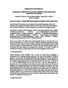
FIG.2. Separation of CRBP(I1) into two forms by chromatography on DEAE-cellulose at pH 6.4. The column was developed with starting buffer, 0.01 M imidazole acetate, until fraction 30 when a linear gradient from 0.01 to 0.04 M imidazole acetate was begun (gradient volume was 550ml). The absorbence of each fraction (5 ml) was monitored at 280 and 348 nm. 1
2
3 4
5
6
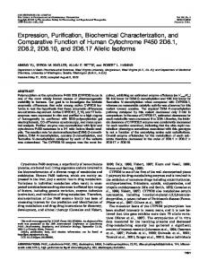
FIG.3. Analysis of CRBP(I1) purification by disc gel electrophoresis. An aliquot from each step of the purification procedure was submitted to electrophoresis in the discontinuous system No. 7, described by Maurer (9). Lane I , pH5extract, 750pg. Lane 2, combined fractions from Sephadex G-75 gel filtration, 80 pg. Lane 3, combined fraction from chromatography at pH 8.3 on DEAE-cellulose, 70 pg. Lane 4, CRBP(II)A, combined fractions from chromatography at pH 6.4on DEAE-cellulose,10 pg. Lane 5, CRBP(II)B, combined as for lane 4, 10 pg. Lane 6, CRBP(I1)A and CRBP(II)B, 10 pg each. Tb 7
TbII Tbl3
'b 14 hbl5
r
0
IO
20
I
I
30
40
I
50
I
60
70
00
Time (min)
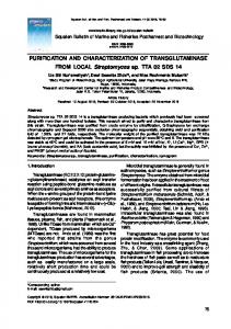
FIG.1. Reverse-phase HPLC separation of peptides produced froma trypsin digestof CRBP(1I)B. Thechromatography was carried out using a Brownlee RP-300 (octyl, 220 X 4.6 mm) column and the following solvent gradient: solvent A, HZ0 with 0.1% trifluoroacetic acid; solvent B, acetonitrile/H20/trifluoroacetic acid (80200.075); 0 min, 0% B; 5 min, 0% B; 45 min, 25% B; 60 min, 28% B; 90 min, 50% B; 100 min, 100% B; flow, 1 ml rnin".
equilibrated in 0.02 M Tris acetate buffer, pH 8.3. The column was eluted with a 550-ml linear gradient of Tris acetate buffer (0.02-0.15 M ) containing 1 mM 8-mercaptoethanol, pH 8.3, a t a flow rate of 1.0 ml/min. Binding activity was monitored by determining absorbance of 5-ml fractions at 348 nm. All binding activity was recovered in a single broad peakat 305 ml ofthe 550 mlgradient. The peak fractions were combined,concentrated, and diafiltered against 0.01 M imidazole acetate, pH 6.4, containing 1 mM 8-mercaptoethanol, and applied to a column (2.6 X 20 cm) of DEAE-cellulose (DE52) equilibrated in 0.01 M imidazole acetate, pH 6.4. Retinol was added before application as above. After application, the column was eluted with 0.01 M imidazole acetate, 1mM 8-mercaptoethanol, pH 6.4, until 30 fractions were collected. A linear gradient of 0.01-0.04 M imidazole acetate, 1 mM 8-mercaptoethanol, pH 6.4, was next applied. The retinol binding activity eluted in two peaks. Peak A eluted in 12 fractions centered a t fractions 62-63 (162 of the 550 ml gradient), and Peak B eluted in 11fractions centered a t fractions 98-99 (342 of the 550 ml gradient). Both peak fractions were pooledseparately and concentrated to about 1 mg/ml. Ratio of absorbance a t 348 nm to absorbance at 280 nm was 1.4 for peak A and 1.49 for peak B. Gel Electrophoresis-Purity of the two peaks obtained was assessed by gel electrophoresis. The first was a discontinuous nondenaturing system (Mauer No. 7) with a 15% gel, separating at pH 4.3 (9). The
4214
of Two Forms of CRBP, Type I1
Purification and Characterization
second gel system used was that of Laemmli (10) with 15% sodium PA) 15 m SPB-5 column and the following gradient: 100 "C for 2 dodecyl sulfate separating gels. min, then 15 "C/min to 320 "C.The column was coupled to a Nermag Protein Cleavage and Peptide Isolation-Purified CRBP(I1)A or lOlOC mass spectrometer that was operated in E1 mode. CRBP(I1)B (20.5 nmol) was reduced in a solution containing 6 mM Authentic Thr-Lys was synthesized using a Beckman 9000 Peptide dithiothreitol, 2.5 mM EDTA, 0.5 M Tris-HCI, pH 7.6 (400 p1 of total Synthesizer (13).N-Ac-Thr-Lys was synthesized by treating aportion volume) for 2 h at room temperature. All solutions were purged with of the Thr-Lys thathad been deblocked, but was still attached to the argon prior to use. Iodoacetic acid (1.25 mg, 15 mM final concentra- resin, with acetic anhydride/pyridine/chloroform (1:l:l) for 4 h a t tion) was added and allowed to react for 20 min in darkness. Excess room temperature prior to cleaving it from the resin with HF. Auiodoacetic acid and dithiothreitol were removed from the protein by thentic Lys-Lys was purchased from Sigma. Extreme care was taken passing it through a Sephadex G-25 column, eluting with 25 mM to preventany cross-contamination between the authentic and NH4HC03 in darkness. After washing the column with 25 ml of 25 CRBP(I1)-derived peptide samples. mM NH4HC03, the void fraction containing the protein was concenPreparation of Immune Reagents-Polyclonal antiserum against trated under vacuum and passed through the G-25 column a second CRBP(I1)B was produced in male New Zealand rabbits as previously time to ensure that excess reagents were removed. The void fraction described (3). IgG fractions were obtained from whole antiserum by was again collected and dried under vacuum. passing the antiserum over a DEAE-column at pH8.0 and combining The carboxymethylated protein was dissolved in 100 pl of 0.1 M fractions containing nonretained protein. This preparation was furNH4HC03 and digested with tosylphenylchloroketone-treatedtrypsin ther fractionated by passing it over a Sepharose-4B column contain(Worthington, Bedford, MA) (1:100, w/w) for 4 h a t 37 "C. Another ing immobilized CRBP(I1)A. The nonretained protein was combined aliquot of trypsin (1:lOO protease/CRBP(II), w/w) was added and the to produce a B-specific preparation. RetainedIgG was eluted with 0.1 mixture allowed to react 5 h more before the digestion was stopped M glycine HC1 buffer at pH 2.5 to produce the immune reagent that by freezing. Samples from the digest were monitored by HPLC to reacted with both form A and B. determine the extent of reaction. Immunohistochemistry-Immunohistochemical localization was Further digestion of specific purified peptides with Staphalococcus accomplished, as previously described, with the Vectastain kit from aureus V8 protease (Miles Laboratories, Naperville, IL) was done in Vector laboratories, which is based on the avidin-biotin-peroxidase 20 p1 of 0.1 M NH4HC03 using 1aliquot of V8 protease (1:25 protease/ system (5). The B-specific IgG fraction was diluted to an ODm of peptide, w/w). The reactions were stopped by freezing after 6.5 h 0.125. The A-B specific IgG fraction was diluted to an ODzw of 0.012. when they appeared to be complete as judged by HPLC. CRBP(I1)A and CRBP(I1)B which had not been carboxymethylRESULTS ated werecleaved with cyanogen bromide as described previously (11). Purification-Previously (3), whole rat pups were used to Peptides generated from the proteolytically and chemically cleaved obtain the two forms of CRBP(II), and five to seven column proteins were separated and isolated by reverse-phase HPLC using a chromatography steps were required to achieve homogenous Brownlee (Santa Clara, CA) RP-300 column (4.6 X 220 mm) eluted with 0.1% trifluoroacetic acid (v/v) in water and acetonitrile/HZO/ preparations. Because the protein is quite abundant in the trifluoroacetic acid (80:200.075) as described in Fig. 1. Fractions small intestine of adult rat, purification from that source was containing the purified peptides were collected, dried under vacuum, accomplished in only three column steps. and redissolved in 20 pl of a mixture of acetonitrile, water, and Briefly, the tissue was homogenized and the supernatant trifluoroacetic acid (25:75:0.1) for structural analyses. liquid collected after centrifugation at 5,000 X g to remove Structural Analyses-Fast atom bombardment-mass spectrometry, the heavier debris. The extract was taken to pH5, precipitatamino acid composition and amino acid sequence analyses were ing particulate material such as mitochondria and microsomes carried out as described previously (11). HPLC fractions that contained dipeptides or mixtures of dipeptides as well as some of the cytosolic protein. The supernatant were analyzed by GC-MS using a modification of the method of liquid from that precipitation was thensubmitted to gel Krutzsch (12). An aliquot containing about 5 nmol of peptides was filtration on Sephadex G-75 andthe elution position of dried under a stream of nitrogen. Acetonitrile (10 pl of acetonitrile CRBP(I1) determined by monitoring the characteristic fluowhich had been dried by passing through a small alumina column) rescence of retinol bound to CRBP(I1). The binding activity and BSTFA (Aldrich) (10 pl) were added to the residue and heated to 70 "C for 15 min to derivatize the peptides for GC-MS analysis. eluted at 0.76 of the totalcolumn volume, consistent with the Components of the mixture were separated on a Varian (Palo Alto, known molecular weight of 15,600.The peak of binding activto ion exchange CA) Vista Series 6000 gas chromatograph with a Supelco (Bellefonte, ity was collected, concentrated, and submitted
TABLE I Amino acid compositionsof CRBP(ZZ)A, CRBP(ZZ)B, and peptides from cyanogen bromide cleavage of both proteins CRBP IIA
Amino acid
ASP Glu Ser GlY His -4% Thr Ala Pro TYr Val Met CYS Ile Leu P he LYs Hser Trp
20.0 (20) 17.6 (18) 1.8 (2) (11) 11.1 1.1 (1) 6.7 (6) 13.1 (14) 3.2 (3) 0.5 ( 0 ) 3.5 (3) 10.5 (11) 1.9 (2) 1.9 (3) 3.9 (4) 10.3 (9) 10.1 6.7 (7) 14.6 (15)
'
IIB CNal CRBP CNa2
1.8 (2) 2.1 (2) 1.2 (1) -
2.1 (2) -
-
-
1.9 (2) 2.9 (3) 0.9 (1) 1.1 (1) 1.2 (1) (1) -
-
1.0 (1) -
-
0.9 0.4 1.0 (1) 1.0 (1) 0.8 (1) 0.8 (1) ND' (4) ND (1) ND ND Molar ratios less than 0.3 not shown. Not determined.
19.5 (20) 16.6 (18) 1.7 (2) 11.4 (11) 1.1 6.5 (6) 12.7 (14) 3.2 (3) 0.4 ( 0 ) 3.6 (3) 10.7 (11) 2.1 (2) 3.7 (3) 3.9 (4) (9) 6.8 (7) 14.9 (15) -
(4)
CNbl
CNb2
2.0 (2) 2.1 (2)
1.7 (2) 2.8 (3) 0.9 (1) 1.1 (1)
-
1.1 (1) -
2.0 (2) -
-
-
-
-
-
1.0 (1) -
1.0 (1)
1.0 (1) 0.6 (1) 0.8 (1) NDND (1) ND ND
CNbl-TP2 CNbl-TP1
-
1.1 (1)
-
1.0 (1) -
1.9 (2) 1.8 (2) 1.6 (1) -
1.0 (1) -
-
-
-
0.7 (1) (1)
Purification and Characterizationof Two Forms of CRBP,Type I1 CRBP
IIB
10 20 30 A c T K D Q N G T W E M E S N E N F E G Y M K A L D I D F A T R
I> > > I -
m
- - - - - - - - - - - - - - - - - -I
I Tn12
m13
I
............. I
I ...........I.......
(--I>>>>>>>”I>>>>>>>>>”I CNDl-!IF1
I
Ib13-sP2 a13-SP3
m13-spl
CNDl-TP2
w 50
40
60
KIAVRLTQTKIIVQDGDNFKTKTNSTFRNY I> >I 1-1-1 I” mlo m5 mn %la lb8
90
80
70
D L D F T V G V E F D E H T K G L D G R N V K T L V T W E G 1-1-1 Tn15 m 3m 6
100 110 120 NTLVCVQKGEKENRGWKQWVEGDKLYLELT I EA
Tb14
m7
ml6
I . ......I.............\... ‘Ibl6-SPl a316-SP2
130
C G D Q V C R Q V F K K K -I-I> >I “ ” ”
Tb9
mlb
............. I Ibl6-SP3
FIG. 4. Amino acid sequenceof CRBP(I1)Bderived from the cDNA sequence of Li et al. (1986).Standard single-letter amino acid abbreviations are used and Ac represents N-acetyl. Residues determined by amino acid sequence analyses, FAB-MS, and amino acid composition are indicated by an arrow (>). Peptides or residues identified by FAB-MS and amino acid composition are shown by a solid line (-). Peptides identified by FAB-MS alone are indicated by a dotted line ( ...) and residues inferred by amino acid composition alone are shown by a dashed line (- - -). A summary of peptides analyzed by FAB-MS is presented in Table 111.
chromatography on DEAE-cellulose, utilizing a gradient of 0.02-0.25 M Tris acetate buffer, pH 8.3. A single broad peak of retinol-CRBP(I1) was observed centered at 305 ml of the 550 ml gradient. No significant resolution of the two forms of CRBP(I1) was observed at this step. The broad peak was combined and concentrated for the final separation step. The resolution and final purification of the two forms of CRBP(I1) was accomplished by chromatography on DEAEcellulose utilizing a shallow gradient of 0.01 + 0.04 M imidazole acetate, pH 6.4, as shown in Fig. 2. Form A was centered at fractions 62-63 (162 ml of the 500-ml gradient) and Form B was centered at fractions 98-99 (342ml of the 500-ml gradient). Previously, these two forms had been demonstrated during the purification of CRBP(I1) from whole rat pups at a developmental stage when both liver and intestine contain CRBP(I1). The possibility had existed that the two forms might be due to an organ-specific distribution. Here it was established that the two forms were, indeed, present in the same organ. Under all conditions employed and for each purification
4215
procedure performed, we obtained essentially equal amounts of the two forms of the binding protein. As an example of a typical preparation, 330 g of small intestine yielded 14.0 mg of CRBP(I1)A and 13.1 mg of CRBP(1I)B. The abundance of total CRBP(I1) in thestartingtissue was determined by radioimmunoassay to be about 0.17 mg/g wet weighttissue or a total of 57 mg of CRBP(I1). The overall yield was 47.5%. The individual steps of the purification, examined by a disc gel electrophoresis system that separated A from B, are shown in Fig. 3. The considerable abundance of CRBP(I1) can be appreciated by the clear demonstration of the two forms in the starting extract. The separated forms were further examined by sodium dodecyl sulfate-electrophoresis as previously described (3). No detectable impurities were observed, a fact confirmed by the suitability of these preparations for the complete structural determination described below. Structural Analyses: General Approach-The amino acid sequence of CRBP(I1)A has been deduced from the cDNA sequence by Li et al. (8).The amino acid sequence of residues 1-20of CRBP(II)A, determined by automated Edman sequencing, was shown to be in complete agreement with the deduced sequence. Attempts to obtain sequence information for the amino-terminal portion of CRBP(I1)B were unsuccessful; CRBP(I1)B was resistant to Edman degradation: suggesting that the protein had a blocked amino terminus. The amino acid compositions of CRBP(I1)A and (1I)B (summarized in TableI) were nearly identical and agreed well with the composition from the derived amino acid sequence. In order to obtain more structural information CRBP(I1)B was cleaved chemically and enzymatically into smaller peptides suitable for structural analyses. CRBP(I1)A was treated similarly to allow further comparison with CRBP(II)B, aswell as to confirm the structure deduced from the DNA sequence. The deduced sequence showed that CRBP(I1)A contained only 2 methionine residues, at positions 10 and 20, in addition to theinitiator methionine which was absent from the isolated protein. Thus, both of the native proteins were cleaved with CNBr, which was expected to produce peptides only from the amino-terminal regions. These peptides were separated by HPLC andcharacterized by FAB-mass spectrometry together with amino acid composition and sequence analyses. The deduced sequence of CRBP(I1)A also indicated that lysine and arginine residues were relatively evenly distributed throughout the protein.Both CRBP(I1)A and (1I)B were reduced and carboxymethylated, then digested with trypsin. The resulting peptides were separated by HPLC (asshown in Fig. 1for CRBP(I1)B) andcollected. The peptides were numbered Tbl to Tb16 (T, trypsin; b, form B of CRBP(I1)) for (T, trypsin; a, form A of CRBP(I1)B andTaltoTal6 CRBP(I1))for CRBP(II)A, based on the order that they eluted from the HPLC column. Several peptides with longer retention times eluted from the column as rather broad peaks. These fractions were chromatographed a second time prior to structural analyses to insure that they were completely resolved. Each peptide was characterized by FAB-mass spectrometry and amino acid composition. Used in combination with molecular weight data, the amino acid compositions of the purified peptides confirm the compositions predicted from the derived sequence and prevent misidentification of amino acids or combinations of amino acids that are isobaric. Furthermore, the molecular weight determined from the FABmass spectrum used in conjunction with the amino acid composition for a peptide is very important for identifying post-translational modifications such as theacetylation of the amino terminus of CRBP(I1)B. Dipeptides produced by the trypsin digest were difficult to analyze by FAB-mass spec-
4216
Purification and Characterizationof Two Forms of CRBP,Type 11 TABLE I1 A m i n o acid compositions of peptides from trypsin digestion of CRBP(II)B Tblc
Tb2a -
Tb2b
Tb3
1.0 (1) 1.0 (1)
Tb4
Tb5
Tb6
Tb7
Tb8
Tb9
TblO
1.0 (1)
-
1.0 (1)
-
1.0 (1)
-
-
Tbll 3.4 (3) 2.4
Tb12 (2)
Tb13 4.1 (4)
Tb15
Tb16
1.1 (1) 4.1 (4)
Tb14
2.1 (2)
2.3 (2) -
1.2 (1) 1.0 (1)
-
1.9 (2)
-
1.0 (1) 2.0 (2) -
1.2 (1) 1.8 (2) 0.9 (1)
Molar ratios less than 0.3 not shown. Recovery is based on 20.5 nmol of CRBP IIB (before reduction and carboxymethylation) and calculated from the yield of each peptide after thefinal chromatography step. a
TABLE 111 Summarv of mass sDectral data for CRBP (IDAand CRBP (IDB CRBP IIA Peptide
1161.5 1171.4 1021.5 586.4 590.4 725.3 2028.9 360.2 1748.9 732.3 390.2
Tal3-SP1 1078.4 CNal 1161.4 CNa2 1171.4 Ta13-SP2 Ta13-SP3 Tal2 1021.4 T a586.2 l0 Ta5 590.3 Tall Ta8 725.3 Tal5 2029.7 Ta6 517.2 Ta3 360.2 Tal4 1748.8 Ta4 732.2 Ta7390.2 Tal6-Spl Ta16-SP2 1110.5 Ta16-SP31110.2 Ta9
CRBP IIB
Position
Observed [M + HI+
Calculated [M + HI'
Peptide
Position
Observed
-
[M + HI+
Calculated [M + H]+
-
1078.5
Tb2 Tbl3-SP1 849.5 CNbl CNb2 Tb13-SP2 Tb13-SP3 Tb12 TblO Tb5 Tbl 1 Tb8 Tb15 Tb6 Tb3 Tb14 Tb4 Tb7 Tbl6-SP1 Tb16-SP2 Tb16-SP3 Tb9
1-2 3-9 1-10 11-20 12-17 18-21 22-30 31-35 36-40 41-50 53-58 59-75 76-80 81-83 84-98 99-104 105-107 108-111 112-118 119-127 128-131
290.2
290.2 849.3 1203.5 1171.4 739.2 498.2 1021.5 586.4 590.4 1148.6 725.3 2028.9 517.3 360.2 1748.9 732.3 390.2 561.3 837.4 1110.5 521.3
1-9 1-10 11-20 12-17 739.2 18-21 498.2 22-30 31-35 36-40 41-50 1148.6 53-58 59-75 76-80 81-83 84-98 99-104 105-107 108-111 112-118 119-127 128-131
739.5 498.2
1148.5 517.3
561.3 837.3
561.3 837.4
521.3
521.3
trometry due to theirlow molecular weight. They were derivatized with BSTFA for GC/EI-MS analysis, which provided amino acid composition, as well as amino acid sequence information. For example, chemical derivatization followed by analysis by GC/EI-MS was used to determine the sequence of the tryptic peptide corresponding to the blocked amino terminus (residues 1-2) of CRBP(II)B, a structure thatcould not be determined by Edman degradation. Using this overall strategy, peptides spanning the entire structure described by the DNA sequence were isolated from the trypsin digests of both CRBP(I1)A and IIB. For simplicity, only those peptides that are essential for determining the structure arediscussed; data for peptides resulting from partial or more extensive cleavage are notshown. The results of these experiments were used to compare CRBP(1I)A andIIBand confirmed the structure deduced from the nucleotide sequence of the cDNA coding for CRBP(I1)A. CRBP(II)B Residues I-21"Treatment of CRBP(I1)B with
1203.4 1171.4 739.5 498.1 1021.7 586.3 590.4 1148.8 725.5 2030.0 517.3 360.3 1748.9 732.1 390.2 561.4 837.6 1110.4 521.4
cyanogen bromide released two small peptides, CNbl and CNb2 (Fig. 4). The amino acid composition of CNbl shown in Table I corresponds to residues 1-10of the derived sequence. This peptide showed an M H+ at m/z 1203 by FABmass spectrometry, 42 mass units higher than expected based on its amino acid composition, suggesting an acetyl moiety as the blocking group at the amino terminus of the peptide. To obtain sequence information, the peptide was further digested with trypsin. Two peptides, CNbl-TP1 and CNbl-TP2,were produced and separated by HPLC. CNbl-TP1 was composed of only threonine and lysine in a 1:l molar ratio consistent with residues 1-2 (Table I).CNbl-TP2 showed an amino acid composition (Table I) and amino acid sequence determined by automatedEdman sequencing (datanot shown) that aligned with residues 3-10. Both the amino acid composition (Table I) and sequence (data not shown) of peptide CNb2 aligned with residues 11-20 of the derived sequence. Peptides aligning with the amino terminus were also re-
+
Purification and Characterization of
Two Forms of CRBP, Type
II
421 7
(Table 11) corresponded to the assignments of Tb12 (residues 22-30), TblO (residues 31-35), Tb5 (residues 36-40), and T b l l (residues 41-50) shown in Fig. 4. Peptide Tbla showed an amino acid composition that was identical to Tb2 (containing only threonine andlysine), but eluted from the HPLC at a shorter retention time than Tb2. Tbla eluted from the HPLC in a fraction that contained a mixture of three small peptides. The FAB spectrum of the mixture showed an M + H' at m/z 248.2 (Table HI), supportingthe presence of ThrLys (Tbla, residues 51-52). The mixture was also derivatized with BSTFA for GC/EI-MS analysis and the spectrum of Tbla is shown in Fig. 6. Tbla has thesame retention time as synthetic Thr-Lys and an identical EI-mass spectrum (Fig. 6). Positions 53-108 of the derived sequence (Fig. 4) correspond 100 200 300 400 500 600 to peptides Tb8 (residues 53-58), Tb15 (residues 59-75), Tb6 m/ z (residues 76-80), Tb3 (residues 81-83), Tb14 (residues 8498), Tb4 (residues 99-104), and Tb7 (residues 105-107). The r3 x 5 FAB-mass spectrum of Tb15 showed an M + H+ at m/z 2030.0, one mass unit higher than was predicted from the cDNA sequence. This could be explained if the asparagine at position 59 were actually an aspartic acid residue. However, the spectrum of this relatively large peptide was rather weak so that such an assignment could not be made with confidence. Furthermore, selective deamidation of this asparagine could have occurred during the isolation of the protein. CRBP(ZI)B Residues 108-133-The amino acid composition of peptide Tb16 (Table 11) was consistent with residues 108-127. This peptide was also too large for FAB analysis and was cleaved into smaller peptides by digestion with S. aureus V8 protease. The V8 protease peptides were separated by HPLC and subjected to FAB-mass spectrometry. The results 100 200 300 400 600 500 for the three peptides, Tb16-SP1 (residues lO%lll),Tb16m/z SP2 (residues 112-118), and Tb16-SP3 (residues 119-127) are FIG. 5. EI-mass spectra and proposed fragmentation scheme €or summarized in Fig. 4 and Table 111. Positions 128-131 correCRBP(I1)B peptide Tb2 ( t o p ) and synthetic Ac-Thr-Lys (bottom), both following derivatization with BSTFA and separation by GC as spond to peptide Tb9(Tables I1 and 111). The sequence derived from the cDNA indicates Lys-Lys at positions 132described under "ExperimentalProcedures." 133. The amino acid composition and theFAB-mass spectrum suggested that this peptide (Tblb) was present in the same covered from digestion of carboxymethylated CRBP(I1)Bwith HPLCfractionas peptide Tbla. When this fraction was trypsin. Peptide Tb2 was composed of threonine and lysine derivatized with BSTFA and analyzed by GC/EI-MS, a peak (Table 11) and showed an M H' at m/z 290 (Table III), eluted from the GC with the same retention time and mass consistent with an acetylated form of the dipeptide. For more complete structuralcharacterization,Tb2 was derivatized spectrum as synthetic Lys-Lys. Thus, Tblb was assigned to with BSTFA and analyzed by GC/EI-MS. The mass spectrum residues 132-133. The EI-mass spectra and proposed fragof Tb2 is shown in Fig. 5 along with a proposed fragmentation mentation patterns for both Tblb and synthetic Lys-Lys are scheme. The spectrum was indistinguishable from the spec- shown in Fig. 6. CRBP(ZZ)A Residues 1-Zl"CRBP(I1)A wascleaved and trum of synthetic N-Ac-Thr-Lys(Fig. 5) and bothcompounds eluted from the GC with identical retention times. fmpor- characterized in the same manner as CRBP(I1)B. Chemical tantly, the observed fragment ions at m/z 260 and 288 con- cleavage of CRBP(1I)A with cyanogen bromide produced two firmed the sequence of residues 1and 2(immediately following peptides, CNal and CNa2 (Fig. 7). The amino acid composithe initiator methionine) that were predicted by the cDNA tions and mass spectral results are summarized in Tables I sequence. The amino acid composition of peptide Tb13 cor- and 111, respectively. Because the protein had an unmodified responded with residues 3-21 of the derived sequence. This amino terminus, the amino acid sequence of CNal (data not peptide was too large for FAB analysis on our instrument and shown) was determined directly without further cleavage. The was further digested with S. aureus V8 protease. The resulting structure (Fig. 7) aligned perfectly with residues 1-10 of the peptides were separated by HPLC and FAB-mass spectra sequence derived from the cDNA and differed from residues were obtained. The results aresummarized in Fig. 4 and Table 1-10of CRBP(I1)B only in the absence of amino-terminal 111. Based on the molecular weights determined from mass acetylation. Similarly, the sequence (datanot shown) and spectra, peptide Tbl3-SP1 corresponded to residues 3-9, structure (Fig. 7) of CNa2 aligned with residues 11-20 and Tb13-SP2 to residues 12-17, and Tb13-SP3 to residues 18- was identical with the analogous peptide of CRBP(I1)B. Peptide Tal3 of CRBP(I1)A showed an amino acid com21. The dipeptide covering positions 10-11 was not identified. However, these residues were confirmed by the cyanogen position (Table IV) that correlated with residues 1-21. This bromide peptides, as well as the amino acid composition of peptide was cleaved into smaller peptides with S. aureus V8 Tb13. protease, and theresulting peptides were separated by reverseCRBP(II)B Residues 22-107-The molecular weight data phase HPLC. The V8 protease peptides were subjected to (Table III), as well as the amino acid composition results FAB analysis, and the results are summarized in Table 111. loo
1 173
x 5
+
Purification and Characterization
4218
of Two Forms 100
s
1
73
of CRBP, Type 218
I1
I 419
75
1
100
200
300
400
500
600
B
7 x 2 0
100
200
300
rnlz
400
500
0
500
600
rnlz
T84
100
200
300 mlz
400
500
600
100
200
300 m lz
400
FIG. 6. El-mass spectra of CRBP(I1)B peptides Tbla (panel A ) and Tblb (panel C) following derivatization of the mixture with BSTFA and separation of the peptides by GC as described under "Experimental Procedures." Panek B and I), respectively, show the E1 spectra and proposed fragmentation schemes for BSTFA derivatives of synthetic Thr-Lys and Lys-Lys. rn IIA
CRBP(II)A Residues 22-133-The peptides spanning residues 22-133 of CRBP(I1)A were all identical to those isolated from the trypsin digest of CRBP(I1)B and were recovered in the same relative yields. Each of the peptides produced from CRBP(I1)A were identified using the same numbering sysI................. 1 I...........(....... 1 tem that was used for the corresponding peptides from Tal3-SP1 Ta13-SP2 Ta13-SP3 CRBP(I1)B. The amino acid compositions for the peptides )>>>>>>>>>"1>>>>>>>>2") are summarized in Table IV andthe FAB-mass spectral CNal cNa2 results are shown in Table 111. Peptides Tala (residues 51FIG. 7 . Amino acid sequence of residues 1-21 of 52) and Talb (residues 132-133) were analyzed by GC/EICRBP(I1)A. Standard single-letter amino acid abbreviations are MS following derivatization with BSTFA and showed GC used. Residues determined by amino acid sequence analyses, FABretention times and mass spectra identical to those of the MS, and amino acid composition are indicated by an arrow (>I. Residues identified by FAB-MS and amino acid composition are synthetic standards (data notshown). Peptides identified by FAB-MS are Cellular Distribution of form A and B-We were interested shown bya solid line (-). indicated by a dotted line (. . .), and residues inferred by amino acid in determining the cell-specific distribution of the two forms composition alone are shown by a dashed line (- - -). A summary of by the technique of immunohistochemistry. Consequently, we peptides analyzed by FAB-MS is presented in Table 111. examined antisera produced from a number of rabbits injected with pure CRBP(I1)B. From one rabbit among six examined, Three peptides were identified Tal3-Spl (residues 1-9), we obtained antiserum that showed an ability to distinguish Ta13-SP2 (residues 12-17), and Tal3 (residues 18-21). Sim- between CRBP(I1)A and CRBP(I1)B. As shown in Fig. 8, that ilar to the findings with CRBP(II)B, the dipeptide aligning antiserum gave greater reaction with CRBP(I1)B. TOenhance with residues 10-11was not identified. Interestingly, the that specificity, the IgG fraction from that antiserum was dipeptide covering residues 1-2of Tal3 was not cleaved passed over a column of immobilized CRBP(I1)A to remove during the trypsin digest of CRBP(II)A, as it was with antibodies recognizing antigenic determinants common to the CRBP(I1)B. Furthermore, apeptide containing threonine and two forms. The nonretained IgG fraction recovered from that lysine and showing the same retention time as N-Ac-Thr-Lys procedure showed considerably enhanced ability to distinguish between the two forms, giving 10-20-fold greater reacwas absent from the tryptic digest of CRBP(I1)A. 20
1 10 TKDQNGTWEMESNENFEGYMK ~"-""""""""_ -I Tal3
Two Forms CRBP, of Type
Purification and Characterization of
11
4219
TABLE IV Amino acid compositions of peptides from trypsin digestion of CRBP(II)A Talc Talb Tala ASP
Clu Ser GlY
His 1.0
Arg Thr Ala
Pro Tyr
Val Met
CYS Ile Leu Phe
LYS 96
Recov-
Ta2
-a -
-
1.0 (1)
1.0(1)
-
-
(1)
-
-
-
-
-
-
-
1.0(1) 81
1.0(1) 33
-
-
-
-
-
2.0 (2) 79
1.0 (1) 1.0 (1)
-
1.0 (1)
-
-
43
Ta7 Ta3 Ta6Ta5Ta4
Ta8 Tal6 Tal5 Ta9 Tal4 Tal3 Tal2 Tall Tal0
1.11.0(1) (1) - 2.0(2) l.O(l)
-
-
-
1.0 (1)
-
-
0.9 (1)
63
-
1.0(1)
-
-
-
1.0 (1)
-
-
0.9 (1)
-
-
2.0(2)
-
-
-
-
-
-
1.0 (1)
-
-
l.O(l) 21
0.9 (1) 65
-
1.2 (1)
-
0.8 (1)
-
2.0 1.0(1) (2)
-
-
-
1.0 (1)
-
-
-
-
-
-
1.0 (1)
-
77
-
-
-
-
l.O(l) 1.9 (2)
-
1.0(1)
1.0 (1) 70
67
1.0(1)
-
- (1)
-
-
-
1.0 (1) 0.9
3.6 (3) 1.0 (1)
-
1.0 (1)
-
-
-
-
1.0 (1)
1.1 (1)
0.9 (1)
-
-
-
1.0(1)
1.2 (2)
-
1.0 (1)
-
-
1.0 (1) 59
1.0(1) 31
1.0 (1) 1.0(1) 70
-
-
-
-
2.3 (2)
-
1.0 (1) 0.9 (1) 1.8 (2)
-
-
l.O(l) 1.0 (1) 1.0(1) 1.0 (1)
-
59
4.1 (4) 5.1 (5) 1.0 (1) 2.1 (2)
l.O(l) 2.0(2)
3.6 (4) 1.9 (2)
2.1 (2) 4.0 (4)
1.1 (1)
2.0(2)
-
-
1.0 (1) 0.9(1)
-
2.1 (2)
2.5 (3)
1.7 (2)
1.0 (1) 1.0 (1)
-
-
-
-
-
l.O(l)
-
-
2.7 (3)
0.9 (1) 1.8 (2)
1.0 (1) 1.9 (2)
-
-
1.7 (2)
-
-
-
-
(1)
-
-
(2)
-
-
-
-
1.9 (2)
3.0 (3)
2.0 (2) 30
1.0 (1) 30
1.0 (1) 1.6 (2) 0.8(1) 50
-
-
1.1 (1) 35
e b
Molar ratios less than 0.3 not shown.
* Recovery is based on 20.5 nmol of CRBP
IIB (before reduction and carboxymethylation) and calculated from the yield of each peptide after the final chromatography step.
P m :
sities of staining. It isknown that theproximal small intestine contains more total CRBP(I1) than the distal intestine (3) and thatmay be the reason for different intensities observed here. Fig. 9B shows a serial section of the same fetus, now 40 stained with the IgG fraction that reacted equally well with both form A and B. No differences in staining pattern could 20 be distinguished from that observed for the B-specific IgG 10 preparation. Consequently, the organ distribution as well as the proximal-distal gradient in the small intestine of form A 5 and B appeared to be identical. Closer examination suggested that the specific cellular distribution of form A and B was 2.5 also similar. Fig. 9C and D show higher powerviewsof a cross-section of fetal small intestine from serial sections 102 stained with the B-specific and A-B-specific IgG preparations, 080 respectively. The intensities observed appeared essentially identical. In particular, no staining was observed in the epithelium of the base of the villi, but the entire villus-associated epithelium showed a relatively homogeneous staining pattern for both immune preparations. Similar results were obtained A B A B A B with adult rat intestine (data not shown). Because the cellular staining pattern was identical for both FIG. 8. Specificity of IgG preparations for form A and B of CRBP(I1). Increasing amounts (0-40 ng) of CRBP(I1) were immo- immune reagents, it could be concludedthat no cells contained bilized on nitrocellulose ( A = form A, B = form B) then reacted with CRBP(I1)A only. Because the staining intensitieswere essenthe different IgG preparations indicated. Panel I , total IgG prepara- tially the same for all cells with each of the immune reagents, tion. Panel II, I$ fraction not retained by immobilized CRBP(1I)A. it could be concluded that both the amount of CRBP(I1)B Panel III, IgG fraction eluted from immobilized CRBP(I1)A. and the total amount of CRBP(I1)A plus CRBP(I1)B were constant for all cells that stained. Given that both A and B tion with CRBP(I1)B compared to CRBP(I1)A (Fig. 8,column ZZ).The retained IgG fraction, eluted from the affinity column were present in uiuo, this indicated all cells contained both by low pH, was examined similarly. As expected, it reacted form A and B. well with both CRBP(I1)A and CRBP(I1)B (Fig. 8,column DISCUSSION
I
ZZZ).
With these immune reagents, we compared the cell-specific distribution of B to that observed for total CRBP(I1). We examined sections of whole rat fetus a t day 21-22 of gestation, immediately prior to birth, in order to observe cross-sections of multiple intestinal segments on the same slide, by a procedure we have previously described (8). Fig. 9A shows the staining patternproduced in the sectioned whole fetus, using B-specific IgG. Strong staining was observed in the epithelial cell layer of the villi of the small intestine. No staining was observed in theepithelium of the stomach or of the colon. A light staining was also evident in the liver, although it is not apparent in this black and white photograph. The different cross-sections of the small intestine showed different inten-
In this report, we have described the isolation of cellular retinol-binding protein type I1 from adult rat small intestine. The protein exists in two resolvable and equally abundant forms, called CRBP(I1)A and CRBP(II)B, and the primary structures of both forms have been characterized. Our results confirm the amino acid sequence for CRBP(I1)A that was deduced from the cDNA sequence (8). Because of the high degree of similarity in primary structure observed between cellular retinoid-binding proteins (e.g. CRBP, cellular retinoic acid-binding protein, and CRBP(I1)A) that have been characterized todate (8, 14, 15), we were concerned that CRBP(I1)B could be yet another similar protein with a primary structure thatwas different from CRBP(I1)A. The only
4220
-
Purification and Characterization Two of
FIG. 9. Immunolocalization of CRBP(I1) in rat.Whole animal sagittal sections (5PM) were obtained from a 20day fetal rat. Sections were stained with the B-specific IgG preparation ( A and C ) or the AB-specific IgG preparation ( B and D).Each section was also lightly stained withhematoxylin to bring out detail. Dark staining indicates presence of the antigen. A, staining for CRBP(I1)B isconfined to the epithelial cell layer of the villi of the small intestine. No staining isobserved in stomach ( S ) or colon (C). B, identical staining pattern observed in a serial section with IgG preparation thatreacted equallywell with both form A and form B. C, staining for CRBP(I1)B in cross-section of small intestine. No staining is observed in the epithelial cell layer at the base of the villi. D, identical stainingpatternobserved in a serial section with IgG preparation that reactedequally well with both form A and B.
Forms CRBP, of Type
11
I
difference observed between the two proteins was acetylation sent in the absorptive cells, is recovered fully acetylated at of the NH2 terminus of CRBP(II)B, which is absent from the NHz terminus (20). It hassequence similarity to CRBP(I1) CRBP(I1)A. Gordon and co-workers (16) have shown that a (8,20) and iseven more abundant, representing about 2% of single gene codes for CRBP(1I)A in rats. Thus, CRBP(1I)A the total soluble protein of the mucosa (21). If acetyl-coA and (1I)B appear to be products of the samegene and are not were limiting, one might expect that the acetylationof fatty products of highly similar but distinctgenes. acid-binding protein would also be affected. The finding that CRBP(I1)A and CRBP(I1)B differed only The specificity of cotranslational processing of proteins, intheacetylation at the NH2 terminus was unexpected. including removal of the initiator methionine and NH2-terPartial acetylation of a purified protein has sometimesbeen minal acetylation, hasbeen studied very recently by examinattributed to an artifact of the isolation procedure. Several ing the expression of a genetically modified protein in yeast lines of evidence indicated artifactual acetylation (ordeacet- (22), as well as the cell-free expression of site-directed muylation) of CRBP(I1) was not involved here. The reaction of tants of hemoglobin (23). The results in the yeast system the B-specific IgG preparationinthe immunolocalization showed that when a threonine residue immediately followed was cleaved and the studies confirmed that the acetylation of the NH2 terminus the initiator methionine, the methionine of CRBP(I1) indeed had occurred in vivo and not during the threonine was N-acetylated. However, cell-free translation of isolation of the protein. Deacetylation of form B after tissue hemoglobins with both a rabbit reticulocyte or wheat germ disruption and during isolation is not likely. Enzymes that system led to only partially (-50 and 40%) acetylation of catalyze removal of NH2-terminal acetyl groups appear to be threonine. This latter finding appears to be consistent with quite rare. Furthermore, CRBP(I1)A and (1I)B are always our results for CRBP(1I)A and (1I)B. However, whether aceisolated in a 1:1 molar ratio from either whole 1-day-old rat tylation of CRBP(1I)B is cotranslationalorpost-translapups or mature rat small intestine, even though the abundance tional, whether CRBP(I1)A is generated from deacetylation of CRBP(II)/g of starting material is considerably lower for of CRBP(II)B, or whether theNH, terminus of CRBP(I1) is whole rat pups. The N-acetyl bond is expected to be chemi- simply a poor substrate for the acetyltransferase resulting in cally stable under the isolation conditionsused so nonenzy- partial acetylation, as suggested by the work with mutant matic hydrolysis of the N-acetyl is also highly unlikely. At no hemoglobins, remains to be established. The extent of acetytime did we observe theappearance of CRBP(I1)A from lation varies amongproteinswiththreonine at the NH2 CRBP(I1)B after thetwo forms had been separated. terminus indicating that, in some instances, structural feaAcetylation of the NH2 terminus of a protein has been tures in addition to the NH2-terminal residue also must play established to bea cotranslational event(17), as is theremoval a role in NH2-terminalprocessing. of the initiator methionine. (Examples, such as &endorphin Few examples of partially acetylated proteins have been and a-melanotropin (18) havebeenidentified, however, in described in the literature. These include yeast ribosomal which acetylation is post-translational, occurring after trans- proteins L7 and L12 (acetylated and nonacetylated forms, lation has been completed.) Because the two proteins have respectively) (24), analcohol dehydrogenase from yeast (19). identical primary structures,differences in NHZ-terminal se- the y-chain of human fetal hemoglobin (25), and a human quence acetylation of CRBP(II)B, CRBP(1I)A.Incomplete hemoglobin variant, SouthFlorida (26).In noneof these cases acetylationduringtranslationduetolimitingamounts of is thefunctional significance of NHz-terminal acetylation acetyl-coA has been suggested in the case of yeast alcohol understood. dehydrogenase(19). However, that seems to beless likely It is unclearif the presence of these two forms of CRBP(I1) here. Intestine-specific fatty acid-binding protein, also pre- has functional significance. A suggestion that it does comes
Purification and Characterization of Two Forms of CRBP, Type I1 from our previous observation that form A was recoveredwith 10-fold more endogenous retinol than form B, when isolated from suckling rat pups (3). As no difference can be detected in binding affinity for retinol between the two forms, this suggests that the nonacetylated form had greater access to the retinol newly absorbed from the milk. No apparent differential distribution of the two forms within the absorptive cell could be detected by immunohistochemistry at the light microscope level. However, acetylation might alter interactions of CRBP(I1) with other cellular components, such as the brush border membrane or enzymes involved in the necessary metabolism of vitamin A that occurs in the absorptive cell. The successful production of antibodies specific for the N acetylated form and that would recognize the native protein indicated that theNH, terminus was accessible, and could be involved in protein-protein recognition. Omnivores, such as the rat, obtain vitamin A from two sources, either from animal tissuesthat contain retinyl esters or from plantmaterial that contains carotenes. Carotene enters the absorptive cell where it is oxidatively cleaved to retinaldehyde, which is thenreduced to retinol. Retinyl esters are cleaved within the lumen of the intestine, and it is the liberated retinol that enters the cell (27). The two forms of CRBP(I1) may participate unequally in these two pathways. Rat milk provides vitamin A as retinyl esters and contains little if any carotene. This then could lead to thedifferential distribution of endogenous retinol between form A and B that was observed with the suckling rat. Future work will explore this possibility. Acknowledgments-We thank Dr. Thomas Lukas for his help in synthesizing the authentic dipeptide standards, Paul Matresian for performing the automatedEdman sequence analyses, and Dr. D. Martin Watterson for access to the protein microchemistry facilities and many helpful discussions. We would also thank Lucie Chytil for assistance in preparation of the antiserum used and Frank Chytil for his interest and support.
REFERENCES 1. Wolbach, R.A., and Howe, P. R. (1925) J. Erp. Med. 4 3 , 753777 2. Morton, R.A. (1972) in Photochemistry of Vision (Dartnall, H. J. A,, ed) pp. 33, Springer-Verlag, New York 3. Ong, D. E. (1984) J. Biol. Chem. 2 6 9 , 1476-1482
4221
4. Levin, M. S., Li, E., Ong, D. E., and Gordon, J. I. (1987) J. Biol. Chem. 262,7118-7124 5. Crow, J. A., and Ong, D. E. (1985) Proc. Natl. Acad. Sci. U. S. A . 82,4707-4711 6. MacDonald. P. N.. and One. D. E. (1987) . , J. Biol. Chem. 262, 10550-10556 ’ 7. MacDonald, P. N., and Ong, D. E. (1988) J. Bwl. Chem. 263, 12478-12482 8. Li, E., Demmer, L. A., Sweetser, D. A., Ong, D. E., and Gordon, J. I. (1986) Proc. Natl. Acad. Sci. U. S. A. 83,5779-5783 9. Maurer, H. R. (1971) Disc Electrophoresis and Related Techniques of Polvacrvlamide Gel Electrophoresis, 2nd Ed., p. 44-45, de druytkr, Berlin 10. Laemmli. U. K. (1970) Nature 227.680-685 11. Schaefer,’ W.H.; Lukas, T. J., Blair, I. A., Schultz, J. E., and Watterson, D. M. (1987) J . Biol. Chem. 262,1025-1029 12. Krutzsch, H.C. (1983) Methods Enzymology 91, 511-524 13. Lukas, T. J., Burgess, W. H., Prendergast, F. G., Lau, W., and Watterson, D. M. (1986) Biochemistry 2 6 , 1458-1464 14. Sundelin, J., Anundi, H., TrHgardh, L., Eriksson, U., Lind, P., Ronne, H., Peterson, P. A., and Rask, L. (1985) J. Biol. Chem. 260,6488-6493 15. Sundelin, J., Das, S. R., Eriksson, U., Rask, L., and Peterson, P. A. (1985) J. Biol. Chem. 260,6494-6499 16. Demmer, L.A., Birkenmeier, E. H., Sweetser, D. A., Levin, M. S., Zollman, S., Sparkes, R. S., Mohandas, T., Lusis, A. J., and Gordon, J. I. (1987) J. Bwl. Chem. 262,245&2467 17. Driessen, H. P. C., deJong, W. W., Tesser, G. I., and Bloemendal, H. (1985) CRC Crit. Reu. Biochem. 18,281-325 18. Glembotski, C . C. (1982) J. Biol. Chem. 2 5 7 , 10493-10500 19. Jornvall, H., Fairwell, T. F., Kratofil, P. K., and Wills, C. (1980) FEBS Lett. 111,214-218 20. Alpers, D. H., Strauss, A.W., Ockner, R. K., Bass, N.M., and Gordon, J. I. (1984) Proc. Natl. Acad. Sci.U. S. A. 81,313-317 21. Bass, N. M., Manning, J. A., Ockner, R. K., Gordon, J. I., Seetharam, S., and Alpers, D. H. (1985) J. Biol. Chem. 260, 1432-1436 22. Huang, S., Elliott, R. C., Liu, P. S., Koduri, R. K., Weickmann, J. L., Lee, J. H., Blair, L. C., Ghosh-Dastidar, P. Bradshaw, R. A., Bryan, K. M., Einarson, B., Kendall, R. L., Kolacz, K. H., and Saito, K. (1987) Biochemistry 26,8242-8246 23. Boissel, J. P., Kasper, T. J., and Bunn, H. F. (1988) J. Biol. Chem. 263,8443-8449 24. Terhorst, C., Moller, W., Laursen, R., and Wittmann-Liebold, B. (1973) Eur. J. Bwchem. 3 4 , 138-152 25. Kasten-Jolly, J., and Abraham, E. C. (1986) Biochim. Biophys. Acta 866,125-134 26. Boissel, J. P., Kasper, T. J., Shah, S. C., Malone, J. I., and Bunn, H. F. (1985) Proc. Natl. Acad. Sci. U. S. A. 82,8448-8452 27. Goodman, D. S., and Blaner, W. S. (1984) in TheRetinoids (Sporn, M. B., Roberts, A. B., and Goodman, D. S., eds) Vol. 2, pp. 2-34, Academic Press, New York. ~
~
~~
I
I