Published: 15 December 2009 © 2009 Faculty of 1000 Ltd
Putative functions of caspase-2 Loretta Dorstyn and Sharad Kumar* Address: Centre for Cancer Biology, SA Pathology, Frome Road, Adelaide, SA 5000, Australia * Corresponding author: Sharad Kumar (
[email protected]) F1000 Biology Reports 2009, 1:96 (doi:10.3410/B1-96)
This is an open-access article distributed under the terms of the Creative Commons Attribution-NonCommercial License (http://creativecommons.org/licenses/by-nc/3.0/legalcode), which permits unrestricted use, distribution, and reproduction in any medium, for non-commercial purposes provided the original work is properly cited. You may not use this work for commercial purposes. The electronic version of this article is the complete one and can be found at: http://F1000.com/Reports/Biology/content/1/96
Abstract Caspase-2 is the most evolutionarily conserved of caspase family members, yet its physiological function has remained unclear and is a matter of considerable debate. Newly published data now suggest that caspase-2 is required for cell cycle regulation, repair of damaged DNA, and in suppressing Myc-induced lymphomagenesis. Additionally, loss of Casp2 in mice leads to features of premature ageing. These findings suggest that caspase-2 has non-apoptotic functions in addition to its context-dependent roles in cell death.
Introduction and context Caspase-2 is one of the closest mammalian homologues of the Caenorhabditis elegans caspase CED-3 and shares significant homology with the Drosophila Nedd2-like caspase (DRONC), both of which are essential for developmentally programmed cell death [1-4]. Although several studies have implicated caspase-2 as a crucial mediator of apoptosis in mammalian cells, its apoptotic function has remained enigmatic, partly due to the fact that Casp2−/− mice are viable and fertile with only minor apoptotic defects in some cell types [2,5-7]. Furthermore, lymphocytes and fibroblasts from mice lacking both initiator caspases, Casp9 and Casp2, are no more resistant to apoptosis than cells from Casp9−/− mice [8]. Together, these findings indicate that the role of caspase-2 in developmental cell death is redundant and can be compensated by other caspases. However, this does not rule out context-dependent and cell-specific caspase-2 functions. For example, one study found an accumulation of oocytes in Casp2−/− mice (although this was not reported in a second Casp2−/− strain) and Casp2−/− mice display premature ageing-related traits [5,9,10]. The activation of caspase-2 has been shown to occur both upstream (by the PIDDosome) and downstream (by caspase-3 or -7) of mitochondrial outer membrane permeabilisation (MOMP) [11,12]. Although this is also controversial since RAIDD (receptor-interacting protein-
associated ICH-1/CED-3 homologous protein with a death domain) and PIDD (p53-inducible protein with a death domain), the protein components of the PIDDosome, are dispensable for caspase-2 activation [13,14]. Interestingly, caspase-2 activation can be mediated by caspase-8-induced cleavage following recruitment to the death receptor-inducing signaling complex (DISC) [15]. However, the importance of the DISC as an activation platform is also unclear since caspase-2 dimerisation and self-processing are sufficient for its activation [14,16]. In addition, cells from Casp2−/− mice are normally sensitive to death receptor-induced apoptosis [6], indicating that caspase-8-mediated cleavage of caspase-2 is not critical for its activation. While there is limited information on physiologically relevant substrates, caspase-2 can cleave and activate the protein Bid, which provides a significant link between caspase-2 and MOMP [15,17]. Furthermore, a unique feature of caspase-2 is its ability to localise to the nucleus in an importin-mediated fashion [18-20]. This nuclear localisation of caspase-2 is likely associated with the recently found functions for caspase-2 in cell cycle regulation and cellular DNA damage response.
Major recent advances In the absence of an overt phenotype in knockout mice, one may speculate that caspase-2 functions under specific contexts, such as under conditions of stress or in the Page 1 of 5 (page number not for citation purposes)
F1000 Biology Reports 2009, 1:96
fine-tuning of stress signaling, resulting in relatively minor aberrations in the whole animal physiology. Several recent studies showing caspase-2 functions in cell cycle regulation, DNA damage response, and tumor suppression seem to be consistent with these predictions. A role for caspase-2 in cell cycle regulation became apparent from observations that Casp2-deficient murine embryonic fibroblasts (MEFs) proliferate faster than their wild-type counterparts and that transformation of Casp2−/− MEFs with E1A/Ras exacerbated this proliferative effect [21]. Another recent study found that caspase-2 is involved in maintaining a G2/M cell cycle checkpoint in response to ionising radiation (IR)-induced DNA damage, with cells lacking Casp2 unable to completely arrest in G2/M [21,22]. In addition, caspase-2 activation has been shown to be inhibited by cyclin-dependent kinase 1 (Cdk1)/cyclin-B1mediated phosphorylation at Ser340 during mitosis to allow for the repair of replication-induced DNA damage [23]. During mitotic arrest, prolonged activation of spindle assembly checkpoint results in apoptosis by ‘mitotic catastrophe’, which may be mediated by caspase-2 [23,24]. When caspase-2 is lacking, arrested cells cannot undergo apoptosis but instead undergo ‘mitotic slippage’ and prematurely exit mitosis with chromosomal abnormalities, resulting in genomically unstable aneuploid cells [23]. In support of this, Casp2−/− MEFs show resistance to cell death induced by microtubule-disrupting drugs and also display increased genomic instability in culture compared with wild-type cells [7,21]. These findings indicate that deregulation of G2/M checkpoint in Casp2−/− cells may contribute to the accumulation of cells with damaged DNA. The study by Shi and colleagues [22] found that caspase2 is involved in DNA damage repair through its interaction with a nuclear complex comprising PIDD and DNA-dependent protein kinase catalytic subunit (DNA-PKc). Following IR-induced DNA damage, this DNA-PKc PIDDosome complex phosphorylates caspase2 at Ser122, leading to its activation. Activated caspase-2 is then required for the repair of double-strand DNA breaks by non-homologous end-joining (NHEJ) with cells lacking Casp2 unable to efficiently repair DNA breaks [22]. Although it is unclear how caspase-2 mediates NHEJ, these important observations establish an additional non-apoptotic nuclear role for caspase-2 in DNA damage signaling (Figure 1). Sidi and colleagues [25] have described an unexpected nuclear function of caspase-2 in an apparently novel pathway of apoptosis in p53-deficient cells. Using zebrafish as a model system, it was found that inhibition or loss of checkpoint kinase 1 (Chk1) restores g-radiation-
http://F1000.com/Reports/Biology/content/1/96
induced apoptosis in p53 mutant fish embryos [25]. A similar ataxia telangiectasia mutated (ATM)/ATM-related (ATR)/caspase-2-dependent pathway seems to be present in Chk1-inhibited p53-deficient human tumor cells and in MEFs following g-irradiation [21,25]. These findings implicate caspase-2 in an apoptosis pathway downstream of ATM/ATR in response to DNA damage, which is independent of p53 (Figure 1). Since ATM/ATR can induce phosphorylation and activation of DNA-PKc, it would be of interest to investigate whether the regulation of caspase-2 phosphorylation in the nucleus finely tunes its function in either apoptosis or DNA damage repair following IR exposure. The role of caspase-2 in specific cell death pathways other than DNA damage is also emerging. In oocytes, the apoptotic activity of caspase-2 has been shown to be inhibited by Ca2+/calmodulin-dependent kinase II (CaMKII)-mediated phosphorylation at Ser135 [26]. The binding of 14-3-3z to phosphorylated caspase-2 prevents Ser135 dephosphorylation, thereby promoting oocyte survival [27]. However, under nutrient-depleted conditions, the dephosphorylation at this site leads to caspase-2 activation and oocyte cell death [26]. These findings suggest that caspase-2 is an important player in metabolic regulation of oocyte cell death and that Ser135 dephosphorylation is a sensor for caspase-2 activation. Caspase-2 has also been implicated in cell death induced by heat shock [28]. A recent study assessed the real-time recruitment of caspase-2 to activation platforms during stress-induced apoptosis, including heat shock, cytoskeletal disruption or DNA damage, and found that caspase-2 activation occurred in the cytosol, not the nucleus [29]. Furthermore, heat shock-induced activation of caspase-2 occurred upstream of MOMP and was RAIDD-dependent and negatively regulated by HSP90a [29]. The caspase-2 functions in oxidative stress-induced apoptosis and ageing are also coming to light. Caspase-2, along with Bid and Bak, were reported to be mediators of superoxide-induced cell death in muscle and in primary neurons [30,31]. In addition, caspase-2 has been shown to be involved in an age-related increase in muscle cell apoptosis in mice [9]. Consistent with these findings, Zhang and colleagues [10] found that Casp2−/− mice show significantly higher levels of oxidised proteins in liver than wild-type mice. This indicates that lack of Casp2 can antagonise apoptosis induced by reactive oxygen species, leading to accumulation of cells with oxidative damage and consequently enhanced ageing phenotypes [10]. The reduced NHEJ activity in Casp2−/− cells may also contribute to the premature ageing phenotype observed in Casp2−/− mice. Page 2 of 5 (page number not for citation purposes)
F1000 Biology Reports 2009, 1:96
http://F1000.com/Reports/Biology/content/1/96
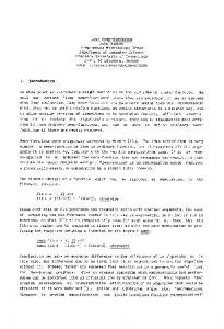
Figure 1. Putative functions of caspase-2
Following double-strand DNA breaks (DSBs), the ataxia telangiectasia mutated (ATM) and ATM-related (ATR) kinases are activated and in turn phosphorylate and activate several target proteins, including checkpoint kinase 1 (Chk1) and Chk2. Chk2 activates the p53 response pathway, which can lead to cell cycle arrest. ATR activation leads to the activation of caspase-2 and apoptosis following irreparable DNA damage. ATR also activates Chk1, which can then act in a feedback loop to negatively regulate ATR and inhibit further activation of nuclear caspase-2. DNA-dependent protein kinase (DNA-PK) is also activated by DSBs, presumably by ATM/ATR, and forms a complex with p53-inducible protein with a death domain (PIDD) and caspase-2 (DNA-PK PIDDosome). This complex serves to phosphorylate and activate caspase-2, which is then required for the initiation of non-homologous end-joining (NHEJ) and DNA repair. Cytosolic caspase-2 is also activated by other stress signals such as reactive oxygen species (ROS), metabolic stress, cytotoxic drugs, heat shock, or endoplasmic reticulum (ER) stress. Following heat shock, RAIDD (receptor-interacting protein-associated ICH-1/CED-3 homologous protein with a death domain) can activate caspase-2, which is inhibited by HSP90a. Ca2+/calmodulin-dependent kinase II (CaMKII) acts to inhibit caspase-2 activation and cell death in oocytes. Activated cytosolic caspase-2 is able to cleave Bid to its truncated form (tBid), which (via Bax/Bak) can induce mitochondrial outer membrane permeability (MOMP), activation of caspase-9 and -3, and cell death. P, phosphorous; PUMA, p53-upregulated mediator of apoptosis.
Our own studies using the Em-Myc transgenic mouse model of B-cell lymphoma found a potential role for caspase-2 in lymphoma suppression [21]. Specifically, the loss of even a single copy of Casp2 resulted in increased tumor susceptibility and markedly accelerated tumor formation in Em-Myc transgenic mice [21]. These studies suggest that caspase-2 can suppress Mycinduced lymphomagenesis. While the precise mechanism of caspase-2-induced tumor suppression remains unclear, it is tempting to speculate that its roles in cell
cycle checkpoint, DNA damage repair, and removal of oxidative damaged cells are important for this function.
Future directions While the recent observations have shed light on possible physiological functions of caspase-2, it remains entirely speculative how caspase-2 might carry out some of the apparently unrelated functions in apoptotic and Page 3 of 5 (page number not for citation purposes)
F1000 Biology Reports 2009, 1:96
non-apoptotic contexts. It is becoming clear that caspase2 may act as a sensor to protect against cellular stress and that regulation of caspase-2 by phosphorylation or nuclear translocation or both may determine its role in cell death, the cell cycle, or NHEJ. It will be important to establish whether these functions also contribute to the tumor suppressor mechanism of caspase-2. The major deficiency in caspase-2 research is that the targets of caspase-2, which may mediate its various functions, remain largely unknown. While it remains a technical challenge to find proteins that are specifically cleaved by caspase-2 in specific contexts, identification of these substrates will be the key to unraveling the functional versatility of caspase-2.
http://F1000.com/Reports/Biology/content/1/96
Moskowitz MA, Li E, Greenberg A, Tilly JL, Yuan J: Defects in regulation of apoptosis in caspase-2-deficient mice. Genes Dev 1998, 12:1304-14. 6.
O'Reilly LA, Ekert P, Harvey N, Marsden V, Cullen L, Vaux DL, Hacker G, Magnusson C, Pakusch M, Cecconi F, Kuida K, Strasser A, Huang DC, Kumar S: Caspase-2 is not required for thymocyte or neuronal apoptosis even though cleavage of caspase-2 is dependent on both Apaf-1 and caspase-9. Cell Death Differ 2002, 9:832-41.
7.
Ho LH, Read SH, Dorstyn L, Lambrusco L, Kumar S: Caspase-2 is required for cell death induced by cytoskeletal disruption. Oncogene 2008, 27:3393-404.
8.
Marsden VS, Ekert PG, Van Delft M, Vaux DL, Adams JM, Strasser A: Bcl-2-regulated apoptosis and cytochrome c release can occur independently of both caspase-2 and caspase-9. J Cell Biol 2004, 165:775-80. F1000 Factor 9.0 Exceptional Evaluated by Christoph Borner 27 Jul 2004
9.
Braga M, Sinha Hikim AP, Datta S, Ferrini MG, Brown D, Kovacheva EL, Gonzalez-Cadavid NF, Sinha-Hikim I: Involvement of oxidative stress and caspase 2-mediated intrinsic pathway signaling in age-related increase in muscle cell apoptosis in mice. Apoptosis 2008, 13:822-32.
10.
Zhang Y, Padalecki SS, Chaudhuri AR, De Waal E, Goins BA, Grubbs B, Ikeno Y, Richardson A, Mundy GR, Herman B: Caspase-2 deficiency enhances aging-related traits in mice. Mech Ageing Dev 2007, 128:213-21.
11.
Tinel A, Tschopp J: The PIDDosome, a protein complex implicated in activation of caspase-2 in response to genotoxic stress. Science 2004, 304:843-6.
Abbreviations ATM, ataxia telangiectasia mutated; ATR, ataxia telangiectasia mutated-related; CED-3, CEll Death abnormality 3; Chk1, checkpoint kinase 1; DISC, death receptorinducing signaling complex; DNA-PKc, DNA-dependent protein kinase catalytic subunit; IR, ionising radiation; MEF, murine embryonic fibroblast; MOMP, mitochondrial outer membrane permeabilisation; NHEJ, nonhomologous end-joining; PIDD, p53-inducible protein with a death domain; RAIDD, receptor-interacting protein-associated ICH-1/CED-3 homologous protein with a death domain.
F1000 Factor 4.9 Must Read Evaluated by Eui-Ju Choi 15 Apr 2004, Sharad Kumar 15 Apr 2004, Surender Kharbanda 18 May 2004 12.
Baptiste-Okoh N, Barsotti AM, Prives C: A role for caspase 2 and PIDD in the process of p53-mediated apoptosis. Proc Natl Acad Sci U S A 2008, 105:1937-42.
13.
Kim IR, Murakami K, Chen NJ, Saibil SD, Matysiak-Zablocki E, Elford AR, Bonnard M, Benchimol S, Jurisicova A, Yeh WC, Ohashi PS: DNA damage- and stress-induced apoptosis occurs independently of PIDD. Apoptosis 2009, 14:1039-49.
14.
The work in our laboratory is funded by the National Health and Medical Research Council and the Cancer Council of South Australia.
Manzl C, Krumschnabel G, Bock F, Sohm B, Labi V, Baumgartner F, Logette E, Tschopp J, Villunger A: Caspase-2 activation in the absence of PIDDosome formation. J Cell Biol 2009, 185:291-303.
15.
Olsson M, Vakifahmetoglu H, Abruzzo PM, Hogstrand K, Grandien A, Zhivotovsky B: DISC-mediated activation of caspase-2 in DNA damage-induced apoptosis. Oncogene 2009, 28:1949-59.
References
16.
Baliga BC, Read SH, Kumar S: The biochemical mechanism of caspase-2 activation. Cell Death Differ 2004, 11:1234-41.
17.
Upton JP, Austgen K, Nishino M, Coakley KM, Hagen A, Han D, Papa FR, Oakes SA: Caspase-2 cleavage of BID is a critical apoptotic signal downstream of endoplasmic reticulum stress. Mol Cell Biol 2008, 28:3943-51.
18.
Baliga BC, Colussi PA, Read SH, Dias MM, Jans DA, Kumar S: Role of prodomain in importin-mediated nuclear localization and activation of caspase-2. J Biol Chem 2003, 278:4899-905.
Competing interests The authors declare that they have no competing interests.
Acknowledgements
1.
Kumar S, Kinoshita M, Noda M, Copeland NG, Jenkins NA: Induction of apoptosis by the mouse Nedd2 gene, which encodes a protein similar to the product of the Caenorhabditis elegans cell death gene ced-3 and the mammalian IL-1 beta-converting enzyme. Genes Dev 1994, 8:1613-26.
2.
Kumar S: Caspase 2 in apoptosis, DNA damage response and tumour suppression: enigma no more? Nature Rev Cancer 2009, 9:897-903.
3.
Kumar S, Tomooka Y, Noda M: Identification of a set of genes with developmentally down-regulated expression in the mouse brain. Biochem Biophys Res Commun 1992, 185:1155-61.
19.
Colussi PA, Harvey NL, Kumar S: Prodomain-dependent nuclear localization of the caspase-2 (Nedd2) precursor. A novel function for a caspase prodomain. J Biol Chem 1998, 273:24535-42.
4.
Wang L, Miura M, Bergeron L, Zhu H, Yuan J: Ich-1, an Ice/ ced-3-related gene, encodes both positive and negative regulators of programmed cell death. Cell 1994, 78:739-50.
20.
Paroni G, Henderson C, Schneider C, Brancolini C: Caspase-2 can trigger cytochrome C release and apoptosis from the nucleus. J Biol Chem 2002, 277:15147-61.
21.
5.
Bergeron L, Perez GI, Macdonald G, Shi L, Sun Y, Jurisicova A, Varmuza S, Latham KE, Flaws JA, Salter JC, Hara H,
Ho LH, Taylor R, Dorstyn L, Cakouros D, Bouillet P, Kumar S: A tumor suppressor function for caspase-2. Proc Natl Acad Sci U S A 2009, 106:5336-41.
Page 4 of 5 (page number not for citation purposes)
F1000 Biology Reports 2009, 1:96
22.
Shi M, Vivian CJ, Lee KJ, Ge C, Morotomi-Yano K, Manzl C, Bock F, Sato S, Tomomori-Sato C, Zhu R, Haug JS, Swanson SK, Washburn MP, Chen DJ, Chen BP, Villunger A, Florens L, Du C: DNA-PKcsPIDDosome: a nuclear caspase-2-activating complex with role in G2/M checkpoint maintenance. Cell 2009, 136:508-20. F1000 Factor 4.9 Must Read Evaluated by Sharad Kumar 26 Feb 2009, Christoph Borner 17 Mar 2009, Hao Wu 06 Apr 2009
23.
24.
http://F1000.com/Reports/Biology/content/1/96
Andersen JL, Johnson CE, Freel CD, Parrish AB, Day JL, Buchakjian MR, Nutt LK, Thompson JW, Moseley MA, Kornbluth S: Restraint of apoptosis during mitosis through interdomain phosphorylation of caspase-2. EMBO J 2009, 28:3216-27. Vakifahmetoglu H, Olsson M, Tamm C, Heidari N, Orrenius S, Zhivotovsky B: DNA damage induces two distinct modes of cell death in ovarian carcinomas. Cell Death Differ 2008, 15:555-66.
death through the CaMKII-mediated phosphorylation of caspase-2. Cell 2005, 123:89-103. F1000 Factor 3.0 Recommended Evaluated by Angel Nebreda 24 Nov 2005 27.
28.
F1000 Factor 6.0 Must Read Evaluated by Christoph Borner 03 Jan 2006 29.
F1000 Factor 3.0 Recommended Evaluated by Carlos Telleria 01 Aug 2008 25.
Sidi S, Sanda T, Kennedy RD, Hagen AT, Jette CA, Hoffmans R, Pascual J, Imamura S, Kishi S, Amatruda JF, Kanki JP, Green DR, D'Andrea AA, Look AT: Chk1 suppresses a caspase-2 apoptotic response to DNA damage that bypasses p53, Bcl-2, and caspase-3. Cell 2008, 133:864-77.
30.
F1000 Factor 6.6 Must Read Evaluated by John Abrams 03 Jun 2008, Sharad Kumar 06 Jun 2008, Matthew O'Connell 11 Jun 2008, Surender Kharbanda 16 Jun 2008 Nutt LK, Margolis SS, Jensen M, Herman CE, Dunphy WG, Rathmell JC, Kornbluth S: Metabolic regulation of oocyte cell
Bouchier-Hayes L, Oberst A, McStay GP, Connell S, Tait SW, Dillon CP, Flanagan JM, Beere HM, Green DR: Characterization of cytoplasmic caspase-2 activation by induced proximity. Mol Cell 2009, 35:830-40. Madesh M, Zong WX, Hawkins BJ, Ramasamy S, Venkatachalam T, Mukhopadhyay P, Doonan PJ, Irrinki KM, Rajesh M, Pacher P, Thompson CB: Execution of superoxide-induced cell death by the proapoptotic Bcl-2-related proteins Bid and Bak. Mol Cell Biol 2009, 29:3099-112. F1000 Factor 3.0 Recommended Evaluated by Atan Gross 20 Jul 2009
31. 26.
Nutt LK, Buchakjian MR, Gan E, Darbandi R, Yoon SY, Wu JQ, Miyamoto YJ, Gibbon JA, Andersen JL, Freel CD, Tang W, He C, Kurokawa M, Wang Y, Margolis SS, Fissore RA, Kornbluth S: Metabolic control of oocyte apoptosis mediated by 14-33zeta-regulated dephosphorylation of caspase-2. Dev Cell 2009, 16:856-66. Tu S, McStay GP, Boucher LM, Mak T, Beere HM, Green DR: In situ trapping of activated initiator caspases reveals a role for caspase-2 in heat shock-induced apoptosis. Nat Cell Biol 2006, 8:72-7.
Tamm C, Zhivotovsky B, Ceccatelli S: Caspase-2 activation in neural stem cells undergoing oxidative stress-induced apoptosis. Apoptosis 2008, 13:354-63.
Page 5 of 5 (page number not for citation purposes)