Human Reproduction Vol.17, No.1 pp. 150–156, 2002
Quantification of the expression level of the gene encoding the catalytic subunit of telomerase in testicular tissue specimens predicts successful sperm recovery M.Schrader1,3, M.Mu¨ller1, W.Schulze2, R.Heicappell1, H.Krause1, B.Straub1 and K.Miller1 1Department 2Department 3To
of Urology, Universita¨tsklinikum Benjamin Franklin, Freie Universita¨t Berlin, Hindenburgdamm 30, 12200 Berlin and of Andrology, University of Hamburg, Hamburg, Germany
whom correspondence should be addressed: E-mail:
[email protected]
BACKGROUND: The objective of the present study was to evaluate the quantitative detection of human telomerase reverse transcriptase (hTERT) mRNA as a new molecular diagnostic parameter in the work-up of testicular tissue specimens from patients presenting with non-obstructive azoospermia. METHODS: hTERT mRNA expression was quantified in 49 cryopreserved testicular tissue specimens by fluorescence real-time RT–PCR in a LightCycler®. This was paralleled by conventional histological work-up in all tissue specimens and additional semithin sectioning preparation in cases with maturation arrest (n ⍧ 20) and Sertoli cell-only syndrome (SCOS; n ⍧ 12). RESULTS: The average normalized hTERT expression (NhTERT) was 136.1 ⍨ 41.7 copies (mean ⍨ standard deviation) in tissue specimens with presence of haploid germs cells, NhTERT ⍧ 48.2 ⍨ 21.0 copies in those with maturation arrest and NhTERT ⍧ 2.7 ⫾ 2.8 copies in those with SCOS. The discriminant analysis showed that detection of NhTERT was able correctly to classify 89.0% of the investigated tissue specimens. CONCLUSIONS: Our results demonstrate that quantitative detection of hTERT mRNA expression in testicular tissue enables a molecular–diagnostic subclassification of spermatogenesis disorders. Quantitative detection of hTERT in testicular biopsies is thus well suited for predicting successful sperm recovery in patients with azoospermia and is a useful molecular diagnostic parameter for supplementing the histopathological evaluation. Key words: fertility/human telomerase reverse transcriptase/human telomerase RNA/spermatogenesis
Introduction Testicular sperm extraction (TESE) with subsequent intracytoplasmic injection of haploid germ cells is the therapy of choice for patients with non-obstructive azoospermia (Palermo et al., 1992; Devroey et al., 1994). One problem with this invasive approach is the lack of prognostic parameters for the presence of haploid testicular germ cells. Even the gold standard for assessing testicular tissue specimens, i.e. diagnostic testicular biopsy with histological work-up, is not a highly accurate parameter for detecting and classifying spermatogenesis (Silber et al., 1997). An approach for improving the diagnostic value of testicular biopsies is the molecular–biological detection of germ cell-specific gene expression. A promising diagnostic parameter is the ribonucleoprotein telomerase, which functions as a cellular reverse transcriptase that catalyzes the synthesis and extension of telomeres (Greider and Blackburn, 1987). Most human somatic cells lose telomeric nucleotides with each cell division, which limits the number of cell divisions (Harley et al., 1990). In contrast, germline, stem and more than 90% of tumour cells are believed to be immortal because telomere length is maintained by the 150
action of telomerase (Kim et al., 1994), which progressively adds hexamer TACGGGs repeats to the end of human chromosomes. Major components of the enzyme are the RNA template human telomerase RNA (hTR) (Feng et al., 1995) and the catalytic subunit, human telomerase reverse transcriptase (hTERT) (Harrington et al., 1997; Kilian et al., 1997; Meyerson et al., 1997; Nakamura et al., 1997). hTERT mRNA is expressed almost exclusively in germ cells, stem cells and malignant tumours and correlates closely with the detection of telomerase activity (Harrington et al., 1997; Kilian et al., 1997; Meyerson et al., 1997; Nakamura et al., 1997). In contrast, hTR is expressed in benign and malignant tissue and correlates only loosely with the detection of telomerase expression (Feng et al., 1995). Several study groups have recently shown that the detection of telomerase activity in testicular biopsies is a helpful parameter for assessing disorders of spermatogenesis (Fujisawa et al., 1998; Yamamoto et al., 1999a; Schrader et al., 2000a). We have also been able to show that the hTERT mRNA encoding for the catalytic subunit of telomerase in testicular tissue is a highly sensitive and specific marker for detecting germ cells in the testicles of men with non-obstructive © European Society of Human Reproduction and Embryology
The catalytic subunit of telomerase in testicular tissue
azoospermia (Schrader et al., 2000a). The objective of the present study was to evaluate the quantitative detection of hTERT mRNA by real-time fluorescence RT–PCR as a new molecular diagnostic parameter in the work-up of testicular tissue specimens from patients presenting with non-obstructive azoospermia. Materials and methods Patients Institutional Review Board approval was obtained for this study. All patients signed a consent form approved by the Committee on Human Rights in Research of the Freie Universita¨ t, Berlin. Fortynine testicular biopsies were taken from patients presenting with infertility of varying aetiology. All had azoospermia. Morning baseline serum concentrations of FSH, LH and testosterone were measured by radioimmunoassay prior to testicular biopsy. Testicular volume was determined by ultrasound. An outpatient testicular biopsy was performed in all cases (n ⫽ 49). A small incision was made in the tunica albuginea to remove samples of the exposed tissue measuring approximately 3⫻3⫻3 mm. Processing of testicular biopsy material The tissue samples were subdivided into seven fractions, and the largest part (three fractions) was immediately placed in 1.0 ml of Sperm-Freeze® solution (Medicult, Hamburg, Germany) and transferred to liquid nitrogen by a computer-controlled system (Planer 10®; Messer-Griesheim, Griesheim, Germany). One sample of testicular tissue from each patient was placed in a Petri dish containing Sperm-Prep® solution (Medicult) and examined within 10 min. Minced tissue was examined by phase-contrast microscopy at ⫻400 magnification to detect cells of spermatogenesis, especially mature spermatids. In the case of negative findings, tissue was treated with collagenase type I (Sigma, Heidelberg, Germany) following a modified form of the protocol published by Schulze (Schulze and Knuth, 1998). In one part of the sample, the expression of hTERT was quantitatively determined by fluorescence real-time RT–PCR. The part intended for this was shock-frozen immediately after removal (3–5 min) and then stored in liquid nitrogen. Another part of the sample was placed in Stieve’s solution [formaldehyde DAB 10 20.0 g, acetic acid 100% DAB 10 4.0 g, aqueous saturated 7% mercuric (II) chloride solution 76.0 g per litre distilled H2O], paraffin-embedded and prepared in 5 µm slices. The slices were stained using haematoxylin–eosin (HE). The biopsy material was histologically evaluated according to the Johnsen score (JS) (Johnsen, 1970). When assessment of the HE slices did not correspond to that of the wet preparation and/or the germ-cell-specific hTERT expression, tissue samples were also prepared using the semithin sectioning technique (Holstein, 1999). This procedure was also performed in all samples with spermatogenetic arrest and SCOS. RNA extraction Total RNA was extracted using the RNAzolB® extraction kit (WAK-Chemie Medical, Bad Homburg, Germany) according to the manufacturer’s instructions, and its quality was assessed as previously described (Schrader et al., 2000a). RNA was treated with DNase (Amersham Pharmacia Biotec, Freiburg, Germany). The RNA yield was quantified by UV spectrophotometry. One microgram of total RNA was subjected to 1% agarose gel electrophoresis. Preservation
of 28S and 18S rRNA species was used to assess RNA integrity. Samples without detection of 28S/18S RNA were excluded from further examination. Quantitative detection of human telomerase catalytic subunit (hTERT) messenger RNA Quantitative detection of hTERT mRNA was performed with the commercially available LightCycler® Telo TAGGG hTERT Quantification Kit® (Rouche Diagnostics GmbH, Mannheim, Germany) using the LightCycler® instrument (Roche Molecular Systems, Alameda, CA, USA) for on-line PCR and all subsequent quantification steps, according to the manufacturer’s instructions. The recently introduced LightCycler® (Wittwer et al., 1997a) is a thermocycler with on-line monitoring of PCR. The amplicon is detected by fluorescence using two short oligonucleotides that hybridize to an internal sequence of the amplified fragment during the annealing phase of the PCR cycles. One probe is labelled with a fluorescent dye at the 5⬘ end, the other with fluorescein at the 3⬘ end. The probes are designed to hybridize to the target strand so that the two dyes are in close proximity and fluorescence resonance energy transfer takes place between the two fluorophores. This leads to the emission of fluorescence, which is detected on-line during the PCR cycles. The linear measuring range of the assay was set at 102–106 copies by the manufacturer in an exemplary system using in-vitro transcribed hTERT mRNA. Our use of the provided in-vitro transcribed hTERT RNA as a reference showed the applied kit to have a sensitivity of approximately 100 hTERT mRNA copies. This high sensitivity of real-time fluorescence RT–PCR with the LightCycler® has also been described by many other groups (Emig et al., 1999; Kreuzer et al., 1999; Nakanishi et al., 1999, 2000; Bolufer et al., 2000; Simpson et al., 2000) and is comparable to results obtained on other real time PCR equipment (Nitsche et al., 1999). hTERT- and the housekeeping gene porphobilinogen deaminase (PBGD)- mRNA specific primers span exon–intron boundaries to prevent co-amplification of genomic DNA. Moreover, the primers have been selected so that one primer of both pairs is also used to prime reverse transcription (‘gene-specific’). Thus only the specific targets are reverse-transcribed, which helps to increase the specificity of the assay. Quantification of hTERT mRNA using the LightCycler® as a ‘onestep’ closed tube system requires no post-amplification manipulation, resulting in short turnover times for data acquisition and analysis and minimizing hands-on time. A typical RT–PCR to quantify hTERT gene expression in 12 patient samples took only 45 min on the LightCycler® instrument. Briefly, hTERT encoding mRNA was reverse-transcribed (10 min at 60°C), followed by denaturation of the RNA/DNA complex (30 s at 95°C) and amplification of a 198 bp fragment of the generated cDNA in 40 PCR cycles (0.5 s at 95°C; 10 s at 60°C; 10 s at 72°C) with specific primers in a one-step RT–PCR reaction. The following were prepared for each PCR assay: 2 µl of hTERT reaction mix, 0.1 µl of reverse transcriptase, 2 µl of hTERT or PBGD detection mix, 13.9 µl of H20 and 2 µl of standard RNA template (1–5)/ 100ng/1 µl of total RNA from an hTERT-positive cell line/H20 or sample RNA. PT–PCR for mRNA encoding the housekeeping gene for porphobilinogen deaminase (PBGD) was equally processed in separate tubes. The reaction product served as a control for RT–PCR and as a reference for relative quantification of hTERT mRNA and hTR. To carry out a positive control and establish an external standard curve, all measurements included the determination of five standards with in-vitro transcribed hTERT mRNA containing 1.3⫻106, 9.8⫻104, 8.0⫻103, 7.2⫻102 and 1.4⫻102 copies/2 µl as well as total RNA
151
M.Schrader et al.
Table I. Serum FSH, LH and testosterone concentrations and testicular volume in patients with azoospermia Histological findings
Patients
OA
n ⫽ 17
MA
n ⫽ 20
SCOS
n ⫽ 12
Hormone concentrations in serum FSH (mIU/ml) (range)
LH (mIU/ml) (range)
Testosterone (ng/ml)(range)
Testicular volume (cm3)(range)
7.1 ⫾ 4.2 (1.2–15.2) 8.2 ⫾ 7.2 (5.8–24.6) 17.0 ⫾ 4.9 (2.2–29.4)
3.5 ⫾ 2.7 (1.9–7.6) 4.3 ⫾ 2.7 (2.4–6.3) 7.2 ⫾ 2.3 (2.0–8.1)
7.1 ⫾ 3.5 (2.6–8.3) 5.0 ⫾ 2.3 (2.4–7.8) 4.3 ⫾ 2.1 (2.2–6.8)
14.5 ⫾ 5.2 (7.6–21.0) 14.0 ⫾ 3.5 (7.5–23.0) 7.5 ⫾ 2.3 (3.5–20.0)
Values are means ⫾ standard deviation (range). MA ⫽ maturation arrest (Johnsen score 3–5); OA ⫽ obstructive azoospermia with normal gametogenesis; SCOS ⫽ Sertoli-cell-only syndrome.
purified from an hTERT mRNA-expressing cell line provided by the detection kit. The graph of the linear regression and calculation of the regression coefficient r served to confirm the accuracy and reproducibility of this approach. The total RNA served as a positive control for detection of PBGD mRNA. Probes without template that otherwise fulfilled the same requirements were examined as negative controls. Each sample was normalized on the basis of its PBGD content according to the formula NhTERT ⫽ hTERT mRNA copies sample/(PBGD mRNA copies sample /1000). Probes were evaluated as hTERT mRNA-positive when the measurement of standard probes and controls yielded adequate results and ⬎400 copies of PBGD mRNA were detected, suggesting an appropriate initial quantity and quality of total RNA. They were assessed as negative when no hTERT mRNA was detected in the presence of ⬎400 copies of PBGD mRNA.
Statistical analysis Statistical analysis was performed using the non-parametric Kruskal– Wallis ANOVA on ranks test to compare the histological subgroups. A discriminant analysis of NhTERT was also performed for the different histological subgroups. Values were expressed as follows: mean, standard deviation, median, 25th percentile, 75th percentile and range. Statistical analysis was performed using SPSS software.
Results Clinical findings in the patient population The average age of patients undergoing testicular biopsy was 34.1 ⫾ 7.2 years (mean ⫾ standard deviation). Serum FSH, LH and testosterone concentrations and testicular volumes are given in Table I for the individual patient groups.
Histopathology of the tissue samples The histopathological examination of testicular biopsy material revealed Sertoli-cell-only syndrome (SCOS) in 12 cases. In 20 others, biopsy findings showed partial tubular atrophy with maturation arrest (MA). These included 16 samples exhibiting spermatogenesis with primary and secondary spermatocytes, corresponding to a Johnsen score of 4–5. Spermatogonia only 152
(Johnsen score 3) were present in four cases. Biopsy results in the remaining 17 cases were within normal histological limits. Testicular biopsies of patients with normal spermatogenesis In 17 patients, testicular biopsy revealed histological evidence of full spermatogenesis. Tissue samples in all these cases were characterized by high hTERT mRNA expression with a mean NhTERT ⫽ 136.1 ⫾ 41.7 copies (range 246.7–80.5). Fifteen of 17 tissue samples with evidence of full spermatogenesis had an hTERT mRNA expression of NhTERT ⬎100 copies. Two tissue samples with only focally full spermatogenesis had an hTERT mRNA expression of NhTERT ⫽ 80.5 and 82.6 copies, which was in the range of tissue samples with maturation arrest. Two specimens with an hTERT expression of NhTERT ⫽ 100.5 and 80.5 showed maturation arrest at the conventional histological work-up. Subsequent assessment by semithin sectioning disclosed focal islands with full spermatogenesis in both of these specimens. Testicular biopsies of patients with maturation arrest Twenty testicular biopsies revealed partial tubular atrophy with maturation arrest. Among these were 16 tissue samples in which semithin sectioning provided histological evidence of spermatogenesis arrest at the primary and secondary spermatocyte level. In these cases, tissue samples showed a mean NhTERT expression of 53.8 ⫾ 16.9 copies (range 83.9–27.1). Four tissue samples evidenced spermatogenesis arrest at the level of the spermatogonia. These samples had a mean NhTERT expression of 16.4 ⫾ 6.2 copies (range 25.0–11.2). hTERT expression (NhTERT range 31.2–11.2 copies) was detected in four tissue specimens even though they evidenced SCOS at the histological work-up. The subsequent work-up by semithin sectioning revealed spermatocytes in two cases (NhTERT ⫽ 31.2 and 30.8 copies) and spermatogonia only in two others (NhTERT ⫽ 11.2 and 14.2 copies). Testicular biopsies of patients with SCOS In 12 patients, the histological work-up revealed germ cell aplasia.
The catalytic subunit of telomerase in testicular tissue
Table II. Quantification of hTERT mRNA in testicular biopsies by real-time fluorescence RT–PCR Histology
hTERT mRNA NhTERT (median) 25th percentile, 75th percentile (range)
OA
n ⫽ 17
MA
n ⫽ 20
SCOS
n ⫽ 12
128.2 112.2, 145.6 (80.5–246.7) 50.1 31.6, 68.9 (11.2–83.9) 2.2 0.0, 5.3 (0.0–7.20)
hTERT ⫽ human telomerase reverse transcriptase; NhTERT ⫽ hTERT mRNA copies sample/[porphobilinogen deaminase (PBGD) mRNA copies sample/1000].
gives a summary of histological findings and the expression of NhTERT in testicular biopsies. Discriminant analysis This disclosed an 89% accuracy of NhTERT for classification in one of the three histological subgroups. The detection technique had a specificity of 88.23% and a sensitivity of 100% with a selected threshold value of NhTERT ⫽ 90 for differentiating tissue specimens with haploid germ cells and those with maturation arrest or SCOS. It had a positive predictive value of 94.12% and a negative predictive value of 100%. The data on the predictive value of the test procedure only apply to the above-mentioned prevalence of the histological findings.
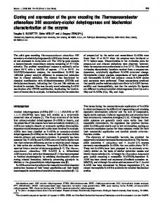
Figure 1. Detection of hTERT mRNA (j) in testicular tissue samples by real-time on-line RT–PCR analysis using the LightCycler®. Two hundred nanograms of total RNA of testicular tissue were analysed. Concomitant detection of PBGD mRNA (x) served as a reference for relative quantification. The copy numbers of the starting template were calculated by comparing the relative fluorescence signals of samples to external hTERT mRNA standards. Relative expression levels were calculated according to the following formula: NhTERT ⫽ hTERT mRNA copies/(PBGD mRNA copies/1000). x axis: cycle number; y axis: fluorescence emission; F2 ⫽ Lightcycler®-Red 640; F1 ⫽ fluorescein. Representative tissue specimens: top: full spermatogenesis, NhTERT ⫽ 127.8 copies; middle: maturation arrest, NhTERT ⫽ 32.8 copies; bottom: Sertoli-cell-only syndrome, NhTERT ⫽ 1.5 copies.
Tissue samples with SCOS showed only minimal hTERT expression (NhTERT ⫽ 2.7 ⫾ 2.8 copies; range 0.00–7.20). Figure 1 shows representative results of normalized hTERT expression in the different histological subgroups. Table II
Discussion A molecular diagnostic parameter that seems suitable for supplementing conventional histopathological diagnostics in the assessment of testicular biopsies is the ribonucleoprotein telomerase, which was first detected in testicular tissue by Kim et al. (Kim et al., 1994). For stem cells and tumour cells, telomerase activity was found to be inhibited with increasing cell differentiation (Sharma et al., 1995). Analogously, a down-regulation of telomerase activity during gametogenesis has been demonstrated for testicular germ cells. Telomerase activity was found to be high during spermatogenesis of spermatogonia to round spermatids, whereas mature spermatids and epididymal sperm were telomerase-negative (Prowse and Greider, 1995; Eisenhauer et al., 1997; Ravindranath et al., 1997; Yamamoto et al., 1999b). Several study groups have recently shown that the detection of telomerase activity in testicular biopsies is helpful for detecting germ cells, particularly in patients with hypergonadotrophic hypogonadism with a predominant SCOS histology (Fujisawa et al., 1998; Yamamoto et al., 1999a; Schrader et al., 2000b). 153
M.Schrader et al.
The procedure applied by the above study groups was methodologically problematic inasmuch as the determination of telomerase activity by the telomeric repeat amplification protocol (TRAP) is influenced by numerous variables that impede an exact quantification. Although the TRAP assay has internal controls for PCR inhibition and telomerase activity, it does not check for variances in the telomerase yield due to cell lysis or instability of the enzyme activity during storage. Moreover, the TRAP assay only partially detects enzyme inhibition by tissue inhibitors and enzyme inactivation with heat or time. Recently, we were able to demonstrate that the hTERT mRNA encoding for the catalytic enzyme component in testicular tissue is highly specific and highly sensitive for the presence of germ cells (Schrader et al., 2000a). This study was the first to quantify hTERT mRNA by realtime fluorescence RT–PCR in testicular tissue specimens from patients with various spermatogenesis disorders for which we intended to establish a molecular diagnostic subclassification. We chose this assay to avoid the above-mentioned problem in achieving an exact telomerase quantification by the TRAP assay. The advantage of this indirect detection procedure is that it accounts for varying tissue degradation with amplification of a housekeeping gene, and primary purification of RNA rules out tissue inhibitors of the PCR. Moreover, it has repeatedly been shown that hTERT mRNA, in contrast to hTR, is rate-limiting for telomerase and that hTERT mRNA expression correlates well with telomerase activity (Harrington et al., 1997; Kilian et al., 1997; Meyerson et al., 1997; Nakamura et al., 1997). Quantification of hTERT mRNA with the LightCycler® shows numerous advantages in this connection. For one thing, real-time online RT–PCR has higher sensitivity compared to other quantification methods such as Northern blotting with in-situ hybridization (Parker and Barnes, 1999) or RNase protection assays (Hod, 1992; Saccomanno et al., 1992). Of practical importance is the fact that this nonradioactive method can start from less than 10 cells to obtain sufficient signals (Wittwer et al., 1997b). A further advantage of the LightCycler® is the ‘real-time’ technique. The fluorescence readings taken at every cycle of the PCR reaction are displayed immediately after each measurement, allowing amplification runs to be terminated or extended, as considered appropriate during individual runs. Real-time RT–PCR also has the advantage of being significantly less variable than conventional RT–PCR procedures (Bustin, 2000). Zhang et al. showed that the coefficient of variation for Ct data is as low as 0.4–0.8% for the LightCycler®, which is significantly better than the 14% reported for conventional RT–PCR (Wittwer et al., 1997b; Zhang and Byrne, 1997; Zhang et al., 1997). In 23 cases, the histological examination disclosed full spermatogenesis. All specimens showed high hTERT expression of NhTERT ⫽ 136.1 ⫾ 41.7 copies. Two of the 17 specimens with reduced spermatogenesis and only focally full spermatogenesis had an hTERT expression of only NhTERT ⫽ 80.5 and 80.7 copies and were thus within the range of tissue samples with maturation arrest. 154
Maturation arrest was diagnosed in 20 specimens. Tissue samples with a JS of 3–5 had a mean hTERT expression of NhTERT ⫽ 48.2 ⫾ 21.0 copies. Four tissue samples with maturation arrest at the level of spermatogonia (JS 3) had a markedly lower hTERT expression (NhTERT ⫽ 17.9 ⫾ 5.3 copies) than those with a JS of 4–5 (NhTERT ⫽ 52.7 ⫾ 10.2 copies). Two tissue specimens with histologically diagnosed maturation arrest (JS 4–5) evidenced an hTERT expression of NhTERT ⫽ 80.5 and 105.0 copies, which was far above the mean value in the other specimens with MA. Other sections of the same tissue examined by the semithin sectioning technique showed focal islands of full spermatogenesis, which indicates that quantitative hTERT determination could contribute to a validation of histopathological findings. Tissue specimens with SCOS showed only minimal hTERT expression with a mean of NhTERT ⫽ 2.7 ⫾ 2.8 copies. In three tissue specimens that evidenced SCOS in the conventional histological work-up but had hTERT mRNA expression (NhTERT ⫽ 11.2–14.5 copies), spermatogonia were found in a renewed histological work-up by the semithin sectioning technique. This indicates that quantitative hTERT determination is highly sensitive and highly specific for detecting germ cells in testicular tissue specimens. Discriminant analysis showed that its determination in tissue samples had a high predictive value (89.0%) for correct classification in one of the three histological subgroups. The markedly higher hTERT mRNA expression we detected in tissue specimens with full spermatogenesis may be due to the different expression level of hTERT mRNA in the telomerasepositive cells, i.e. spermatogonia, primary and secondary spermatocytes and round spermatids, depending on the spermatogenesis disorder. The considerably higher hTERT gene expression in tissue specimens with full spermatogenesis would thus be partially attributable to the fact that comparable germ cells, e.g. spermatocytes, have markedly higher hTERT expression in specimens with full spermatogenesis than in those with spermatogenesis arrest at the level of primary or secondary spermatocytes. This is supported by studies of Yamamoto et al. (Yamamoto et al., 1999b) showing that telomerase profiles tend to be smaller in highly purified fractions of primary spermatocytes recovered from mice with primary testicular damage than in those recovered from healthy mice. Moreover, a number of studies have demonstrated that hTERT mRNA expression shows marked variation in telomerase-positive cells, the different regulatory mechanisms including the cell cycle (Greenberg et al., 1998; Koyanagi et al., 2000), hormones (Kyo et al., 1999), cytokines (Uchida et al., 1999; Xu et al., 2000) and many other control mechanisms (Liu et al., 1999; Nakamura et al., 1999; Wick et al., 1999; Harada et al., 2000; Latil et al., 2000). This suggests that similar regulatory mechanisms may be responsible for the different expression levels we observed in relation to the histological findings. The increased hTERT expression in testicular tissue specimens may also be explained by the larger number of telomerase positive germ cells (e.g. spermatogonia, primary and secondary
The catalytic subunit of telomerase in testicular tissue
spermatocytes and round spermatids) per testis weight unit in patients with full spermatogenesis than in those with maturation. This hypothesis is supported by the study of Mio et al., which demonstrated a larger number of primary spermatocytes per testicular weight unit in Klinefelter men who are positive for haploid germ cells than in those who are positive for primary spermatocytes but negative for haploid germ cells (Mio et al., 1998). The results of the present study show that hTERT mRNA expression in testicular tissue is highly sensitive and specific for germ cell activity and that its quantitative determination by real-time fluorescence RT–PCR enables a molecular-diagnostic classification of spermatogenesis disorders. Thus quantitative determination of hTERT mRNA expression in testicular tissue appears to be well suited for predicting successful sperm recovery in patients with non-obstructive azoospermia and is a useful objective molecular diagnostic parameter for supplementing the histopathological evaluation. Our investigations show that an hTERT expression of NhTERT ⬍ 90 copies indicates full spermatogenesis, while maturation arrest without full spermatogenesis may be assumed at values of NhTERT ⬍ 70 copies. In the grey range of NhTERT ⫽ 70 ⬍ 90 copies, the diagnosis of maturation arrest should be checked by a further work-up of the specimens.
Acknowledgements The authors wish to thank Ms Angelika Schneller, Ms Petra von Kwiatkowski and Ms Antonia Maas from the Department of Urology for their excellent technical support in assessing the samples. They are also grateful to Thomas Emrich (Roche Diagnostic, Penzberg, Germany) for technical assistance with the LightCycler® and to Werner Hopfenmu¨ ller (Department of Medical Statistics) for help in performing the statistical analysis.
References Bolufer, P., Sanz, G.F., Barragan, E. et al. (2000) Rapid quantitative detection of BCR–ABL transcripts in chronic myeloid leukemia patients by real-time reverse transcriptase polymerase-chain reaction using fluorescently labelled probes. Haematologica, 85, 1248–1254. Bustin, S.A. (2000) Absolute quantification of mRNA using real-time reverse transcription polymerase chain reaction assays. J. Mol. Endocrinol., 25, 169–193. Devroey, P., Liu, J., Nagy, Z. et al. (1994) Normal fertilization of human oocytes after testicular sperm extraction and intracytoplasmic sperm injection. Fertil. Steril., 62, 639–641. Eisenhauer, K.M., Gerstein, R.M., Chiu, C.P. et al. (1997) Telomerase activity in female and male rat germ cells undergoing meiosis and in early embryos. Biol. Reprod., 56, 1120–1125. Emig, M., Saussele, S., Wittor, H. et al. (1999) Accurate and rapid analysis of residual disease in patients with CML using specific fluorescent hybridization probes for real time quantitative RT–PCR. Leukemia, 13, 1825–1832. Feng, J., Funk, W.D., Wang, S.S. et al. (1995) The RNA component of human telomerase. Science, 269, 1236–1241. Fujisawa, M., Tanaka, H., Tatsumi, N. et al. (1998) Telomerase activity in the testis of infertile patients with selected causes. Hum. Reprod., 13, 1476–1479. Greenberg, R.A., Allsopp, R.C., Chin, L. et al. (1998) Expression of mouse telomerase reverse transcriptase during development, differentiation and proliferation. Oncogene, 16, 1723–1730. Greider, C.W. and Blackburn, E.H. (1987) The telomere terminal transferase of tetrahymena is a ribonucleoprotein enzyme with two kinds of primer specificity. Cell, 51, 887–898.
Harada, K., Kurisu, K., Sadatomo, T. et al. (2000) Growth inhibition of human glioma cells by transfection-induced P21 and its effects on telomerase activity. J. Neurooncol., 47, 39–46. Harley, C.B., Futcher, A.B. and Greider, C.W. (1990) Telomeres shorten during ageing of human fibroblasts. Nature, 345, 458–460. Harrington, L., Zhou, W., McPhail, T. et al. (1997) Human telomerase contains evolutionarily conserved catalytic and structural subunits. Genes Dev., 11, 3109–3115. Hod, Y. (1992) A simplified ribonuclease protection assay. Biotechniques, 13, 852–854. Holstein, A.F. (1999) Human spermatogenesis: basic research and clinical issues. Anat. Anz., 181, 427–436. Johnsen, S.G. (1970) Testicular biopsy score count – a method for registration of spermatogenesis in human testes: normal values and results in 335 hypogonadal males. Hormones, 1, 2–25. Kilian, A., Bowtell, D.D., Abud, H.E. et al. (1997) Isolation of a candidate human telomerase catalytic subunit gene, which reveals complex splicing patterns in different cell types. Hum. Mol. Genet., 6, 2011–2019. Kim, N.W., Piatyszek, M.A., Prowse, K.R. et al. (1994) Specific association of human telomerase activity with immortal cells and cancer. Science, 266, 2011–2015. Koyanagi, Y., Kobayashi, D., Yajima, T. et al. (2000) Telomerase activity is down regulated via decreases in hTERT mRNA but not TEP1 mRNA or hTERC during the differentiation of leukemic cells. Anticancer Res., 20, 773–778. Kreuzer, K.A., Lass, U., Bohn, A. et al. (1999) LightCycler technology for the quantitation of bcr/abl fusion transcripts. Cancer Res., 59, 3171–3174. Kyo, S., Takakura, M., Kanaya, T. et al. (1999) Estrogen activates telomerase. Cancer Res., 59, 5917–5921. Latil, A., Vidaud, D., Valeri, A. et al. (2000) hTERT expression correlates with MYC over-expression in human prostate cancer. Int. J. Cancer, 89, 172–176. Liu, K., Schoonmaker, M.M., Levine, B.L. et al. (1999) Constitutive and regulated expression of telomerase reverse transcriptase (hTERT) in human lymphocytes. Proc. Natl Acad. Sci. USA, 96, 5147–5152. Meyerson, M., Counter, C.M., Eaton, E.N. et al. (1997) hEST2, the putative human telomerase catalytic subunit gene, is up-regulated in tumor cells and during immortalization. Cell, 90, 785–795. Mio, Y., Yamamoto, Y., Sofikitis, N. et al. (1998) Employment of a highly sensitive quantitative telomerase assay in intracytoplasmic sperm injection programs for the treatment of 4, XXY non-mosaic Klinefelter men. Fertil. Steril. (Suppl.), S19. Nakamura, T.M., Morin, G.B., Chapman, K.B. et al. (1997) Telomerase catalytic subunit homologs from fission yeast and human. Science, 277, 955–959. Nakamura, Y., Tahara, E., Tahara, H. et al. (1999) Quantitative reevaluation of telomerase activity in cancerous and noncancerous gastrointestinal tissues. Mol. Carcinog., 26, 312–320. Nakanishi, H., Kodera, Y., Yamamura, Y. et al. (1999) Molecular diagnostic detection of free cancer cells in the peritoneal cavity of patients with gastrointestinal and gynecologic malignancies. Cancer Chemother. Pharmacol., 43, S32–S36. Nakanishi, H., Kodera, Y., Yamamura, Y. et al. (2000) Rapid quantitative detection of carcinoembryonic antigen-expressing free tumor cells in the peritoneal cavity of gastric-cancer patients with real-time RT–PCR on the lightcycler. Int. J. Cancer, 89, 411–417. Nitsche, A., Steuer, N., Schmidt, C.A. et al. (1999) Different real-time PCR formats compared for the quantitative detection of human cytomegalovirus DNA. Clin. Chem., 45, 1932–1937. Palermo, G., Joris, H., Devroey, P. et al. (1992) Pregnancies after intracytoplasmic injection of single spermatozoon into an oocyte. Lancet, 340, 17–18. Parker, R.M., and Barnes, N.M. (1999) mRNA: detection by in situ and northern hybridization. Methods Mol. Biol., 106, 247–283. Prowse, K.R. and Greider, C.W. (1995) Developmental and tissue-specific regulation of mouse telomerase and telomere length. Proc. Natl Acad. Sci. USA, 92, 4818–4822. Ravindranath, N., Dalal, R., Solomon, B. et al. (1997) Loss of telomerase activity during male germ cell differentiation. Endocrinology, 138, 4026– 4029. Saccomanno, C.F., Bordonaro, M., Chen, J.S. et al. (1992) A faster ribonuclease protection assay. Biotechniques, 13, 846–850.
155
M.Schrader et al. Schrader, M., Muller, M., Heicappell, R. et al. (2000a) Telomerase activity and expression of telomerase subunits in the testicular tissue of infertile patients. Fertil. Steril., 73, 706–711. Schrader, M., Muller, M., Sachsinger, J. et al. (2000b) Telomerase activity in testicular biopsy material (letter). Hum. Reprod., 15, 2057–2059. Schulze, W.H.H. and Knuth, U.A. (1998) Cryopreservation of testicular tissue: a highly effective method to provide sperm for successful TESE/ICSI procedures. In Fertility and reproductive medicine. Elsevier, Amsterdam, pp. 621–626. Sharma, H.W., Sokoloski, J.A., Perez, J.R. et al. (1995) Differentiation of immortal cells inhibits telomerase activity. Proc. Natl Acad. Sci. USA, 92, 12343–12346. Silber, S.J., Nagy, Z., Devroey, P. et al. (1997) Distribution of spermatogenesis in the testicles of azoospermic men: the presence or absence of spermatids in the testes of men with germinal failure. Hum. Reprod., 12, 2422–2428. Simpson, D.A., Feeney, S., Boyle, C. et al. (2000) Retinal VEGF mRNA measured by SYBR green I fluorescence: a versatile approach to quantitative PCR. Mol. Vis., 6, 178–183. Uchida, N., Otsuka, T., Shigematsu, H. et al. (1999) Differential gene expression of human telomerase-associated protein hTERT and TEP1 in human hematopoietic cells. Leuk. Res., 23, 1127–1132. Wick, M., Zubov, D. and Hagen, G. (1999) Genomic organization and promoter characterization of the gene encoding the human telomerase reverse transcriptase (hTERT). Gene, 232, 97–106.
156
Wittwer, C.T., Herrmann, M.G., Moss, A.A. et al. (1997a) Continuous fluorescence monitoring of rapid cycle DNA amplification. Biotechniques, 22, 130–131, 134–138. Wittwer, C.T., Ririe, K.M., Andrew, R.V. et al. (1997b) The LightCycler: a microvolume multisample fluorimeter with rapid temperature control. Biotechniques, 22, 176–181. Xu, D., Wang, Q., Gruber, A. et al. (2000) Downregulation of telomerase reverse transcriptase mRNA expression by wild type p53 in human tumor cells. Oncogene, 19, 5123–5133. Yamamoto, Y., Sofikitis, N., Mio, Y. et al. (1999a) Highly sensitive quantitative telomerase assay of diagnostic testicular biopsy material predicts the presence of haploid spermatogenic cells in therapeutic testicular biopsy in men with Sertoli cell-only syndrome. Hum. Reprod., 14, 3041–3047. Yamamoto, Y., Sofikitis, N., Ono, K. et al. (1999b) Postmeiotic modifications of spermatogenic cells are accompanied by inhibition of telomerase activity. Urol. Res, 27, 336–345. Zhang, J. and Byrne, C.D. (1997) A novel highly reproducible quantitative competitive RT–PCR system. J. Mol. Biol., 274, 338–352. Zhang, J., Desai, M., Ozanne, S.E. et al. (1997) Two variants of quantitative reverse transcriptase PCR used to show differential expression of alpha-, beta- and gamma-fibrinogen genes in rat liver lobes. Biochem. J., 321, 769–775. Received on May 21, 2001; accepted on October 1, 2001