Journal of Gerontology: BIOLOGICAL SCIENCES Cite journal as: J Gerontol A Biol Sci Med Sci Sci. 2012 October;67(10):1022–1035 doi:10.1093/gerona/glr263
© The Author 2012. Published by Oxford University Press on behalf of The Gerontological Society of America. All rights reserved. For permissions, please e-mail:
[email protected]. Advance Access published on March 29, 2012
Review: Quantifying Mitochondrial Dysfunction in Complex Diseases of Aging Martin P. Horan, Nicolas Pichaud, and J. William O. Ballard Faculty of Sciences, School of Biotechnology and Biomolecular Sciences, University of New South Wales, Sydney, Australia. Address correspondence to J. William O. Ballard, PhD, Faculty of Sciences, School of Biotechnology and Biomolecular Sciences, University of New South Wales, Sydney, Australia. Email:
[email protected]
Key Words: Mitochondrial dysfunction—Aging. Received November 14, 2011; Accepted December 28, 2011 Decision Editor: Rafael de Cabo, PhD
O
LD age is associated with impaired mitochondrial function, and this has been proposed as a proximal mechanism for aging. In 1980, Miquel and colleagues (1) first proposed the mitochondrial theory of aging and suggested that mitochondria were key sources for the production and targets of free radicals and that these are associated with aging and a number of age-related diseases (1). From that time onward, much evidence implicating mitochondria in the aging process has emerged (2,3,4,5,6,7). Mitochondrial respiratory malfunction is also intimately associated with aging-associated complex diseases. But not everyone agrees that mitochondria are central to the development of these complex diseases. Complex diseases are just that, complex, and the mechanistic role of mitochondrial dysfunction in the causality of these diseases is highly debated. Likely, however, the study of cellular metabolism and specifically the functional properties of mitochondria will provide a greater understanding of the underlying metabolic malfunctions associated with the progression of many complex diseases. The frequency of mitochondrial DNA mutations in disease may be as high as 1/5,000 (8,9), and the involvement of mildly deleterious mitochondrial mutations in complex diseases of aging are almost certainly much higher. In this review, we describe two new methodologies and contrast the utility of each system in studying respiration in isolated mitochondria, in cultured cells, and in cell fibers and tissues. We then discuss the application of each methodology in the study of mitochondrial malfunction in two common aging-associated complex diseases: Alzheimer’s 1022
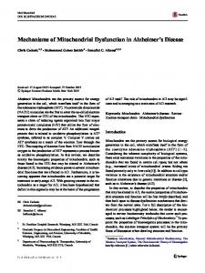
disease and type 2 diabetes mellitus. We finally discuss the technology and its role in elucidating mitochondrial malfunction in aging-associated autophagy impairment. We conclude that the methodologies and biological systems have unique strengths and weaknesses that have the potential to complement each other for the evaluation of respiratory malfunction in complex disorders of aging. Description of the Methodologies for Assaying Mitochondrial Function Mitochondrial respiration occurs on the inner mitochondrial membrane and is achieved by the flow of electrons through the electron transport system, which consists of four complexes (complex I, II, III, and IV) with a further complex (complex V) serving as a site for ATP synthesis (ATP synthase; Figure 1). Impairment of any complex disrupts electron flow and may cause mitochondrial respiratory dysfunction (10,11). Analysis of mitochondrial metabolic dysfunction has, to date, predominantly relied upon the classical oxygen electrode (12). This system has been in use over the past 50 years and has led to an intimate understanding of mitochondrial respiratory function. Although the oxygen electrode remains an inexpensive and useful tool for the evaluation of mitochondrial respiration, it nonetheless has notable limitations with regard to stability of the signal, instrumental background noise, resolution, and throughput (13). There are a multitude of biochemical assays that are specific for the determination of mitochondrial malfunction. These include assays for cytochrome c oxidase and succinate 1
Downloaded from http://biomedgerontology.oxfordjournals.org/ at University of New South Wales on October 2, 2012
There is accumulating evidence that mitochondrial respiratory malfunction is associated with aging-associated complex diseases. However, progress in our understanding of these diseases has been hampered by the sensitivity and throughput of systems employed to quantify dysfunction and inherent limitations of the biological systems studied. In this review, we describe and contrast two methodologies that have been developed for measuring mitochondrial function to address the need for improved sensitivity and increased throughput. We then consider the utility of each methodology in studying three biological systems: isolated mitochondria, cultured cells, and cell fibers and tissues. Finally, we discuss the application of each methodology in the study of mitochondrial dysfunction in Alzheimer’s disease, type 2 diabetes mellitus, and aging-associated autophagy impairment and mitochondrial malfunction. We conclude that the methodologies are complementary, and researchers may need to examine multiple biological systems to unravel complex diseases of aging.
2
BIOENERGETIC ANALYSIS INAL. AGING DISORDERS HORAN ET
1023
dehydrogenase activity (14), membrane potential activity, Western blot, blue native polyacrylamide gel electrophoresis, reactive oxygen species assays, and ATP content assays (15). However, these assays are destructive to cells and tissues, and none can be performed in real time. Considering the need to more accurately assess mitochondrial oxidative phosphorylation function, two new systems have been developed that enable the detection of subtle changes in the respiration rate of mitochondria as a consequence of gene mutation, diet, and environmental insults. These systems are the high-resolution Oxygraph-2k (O2k, Oroboros Instruments, Austria) and the sensitive high-throughput Seahorse XF Extracellular Flux Analyzer (Seahorse XF, Seahorse Bioscience Inc.). The specific measurement of mitochondrial bioenergetics can be performed with high accuracy using the O2k and Seahorse XF systems. Both can analyze, in real time, the mitochondrial respiratory function in the classic in vitro experiments on isolated mitochondria as well as in cultured cells and tissues. Wang and Moraes (16) found that by using three different human cancer cell lines (HeLa, 143B, and MDA-MB-231) and experimentally increasing mitochondrial biogenesis, both the O2k and Seahorse XF produced equivalent data when assaying for total respiration. Thus, the use of these systems in bioenergetic profiling of disease tissue is likely to offer novel insights into aging and complex disease development and progression. Furthermore, the application of these systems in the analysis of aging-associated complex diseases may
potentially aid in the development of novel therapeutic intervention strategies. The O2k and Seahorse XF have notable advantages and limitations for respiratory analysis (Table 1). Nonetheless, accurate and robust quantification of mitochondrial bioenergetic malfunction can be determined with high resolution and high sensitivity in both systems using either isolated mitochondria or intact cultured cells. Indeed, since the introduction of both these technologies, there has been an increase in the study of underlying respiratory malfunction in complex diseases (Table 2). Oroboros O2k The O2k was developed during the mid-1990s (50) and has been used extensively for respiratory analysis. The O2k is a development over the traditional oxygen electrode for measuring mitochondrial respiration (50). The system possesses two separate 2-mL chambers equipped with polarographic oxygen sensors that can measure in real time both the oxygen concentration (nanomoles/milliliter) and the oxygen consumption (picomoles/second/milliliter) within each chamber. The basic principle of the system is to measure the concentration and consumption of oxygen by injecting substrates directly to isolated mitochondria or cells that are suspended in solution within the chamber (50). The high-resolution detection capability of the O2k is enhanced by a temperature controlling system allowing respiratory measurements to be made from 2°C to 45°C
Downloaded from http://biomedgerontology.oxfordjournals.org/ at University of New South Wales on October 2, 2012
Figure 1. Oxidative phosphorylation in the mitochondrial inner membrane. Q = ubiquinone; Cyt = cytochrome; FMN = flavin mononucleotide; FeS = ironsulfur cluster.
1024
HORAN ETINAL. BIOENERGETIC ANALYSIS AGING DISORDERS
3
Table 1. Comparison of the Oroboros Oxygraph-2k (O2k) and the Seahorse XF Extracellular Flux Analyzer for Technical Specifications and Respiratory Specifics Equipment Specifics
Seahorse XF Extracellular Flux Analyzer
$50k No High (needs constant operator input) No OXPHOS† 2-Chamber based Yes (polarographic sensors) No (two samples per run) Slow—approximately 1 h for two samples Yes (4–45°C) No (separate assay required per chamber) Mitochondria/cells/permeabilized fibers Yes Unlimited Yes Cheap—reagents/substrates required
$200k Yes Minimal (system is fully automated) Yes OXPHOS/glycolysis/CO2 24- or 96-Well plate based Yes (fluorescence sensors) Yes (24–96 samples per run) Fast—approximately 90 min for up to 96 samples Yes, but ambient temperature needs to be 8°C below desired temperature Yes (background control wells on plate) Mitochondria/cells/pancreatic islets/muscle fibers/hippocampal slices No Limited to four No Expensive—reagents/substrates plus new fluorescent plate for each assay
Notes: *Approximate U.S. dollars. phosphorylation. ‡ Mitochondrial membrane potential analysis. § Analysis to determine if the respiration rate alters between high and low oxygen concentrations. || Cost is relative because the experimental design using the Oroboros O2k may prove to be equally or more expensive in the longer term if the cost of operator usage constantly monitoring an equivalent Seahorse XF sample output over time is taken into account. † Oxidative
(13). A further advantage of the O2k system over the traditional Clark-type oxygen electrode is that only small quantities of sample are required for metabolic analysis. However, a particular advantage specific to the O2k system is that oxygen concentration in the measurement chambers can be allowed to remain elevated during an assay or until the functional stability of the sample is impaired, enabling extended substrate–uncoupler–inhibitor–titration protocols to be applied (51). This is important because these protocols enable the evaluation of all the mitochondrial coupling and respiratory control measurements to be accurately performed. Because substrate injections are performed manually, each complex of the electron transport system can be studied independently using different substrate combinations. For example, sequential injections of glutamate, malate, and pyruvate can be used to determine the effect of each of these substrates on mitochondrial respiration when electrons are provided to complex I. A further injection of succinate can be used to assess the effect of electron input through complex I and complex II. Rotenone can then be added to specifically inhibit complex I to evaluate mitochondrial respiration from complex II. Thus, the ability to manually inject specific substrates to test different complexes for malfunction or to determine the effect of multiple substrates or drugs on the rate of mitochondrial respiration allows for an analysis of a multitude of injectable reagents. Limitations of the O2k There are three primary limitations to the O2k. First, operator input and labor intensity are high because constant monitoring and adjustments of concentrations of injectable
reagents are required to obtain optimal signal intensity. Second, the O2k is not capable of high-throughput capability as only two samples can be performed at any one time. Because analysis using substrate–uncoupler–inhibitor– titration protocols take approximately 1 hour to complete, it is only possible to analyze 16 samples over an average 8-hour day. This can prove to be expensive for large-scale studies (ie, the testing of multiple drug compounds over multiple cell types) when the cost of an operator constantly monitoring a large number of mitochondrial assays is taken into account. A third limitation is that the O2k is incapable of recording extracellular pH levels. This represents an important measurement because it allows for a determination of glycolytic pathway utilization. Because cancer cells preferentially use glycolysis as their main source of energy (52), the testing of compounds that revert cells to oxidative phosphorylation would be beneficial for monitoring any potential therapeutic effect. Seahorse XF Extracellular Flux Analyzer The Seahorse XF was introduced during 2006/2007 (53) and was developed to address the need for a high-throughput system for the determination of mitochondrial malfunction. The basic principle of the system is to measure the rate of mitochondrial oxidative phosphorylation (through determination of the oxygen consumption rate measured in picomoles/minutes) and glycolysis (through determination of the extracellular acidification rate measured in milli-pH units/minutes; 54). The Seahorse XF operates on a 24 or 96 microplate-based assay platform. A feature of the microplate-based assay is that a single plate containing multiple samples can be
Downloaded from http://biomedgerontology.oxfordjournals.org/ at University of New South Wales on October 2, 2012
Cost of equipment* Automated Labor intensity Internal calibration Respiratory measurements Assay format High-resolution sensitivity High-throughput capability Time per sample assay Controlled temperature range Background correction per assay Tissue types analyzed MMP‡ Substrates injected per assay Injection of oxygen§ Cost per assay||
Oroboros O2k
4
BIOENERGETIC ANALYSIS INAL. AGING DISORDERS HORAN ET
1025
Table 2. Examples of Complex Human Diseases Analyzed for Mitochondrial Malfunction Using the Oroboros Oxygraph-2k (O2k) and the Seahorse XF Extracellular Flux Analyzer Disease Aging of colonic crypts Aging
Gene
Autosomal dominant optic atrophy Breast cancer Cardiovascular disease
APO Tau OPA1 KLRK1 Esrrg
Charcot–Marie–Tooth disease Colorectal cancer
MFN2 BAX
Diabetes
Tbx15 Slc25a25 mdx TTC19 MTCOI FH ATPIF1 Atg7 EGLN2, EGLN3 SURF1 Phd1 TK2 Tim23 Ipla2γ TNFR1 SNCA, Gss DJ-1 PINK1 TNFR1 PARL-β Tfb1m
Duchene muscular dystrophy Encephalopathy Epilepsia partialis continua HLRCC‡ Human carcinomas Impaired autophagy in aging Ischemic cardiomyopathy Leigh syndrome Liver ischemia/reperfusion injury MDS§ Mohr-Tranebjaerg syndrome Obesity Parkinson’s disease
TRAPS|| Type 2 diabetes mellitus
p.Y19H Sod2 knockout mouse Heterozygous Sod2 mouse Bmi1 knockout mouse Triple-transgenic knockout mutant mouse Isoform E4 expression p.P301L (transgenic mouse) p.R932C Forced expression of KLRK1 Small interfering RNA gene knockdown of Esrrg p.R468H Homologous recombinant BAX knockout cells Forced expression of Tbx15 Slc25a25 knockout mouse p.Q995X (mdx mouse) c.517C>T (p.Gln173X) p.L196I p.Q396P siRNA gene knockdown of ATPIF1 Atg7 knockout mouse EGLN2, EGLN3 Knockout mouse p.R230X Phd1 knockout mouse TK2 knockout mouse Tim23 knockout mouse Ipla2γ knockout mouse TNFR1 knockout mouse Dual transgenic inducible mouse DJ-1 knockout mouse p.G309D p.C33Y, p.T50M p.R54G + p.K55S + p.R58G + p.K59S Heterozygous Tfb1m mice
System (O2k*, S†)
Cells/mitochondria studied
Reference
O2k S O2k S S
Intact/permeabilized colonocytes Embryonic fibroblasts Permeabilized muscle fibers Thymocytes Embryonic neuronal cells
(17) (18) (19) (20) (21)
S O2k O2k S S
Neuronal cells Cerebellar mitochondria Permeabilized fibroblasts MCF-7 cells Cardiomyocytes
(22) (23) (24) (25) (26)
O2k S
Permeabilized fibroblasts HCT-116 cells
(27) (28)
S S O2k S O2k S S S S O2k O2k S O2k O2k O2k S O2k O2k S S S
3T3-L1 preadipocytes Embryonic fibroblasts Myoblasts Fibroblasts/myoblasts Permeabilized muscle fibers UOK 262 cells HeLa cells Embryonic mitochondria/fibroblasts Cardiomyocytes Intact/permeabilized fibroblasts Hepatocytes Neuronal cells Permeabilized embryonic stem cells Muscle/adipose tissue mitochondria Permeabilized muscle fibers Olfactory bulb cells Embryonic fibroblasts Whole-brain mitochondria Embryonic fibroblasts RMS-13 human myoblasts INS-832/13 beta cells
(29) (30) (31) (32) (33) (34) (35) (36) (37) (38) (39) (40) (41) (42) (43) (44) (45) (46) (47) (48) (49)
Notes: *Oroboros O2k. XF Extracellular Flux Analyzer. ‡ Hereditary leiomyomatosis renal cell carcinoma. § Mitochondrial depletion syndrome. || Tumor necrosis factor receptor-associated periodic syndrome. † Seahorse
analyzed with high resolution, thus providing sensitive highthroughput capability. The system utilizes a novel fluorescent sensor-containing biocartridge, which fits over a cell culture microplate. The biocartridge is analogous to a piston and is used for reagent mixing and measuring extracellular acidification and oxygen consumption. Pharmaceutical reagents or mitochondrial respiratory electron transport system inhibitors distributed in ports surrounding the sensor are then sequentially injected to each well. Background correction wells (ie, wells that have not been seeded with cells or mitochondria) are included in the assay to normalize the data to background plate noise. Limitations of the Seahorse XF Extracellular Flux Analyzer There are three main limitations to consider when analyzing mitochondrial respiration using the Seahorse XF. First,
the cost of optimizing injectable concentrations and the subsequent cost per respiratory assay are high in comparison to the Clark-type electrode and the O2k. The Seahorse XF fluorescent plates are costly and need replacing on completion of each assay. This can become expensive, as it is essential to optimize the concentration of each injectable reagent prior to any respiratory assay. Furthermore, on completion of mitochondrial and cell optimization steps, further optimization will often be required when testing for the effect of cultured cells grown in the presence of pharmaceutical compounds. This optimization is required to ensure that pharmaceutical reagents do not interfere with the electron transport system inhibitors and produce misleading data. A second limitation is that only four injectable compounds can be used to detect mitochondrial respiratory malfunction in any single assay. Thus, it will be necessary to use multiple plates when the testing of more than four injectable reagents is required
Downloaded from http://biomedgerontology.oxfordjournals.org/ at University of New South Wales on October 2, 2012
Alzheimer’s disease
MTCOI Sod2 Sod2 Bmi1 App, Tau, PS1
Mutation/function studied
1026
HORAN ETINAL. BIOENERGETIC ANALYSIS AGING DISORDERS
(ie, identifying the effect of multiple compounds on mitochondrial respiration for novel therapeutic identification), which again increases cost. A third limitation is that injectable compounds may potentially interfere with sensor fluorescence or the plastic plate and produce misleading data (55).
Oroboros O2k Determination of mitochondrial functional integrity and the maximal capacity of oxidative phosphorylation has been demonstrated to be advantageous through in vitro–based assays using the O2k (58). Studies have also used isolated mitochondria for metabolic flux control analysis (59,60,61). The control of oxidative phosphorylation is shared between all the enzymes of the metabolic pathway that incorporates the phosphorylation system (ATP synthase and adenine nucleotide translocase), the electron transport system (complex I to complex IV), and the reactions that provide substrates and electrons to the system (tricarboxylic acid cycle and metabolite transporters). This represents the global flux of oxidative phosphorylation, and the manipulation of flux enables the investigation of specific metabolic pathways. The analysis of flux requires that all enzymatic steps involved in the electron transport system be identified with the importance of each step being quantified (62). There are several approaches to achieve this goal (56,63,64), but all require the titration of the whole respiratory chain (global flux). These protocols are time consuming, quite complicated, and often limited due to functional instability after isolation. In contrast, the use of substrate–uncoupler–inhibitor–titration protocols using the O2k system greatly simplifies analysis and may allow the specific steps implicated in disease to be determined (51). Substrate–uncoupler–inhibitor–titration protocols enable the accurate determination of all the mitochondrial coupling and respiratory control parameters. For
example, these protocols can control the rate of complex IV flux on oxygen dependence and can help explain the pathological phenotype of mild complex IV deficiencies observed in some mitochondrial myopathies (65). Moreover, the application of the flux control analysis using the O2k system has demonstrated that complexes I and III in mammals behave as single units, suggesting the existence of substrate channeling and supercomplex organization (66). In addition to providing a quantitative analysis of oxidative phosphorylation control (62,67), it has been further suggested that the disruption of supercomplex organization may lead to mitochondrial functional derangements and the progression of disease development (68). Seahorse XF Extracellular Flux Analyzer Mitochondrial analysis on the Seahorse XF can be performed using very small quantities of mitochondria (1–10 μg of mitochondrial protein per well; 69). Prior to the addition of electron transport system reagents, the basal levels of oxygen consumption by the mitochondria are first determined to establish steady state levels. The Seahorse XF incorporates a further measurement to determine oxygen tension (a similar measurement to oxygen concentration in the traditional oxygen electrode). This is a useful function that can help determine the correct concentration of mitochondria being loaded directly into each well. Once optimal concentrations are established, all the mitochondrial-measured parameters can be determined with electron transport system inhibitors. Comparisons can then be made between experimental samples. However, the results obtained from the Seahorse XF may vary in replicate wells due to clumping of mitochondria as a consequence of the isolation procedure. Thorough suspension and careful dilutions of extracted mitochondria are necessary to give robust results. An example of the high-throughput capability of the Seahorse XF was provided by Abadir and colleagues (70) who identified respiratory malfunction in the mitochondrial rennin–angiotensin system. In addition, they present evidence of age-related changes in mitochondrial rennin–angiotensin receptor expression. Isolated rat heart or liver mitochondria from mice were plated into a Seahorse XF 96-well plate, and the ADP-activated state was determined using substratesupported respiration. A single assay was capable of determining the effect of different substrates and mitochondrial angiotensin 2 receptor agonists and antagonists on the activation of mitochondrial respiration in multiple replicates from different mitochondrial isolations. The data from this study suggest that a mitochondrial angiotensin 2 receptor agonist was capable of reducing ADP-activated respiration in parallel with nitric oxide production. Nitric oxide production inhibits mitochondrial respiration through binding to the electron transport system complex IV (71). These data strongly suggest that inadvertent mitochondrial angiotensin 2 receptor agonist activation could lead to disease development.
Downloaded from http://biomedgerontology.oxfordjournals.org/ at University of New South Wales on October 2, 2012
Analysis of Isolated Mitochondria The definitions of the mitochondrial respiratory measured states first proposed by Chance and Williams (10) have evolved, and new conventions regarding the active measurement of mitochondrial function can be derived by the sequential injection of different substrates to isolated mitochondria (56; Supplementary Table 1). The addition of different substrates is important because it stimulates the flow of electrons from different complexes of the electron transport system, thus allowing the identification of a specific malfunctioning complex. Mitochondrial parameters are measured over five principle respiratory states, which in combination with different substrates are used to determine the functional activity of the electron transport system (Supplementary Table 1). Malfunctioning mitochondria isolated from disease tissue can therefore be identified in comparison to the normal respiratory activity of isolated control mitochondria. More detailed information on the measurement of respiratory states can be found elsewhere (10,51,57).
5
6
BIOENERGETIC ANALYSIS INAL. AGING DISORDERS HORAN ET
Analysis of Intact Cultured Cells The respiratory rates measured in intact cultured cells vary slightly in comparison to measured rates from isolated mitochondria. For example, the coupled ADP-activated state 3 (state 3ADP) rate is not measured in cultured cells because ADP is incapable of penetrating the outer membrane of cultured cells (13,58). However, the real-time analysis of in vivo basal respiration, oxygen consumption, ATP turnover, coupling efficiency, proton leak, maximum respiration rate, respiratory control ratio, spare respiratory capacity, and non-mitochondrial respiratory parameters can all be determined by the sequential addition of specific electron transport system inhibitors (eg, oligomycin, carbonylcyanidep-trifluoromethoxyphenylhydrazone, rotenone, and antimycin A, Supplementary Table 1). A more detailed description
and analysis of these measured parameters can be found in the review by Brand and Nicholls (57). Oroboros O2k The assessment of mitochondrial activity in suspension cells such as skin fibroblasts (38), lymphocytes (83), HEK293 cells (84), and 32D cells (85) has been performed in real time using the O2k system to determine respiratory malfunction. Cells may be suspended in a culture medium supporting physiological in vivo oxygen consumption and growth by exogenous substrates (advantageous when aiming at near-physiological conditions for cultured cells). Alternatively, cells may be in crystalloid medium without energy substrates (eg, mitochondrial respiration medium) yielding physiological in vivo oxygen consumption with endogenous substrates (86). Inhibitors and an uncoupler are then added to evaluate the different respiratory rates. Analysis of disease-associated mutations can readily be analyzed by the O2k for respiratory malfunction. For example, cultured COS cells (derived from kidney cells of the African green monkey) have been used to analyze the respiratory response to increased expression of the Parkinson’s diseaseassociated α-synuclein protein (87). Furthermore, duplications and triplications of the α-synuclein gene, SNCA, have been associated with more severe forms of the Parkinson’s disorder (88,89). However, the underlying functional consequences of these mutations remain to be determined. Nakamura and colleagues (87) found that the overexpression of α-synuclein produced an increase in mitochondrial fragmentation and reduced both the basal and maximum levels of mitochondrial respiration, thus indicating that the affected cells could no longer respond efficiently to cell stress. This observation was further confirmed in neuronal cells where an increase in α-synuclein also produced mitochondrial fragmentation and decreased cell survival (87). These data indicate that mitochondrial malfunction as a consequence of the overexpression of α-synuclein may help explain some of the underlying mechanisms responsible for the Parkinson’s disease severity and progression. Seahorse XF Extracellular Flux Analyzer The Seahorse XF can measure cellular mitochondrial metabolism in real time and is capable of analyzing both adherent cells and, in some cases, cells in suspension (however, suspension cells first need to be centrifuged to form a monolayer on the bottom of the well prior to analysis). For cell analysis, cells are grown to 70%–80% confluency. Growth media is removed and replaced with cell assay media. The derived measured parameters in cells are then determined after the addition of electron transport system inhibitors (57). Novel bioenergetic properties of currently available pharmaceutical compounds have been identified with the Seahorse XF, and these have the potential to treat complex
Downloaded from http://biomedgerontology.oxfordjournals.org/ at University of New South Wales on October 2, 2012
Limitations of Quantifying Mitochondrial Dysfunction in Isolated Mitochondria Extraction of mitochondria disrupts the intricate mitochondrial network integrity leading to differences in in vitro and in vivo data (51,58,72). Mitochondria exist as large tubular assemblies in living cells, are in close contact with other organelles in the cell matrix (73,74), and undergo fusion and fission events (75). Disruption of these networks (during isolation) may have important ramifications in determining the specific mutations that cause disease. For example, previous studies have demonstrated that mutations in proteins involved in mitochondrial fusion, fission, motility, or degradation cause neurological disorders including hereditary optic neuropathies (76) and Charcot–Marie–Tooth type 2A disease (77). Thus, if the mitochondrial network integrity is disrupted during the isolation procedure, it is possible that false-negative respiratory data may be produced. In addition to mitochondrial network disruption (78), there are other limitations of studying isolated mitochondrial preparations that may cause in vitro properties of isolated mitochondria to be different from mitochondria maintained in vivo (58). These limitations equally affect data collected from the Clark-type electrode, O2k, as well as the Seahorse XF. Mitochondrial isolation from normal tissues is performed under optimal centrifugation speeds that have been demonstrated not to damage mitochondria and thus provide the highest quality of mitochondrial integrity (intactness of outer and inner membranes). However, different pathologies may affect the membrane intactness and therefore the sensitivity of mitochondria to centrifugation (79). If this happens, centrifugation methods may result in a biased selection of a particular mitochondrial population (80). Moreover, large quantities of cells or tissues are required for the optimal yield and quality of isolated mitochondria (81). The measurement of respiratory control in cell populations has the benefit of maintaining the intricate structural network of mitochondrial connections, and the study of bioenergetic function in cultured cells may be preferable in some situations (57,82).
1027
1028
HORAN ETINAL. BIOENERGETIC ANALYSIS AGING DISORDERS
7
Oroboros O2k The O2k can quantify mitochondrial dysfunction in situ in permeabilized muscle fibers and in tissues. The
Seahorse XF Extracellular Flux Analyzer An adaptation of the Seahorse XF plate assay has been the development of microplates that are capable of analyzing
Limitations of Quantifying Mitochondrial Dysfunction in Cultured Cells Although cultured cells maintain a cellular architecture and are physiologically relevant for mitochondrial bioenergetic analysis, there are, however, several problems associated with the construction and maintenance of such cell lines. These problems include contamination by viruses, mycoplasma, and other cell lines (91). The generation of immortalized cell lines are also limited due to loss of differentiated functions, high glycolytic rates, and reduced respiration (92). Furthermore, most often individual cell populations are analyzed, and they lack direct comparisons with whole-tissue measurements that may contain multiple cell types. Another major issue is that cultured cells do not allow certain important functional parameters of mitochondria to be studied. For example, mitochondrial responses to various substrates such as ADP, creatine, or cytochrome c cannot be investigated in intact preparations because they are unable to penetrate the cellular membrane (13,58). However, these issues are not specific to the O2k and the Seahorse XF but rather a general limitation to cell growth–based research.
Downloaded from http://biomedgerontology.oxfordjournals.org/ at University of New South Wales on October 2, 2012
Analysis of Cell Fibers and Tissues One strategy to overcome the limitations found in both mitochondrial isolations and in cultured cells is to study mitochondrial function using in situ–based approaches. Indeed, novel methods have been developed with the O2k to remove cell membrane inhibition and allow mitochondrial substrates to penetrate the cellular membrane (ie, permeabilization; 58). Methods have also been developed for the Seahorse XF that allow for the respiratory analysis of wholetissue samples (ie, hippocampal slices; 93). A continued development of both systems to study cell fibers and tissues is expected in the next few years and will likely provide additional insight into complex diseases of aging.
permeabilized in situ approach is also suitable for the application of substrate–uncoupler–inhibitor–titration protocols for the analysis of ADP affinity (78,94) and for all functional studies of the electron transport system complexes (51,95). Although the in situ approach can be used with the Clark-type oxygen electrode (96), the O2k system has provided an important advance because of the stability of the signal, low instrumental background noise, and increased resolution (13,97,98,99,100). The in situ approach weakens or permeabilizes the plasma membrane with a chemical compound so that mitochondrial function can be analyzed (96,97,98,101). The plasma membrane contains almost five times more cholesterol than the endoplasmic reticulum and 7–10 times more than the outer and inner mitochondrial membrane (102,103). Permeabilizing agents like saponin or digitonin interact specifically with cholesterol molecules abundant in the plasma membrane resulting in pore formation without affecting other cellular components like the endoplasmic reticulum, mitochondria, myofilaments, and cytoskeleton (58,78). This approach has been demonstrated to be physiologically relevant because it enables analysis of mitochondria in their normal intracellular assembly (58). Furthermore, preparations of permeabilized fibers have been shown to display mitochondrial functional stability for up to 24 hours when stored under cold conditions (104). The in situ approach is also appropriate for the assessment of mitochondrial function in model species of aging (105). For example, a constraint of many aging studies in Drosophila is that mitochondria increase in size as the fly ages. As a consequence, it has not been possible to study intact mitochondrial function in flies over 25 days of age (80). The O2k has been used to quantify specific changes in mitochondrial function associated with aging disorders using permeabilized muscle fibers that was not detected using mitochondrial isolations. For example, using isolated mitochondria from skeletal muscle, it has been shown that sarcopenia, the decline of muscle mass and function with aging, is correlated with age-related changes in mitochondrial function (106,107). Sarcopenia is an important contributor to frailty and age-related disability. Using the O2k system, Picard and colleagues (72) performed a comparison of mitochondrial functions in mitochondrial isolations and in permeabilized myofibers from skeletal muscle of young adults and senescent rats. This study demonstrated that mitochondrial isolations employed to study aged muscles expose functional impairments not seen in situ in the permeabilized myofibers. Therefore, it was concluded that aging is associated with more modest changes in mitochondrial function in sarcopenic muscle using a physiologically relevant approach than previously thought using isolated mitochondria.
diseases of aging. Gohil and colleagues (90) included >3,500 chemical compounds assayed across multiple cell types identified that various food and drug administration–approved compounds shifted mitochondrial respiration to glycolysis. One of these compounds was meclizine, a nausea and vertigo approved compound that is readily available over the counter. Meclizine was found to reduce oxygen consumption and increase extracellular acidification in all cell types studied. This newly identified metabolic activity for meclizine was further found to confer protection in models of cardiac ischemia–reperfusion injury and significantly reduced infarct volume in an in vivo model of cerebral ischemia when used as a prophylactic (90).
8
BIOENERGETIC ANALYSIS INAL. AGING DISORDERS HORAN ET
Limitations of Quantifying Mitochondrial Dysfunction in Cell Fibers and Tissues Analysis of cell fibers and tissues for mitochondrial functional determination has at least three major limitations that should be considered before this approach is initiated. First, for permeabilized cells and tissues, respiratory parameters of different mitochondrial subpopulations cannot be distinguished. This may be a serious problem in cases of mitochondrial myopathy where mitochondrial DNA heteroplasmy rates can differ within and between cells. Second, the impact of cytosolic factors cannot be studied because the cytoplasm and all cellular soluble components are lost during the permeabilization procedure (58). This is particularly important because normal mitochondrial function requires crosstalk with cytosolic factors to maintain cellular homeostasis. Thus, misleading respiratory data may be produced when using permeabilized cells. Third, cell fibers and tissues cannot interact with neighboring tissues. This means that the influence of multiple tissue/organ types cannot be analyzed for mitochondrial functional analysis. These limitations are equally applicable to all biochemical and gene expression– based assays.
Respiratory Analysis in Age-Associated Complex Human Disease Respiratory analysis of mitochondrial function has been performed on multiple tissue types across various complex human diseases (Table 2). These studies offer insights into the underlying mechanisms of disease progression and development. Here we consider the contributions that the O2k and Seahorse XF systems have made in our understanding of two aging-associated complex diseases: Alzheimer’s disease and type 2 diabetes mellitus. We further discuss the role of mitochondrial malfunction as a consequence of autophagy dysfunction in aging. In each section, we briefly review the role of each disorder and conclude by considering possible future directions. More complete reviews of Alzheimer’s disease, type 2 diabetes mellitus, and autophagy dysfunction in disease can be found elsewhere (111,112,113). Alzheimer’s Disease Alzheimer’s disease is the most common form of dementia in the elderly and is characterized by a decline in memory function and cognitive ability. It tends to occur in individuals older than 65 years of age. The underlying mechanisms of Alzheimer’s disease are degeneration of brain tissue with extensive neuronal loss, accumulation of senile plaques (caused by aggregates of the amyloid-β protein), and the presence of filamentous inclusions known as neurofibrillary tangles due to hyperphosphorylation of Tau proteins (114). The amyloid-β protein is derived from the amyloid precursor protein (APP) through an initial β-secretase cleavage, followed by an intramembranous cleavage by the presinilin 1 (PS1) and presinilin 2 (PS2) subunits of the α-secretase complex. The amyloid-β protein is also known to be a potent inhibitor of mitochondrial enzyme activity and therefore serves to reduce the flow of electrons through the electron transport system and decreases the rate of ATP production and oxygen consumption (115,116). Moreover, mitochondrial dysfunction is one of the earliest features identified in the pathology of Alzheimer’s disease (117,118). Mutations in mitochondrial DNA in the brains of Alzheimer’s disease patients are also detected with higher frequency than agematched controls, which is suggestive of a mitochondrial genetic link in Alzheimer’s disease progression (119,120). Respiratory analysis of extracted mitochondria using the O2k system has demonstrated that both double- (APPSW/ PS2N141I) and triple (APPSW/PS2N141I/TauP301L)-transgenic Alzheimer’s disease mice exhibit a pronounced decrease in coupling efficiency and in the uncoupled state relative to non-mitochondrial respiration by the age of 8 months (121). Furthermore, the ADP-activated state and the uncoupled state are decreased at 8 months of age in the double-transgenic mice and at 12 months of age in the triple-transgenic mice (121). In combination, these data suggest that amyloid-β protein aggregation in the transgenic mice has a stronger biochemical effect in the double- (APPSW/PS2N141I) mutant
Downloaded from http://biomedgerontology.oxfordjournals.org/ at University of New South Wales on October 2, 2012
pancreatic islets for the study of insulin secretion (108). Pancreatic islets are scarce, and isolating sufficient mitochondria from them is difficult. Analyzing whole islets has the advantage of maintaining the cellular architecture. For respiratory analysis, pancreatic islets are isolated and plated into a recess in the bottom of the Seahorse XF islet capture microplate and immobilized with a securing screen. The pancreatic islets are analyzed in the same way as cultured cells to determine metabolic function. Indeed, it has been established that the reduction of cytochrome c in isolated pancreatic islets is necessary for insulin secretion and is unaffected by a decrease in oxygen consumption (108). A further development of the Seahorse XF islet capture microplate has enabled the analysis of hippocampal tissue slice preparations (93) and the bioenergetic profiling of whole zebra fish embryos (109). The zebra fish study demonstrated that total respiration increased with age of the embryo and was associated with an increase in mitochondrial content. Because whole embryos can be studied, there is a potential to determine respiratory malfunction in aging disorders and to test the effects of therapeutic agents (109). A second adaptation of the Seahorse XF analyzer is the measurement of mitochondrial bioenergetics in intact skeletal muscle fibers (110). This protocol has implications for studying genotype-specific decline of function with increasing age as well as neuromuscular disorders including muscular dystrophy and cachexia. For intact muscle analysis, single muscle fibers are enzymatically isolated and seeded on to a culture microplate to cover approximately 50%–60% of the well bottom. Cellular measurements are then performed following a similar analysis as for cultured cells (110).
1029
1030
HORAN ETINAL. BIOENERGETIC ANALYSIS AGING DISORDERS
Type 2 Diabetes Mellitus Type 2 diabetes mellitus is the most common metabolic disease found in an aging population (127). Although the primary cause of type 2 diabetes remains unknown, the development of insulin resistance in both skeletal muscle and liver is pivotal in the disease progression prior to the onset of pancreatic β cell dysfunction (128). Several studies have further implicated type 2 diabetes with mitochondrial malfunction including reduced mitochondrial density (129), reduced mitochondrial oxidative phosphorylation (130,131), and increased levels of reactive oxygen species (132,133).
However, the underlying molecular mechanisms responsible for mitochondrial malfunction remain to be identified. Decreased mitochondrial function has been suggested to play an important role in the pathogenesis of insulin resistance (134). However, it is not clear whether the observed reduction in oxidative capacity is due to impairment in the functional properties of mitochondria or to a reduction in mitochondrial DNA copy number. The O2k system has demonstrated that mitochondrial respiration is altered in type 2 diabetes patients (135,136). Using the in situ approach on permeabilized muscle fibers, a decrease in the ADPactivated and in the uncoupled state was observed in type 2 diabetes patients compared with age- and body mass index–matched control subjects (136). However, oxygen consumption was the same for both type 2 diabetes patients and healthy control subjects after normalization for citrate synthase activity (a marker of mitochondrial content; 135, 136). The O2k data therefore suggest that mitochondrial malfunction is not an underlying mechanism involved with type 2 diabetes. In contrast, data from the Seahorse XF suggest that mitochondrial bioenergetics may be altered in type 2 diabetes as a consequence of the decreased expression of the human presenilin-associated rhomboid-like (PARL) gene (48). PARL malfunction is associated with type 2 diabetes (72,137), and analysis of skeletal muscle biopsies taken from type 2 diabetes patients has revealed that PARL messenger RNA and mitochondrial DNA content is reduced in comparison to healthy controls (48). The enzymatic cleaved product of PARL (PARL-β) is thought to mediate mitochondria-to-nucleus crosstalk and regulate the expression of mitochondrial-specific genes (138). The Seahorse XF study by Civitarese and colleagues (48), using human RMS-13 myoblast cells transfected with either normal PARL-β or mutant PARL-β constructs, reported that oxygen consumption is reduced in the mutanttransfected cells after normalization to citrate synthase (48). Furthermore, the RMS-13 myoblast cells expressing the synthetic normal PARL-β peptide exhibited increased levels of sirtuin-1 (48). This is an interesting finding because sirtuin-1 is an activator of peroxisome proliferator–activated receptor gamma, coactivator 1 alpha, a transcriptional coactivator responsible for mitochondrial biogenesis (139,140). These data therefore suggest that PARL activates the sirtuin pathway and that a decrease in the expression of PARL may reduce mitochondrial biogenesis. The data generated from the O2k and Seahorse XF appear to conflict, and three explanations would appear possible. One explanation for the differences is that the O2K study assayed the parameters of ADP-activated state and uncoupled state using excess concentration of substrates where the Seahorse XF study assayed for oxygen consumption using the endogenously expressed substrates within the cells. A second explanation is that the O2k used permeabilized fibers that lack nuclear factors and the cytosol, but the Seahorse XF study used intact cultured cells. A third possible
Downloaded from http://biomedgerontology.oxfordjournals.org/ at University of New South Wales on October 2, 2012
pathway than the (TauP301L) pathway (121). This example shows that different genetic pathways altering cellular bioenergetics can be determined using the O2k system and can help improve our understanding of the underlying mechanisms leading to mitochondrial dysfunction in Alzheimer’s disease. Data generated from Seahorse XF also indicate that mitochondrial dysfunction may be a causal factor in Alzheimer’s disease pathology (21). Studies have established that mitochondrial malfunction is severely compromised in a triple-transgenic (3xTg, APPSW/PS1M146V/TauP301L) mutant Alzheimer’s disease mouse model (21). Yao and colleagues (21) determined that mitochondrial respiration is decreased in primary hippocampal neurons cultured from female transgenic (3xTg) Alzheimer’s disease mice compared with neurons in age-matched nontransgenic (non-Tg) controls (21). The Seahorse XF data derived from the 3xTg Alzheimer’s disease mice identified reduced levels of steady state basal respiration, ATP turnover (uncoupled), respiratory control ratio, uncoupled state, and spare respiratory capacity in comparison to the non-Tg controls. Furthermore, the primary neuronal cultures used in the study derived from embryonic day 14 mice indicate that the mitochondrial dysfunction can be detected early in the pathogenesis of Alzheimer’s disease. These data strongly suggest that mitochondrial malfunction precedes the development of plaque formation, which is the classical histopathological marker of Alzheimer’s disease. The data generated from both the O2k and the Seahorse XF have identified altered bioenergetics in mouse models of Alzheimer’s disease. Both sets of data suggest that genetic risk factors for mitochondrial bioenergetics could be critical in the development of Alzheimer’s disease. This is supported by studies that found a relationship between a brain hypometabolic profile and an increased risk of Alzheimer’s disease development in offspring from females with a family history of Alzheimer’s disease (122,123). Combined, these studies suggest that a potential therapeutic strategy in the prevention of Alzheimer’s disease would be to enhance mitochondrial metabolic function in individuals with Alzheimer’s disease. Estrogen is one such metabolite that has been found to enhance mitochondrial function by upregulating key enzymes in brain metabolism (124,125,126).
9
10
BIOENERGETIC ANALYSIS INAL. AGING DISORDERS HORAN ET
explanation is that O2k is more physiologically relevant because human biopsies were directly assayed (136). In contrast, the Seahorse XF study represents an artificial system in that synthetic constructs were forcibly expressed within cultured cells (48). What is clear is that further analyses using the same cells and measured parameters are required to determine system sensitivity and complementarity.
associated mitochondrial malfunction in aging mice (36). The bioenergetic profile of isolated mitochondria from Atg7−/− skeletal cells and from embryonic fibroblasts derived from Atg7−/− mice was determined against normal control mice. The isolated mitochondria from Atg7−/− skeletal cells revealed that basal mitochondrial respiration levels were significantly reduced in comparison to normal controls, and further, the ADP-activated state failed to induce respiration rates above the basal level. The derived data thus indicate that the respiratory control ratio and the spare respiratory capacity are also decreased. Similarly, the embryonic fibroblasts derived from the Atg7−/− mice also demonstrated reduced basal levels of oxygen consumption with associated increased levels of extracellular acidification on addition of oligomycin indicating a preference for the glycolytic pathway for ATP generation (36). The mitochondria were also found to be damaged and produced increased levels of reactive oxygen species. Increases in reactive oxygen species are associated with further damage to mitochondria, which in turn produce yet more reactive oxygen species in a damaging cycle (120). In addition, Wu and colleagues (36) found that treatment of Atg7−/− cells with the antioxidant N-acetylcysteine, a reactive oxygen species inhibitor, helped stimulate mitochondrial respiration in Atg7−/− cells. This suggests that antioxidanttargeted therapy might be beneficial in pathologies with impaired autophagy. Indeed, an autophagic defect associated with mitochondrial malfunction has also recently been identified in Huntington’s disease patients (151). The data generated from both the O2k and the Seahorse XF studies indicate that mutations in nuclear genes lead to malfunctioning mitochondria and that this is further associated with impaired autophagy. The Atg7−/− murine studies also suggest that increasing the autophagic flux cycle may offer a protective strategy in ameliorating the onset of disease (36). However, She and colleagues (152) found elevated levels of Atg7 in the skeletal muscle of a mouse model of Huntington’s disease and that this is associated with severe muscle wasting. This further suggests that therapies aimed at increasing the ATG7-associated autophagy system in the skeletal muscle of patients with Huntington’s disease could be severely damaging. Further studies are therefore warranted to determine the functional role of autophagy in the Huntington’s disease phenotype. Conclusion The study of cellular metabolism and specifically the functional properties of mitochondria is likely to provide a greater understanding of the underlying metabolic malfunctions associated with the progression of many complex diseases of aging. Central in this increased understanding of basic bioenergetics is the development of two parallel systems for studying mitochondrial dysfunction. The O2k and Seahorse XF represent technological advances in comparison to the traditional oxygen electrode. Determination
Downloaded from http://biomedgerontology.oxfordjournals.org/ at University of New South Wales on October 2, 2012
Aging-Associated Autophagy Impairment and Mitochondrial Malfunction Autophagy is a process whereby components of a cell that are no longer required for cell maintenance are lysed and digested by lysosomes and recycled for essential cellular activities (141). A further autophagic mechanism leading to the specific elimination of mitochondria is mitophagy (142). These form part of the normal homeostatic mechanism within cells, but evidence is growing that impairment of the autophagy system may be associated with disease progression (137,143,144). Indeed, impaired autophagy is associated with the pathologies of Alzheimer’s, Huntington’s, and Parkinson’s disease (145). Of particular relevance was the observation that a decrease in autophagy is associated with an increase in age and age-related disorders (146). Moreover, the aging process is linked with accumulated morphological and enzymatic mitochondrial defects (147,148,149). Thus, impairment of mitochondrial function may be a major contributing factor in the aging process and age-associated diseases. The application of the O2k system has demonstrated that loss of the Parkinson’s associated DJ-1 (Parkinson protein 7, PARK7) gene (in a DJ-1 knockout mouse model) is associated with both reduced autophagy levels and mitochondrial respiratory rates (45). Initial bioenergetic analysis on embryonal fibroblasts derived from the DJ-1 knockout mice demonstrated that endogenous respiration was reduced by 33% in comparison to normal control cells. Furthermore, rates of uncoupled respiration were also reduced by 34%. O2k substrate–uncoupler–inhibitor–titration protocols using multiple substrates were further applied to determine complex I and complex II electron transport system malfunction. The ADP-activated state for complex I was found to be reduced by 50% in the DJ-1 knockout embryonal fibroblasts compared with the normal controls. Analysis of complex II was, however, found to be normal in both cell types. These data indicate that mitochondrial respiration is altered in the absence of DJ-1 expression and that mitochondrial metabolic malfunction is restricted to complex I (45). Together, these data suggest that impaired mitochondrial respiration is associated with reduced levels of autophagy in a Parkinson’s disease model and indicates that DJ-1 functions in maintaining mitochondrial homeostasis. A Seahorse XF study on mouse models expressing a mutant form of Atg7, an essential gene required for autophagosome formation (150), has also revealed impaired autophagy with
1031
1032
HORAN ETINAL. BIOENERGETIC ANALYSIS AGING DISORDERS
Supplementary Material Supplementary material can be found at: http://biomedgerontology. oxfordjournals.org/ Acknowledgments We wish to thank George Rogers and members of the Ballard/Wilton group for comments on the manuscript. References 1. Miquel J, Economos AC, Fleming J, Johnson JE Jr. Mitochondrial role in cell aging. Exp Gerontol. 1980;15:575–591. 2. Pallardo FV, Lloret A, Lebel M, et al. Mitochondrial dysfunction in some oxidative stress-related genetic diseases: Ataxia-Telangiectasia, Down Syndrome, Fanconi Anaemia and Werner Syndrome. Biogerontology. 2010;11:401–419. 3. McLean AJ, Le Couteur DG. Aging biology and geriatric clinical pharmacology. Pharmacol Rev. 2004;56:163–184. 4. Kujoth GC, Hiona A, Pugh TD, et al. Mitochondrial DNA mutations, oxidative stress, and apoptosis in mammalian aging. Science. 2005; 309:481–484. 5. Guarente L. Mitochondria—a nexus for aging, calorie restriction, and sirtuins? Cell. 2008;132:171–176. 6. Lopez-Lluch G, Irusta PM, Navas P, de Cabo R. Mitochondrial biogenesis and healthy aging. Exp Gerontol. 2008;43:813–819. 7. Shigenaga MK, Hagen TM, Ames BN. Oxidative damage and mitochondrial decay in aging. Proc Natl Acad Sci U S A. 1994;91: 10771–10778. 8. Schaefer AM, Taylor RW, Turnbull DM, Chinnery PF. The epidemiology of mitochondrial disorders—past, present and future. Biochim Biophys Acta. 2004;1659:115–120. 9. Schaefer AM, McFarland R, Blakely EL, et al. Prevalence of mitochondrial DNA disease in adults. Ann Neurol. 2008;63:35–39.
10. Schildgen V, Wulfert M, Gattermann N. Impaired mitochondrial gene transcription in myelodysplastic syndromes and acute myeloid leukemia with myelodysplasia-related changes. Exp Hematol. 2011; 39:666–675. 11. Arthur CR, Morton SL, Dunham LD, Keeney PM, Bennett JP Jr. Parkinson’s disease brain mitochondria have impaired respirasome assembly, age-related increases in distribution of oxidative damage to mtDNA and no differences in heteroplasmic mtDNA mutation abundance. Mol Neurodegener. 2009;4:37. 12. Chance B, Williams GR. Respiratory enzymes in oxidative phosphorylation. I. Kinetics of oxygen utilization. J Biol Chem. 1955; 217:383–393. 13. Gnaiger E. Polarographic oxygen sensors, the oxygraph, and highresolution respirometry to assess mitochondrial function. In: Dykens JA, Will Y, eds. Drug-Induced Mitochondrial Dysfunction. Hoboken, NJ: John Wiley & Sons; 2008:327–352. 14. Ross JM. Visualization of mitochondrial respiratory function using cytochrome C oxidase/succinate dehydrogenase (COX/SDH) doublelabeling histochemistry. J Vis Exp. 2011;57. 15. Hiona A, Sanz A, Kujoth GC, et al. Mitochondrial DNA mutations induce mitochondrial dysfunction, apoptosis and sarcopenia in skeletal muscle of mitochondrial DNA mutator mice. PLoS One. 2010;5: e11468. 16. Wang X, Moraes CT. Increases in mitochondrial biogenesis impair carcinogenesis at multiple levels. Mol Oncol. 2011;5:399–409. 17. Pye D, Kyriakouli DS, Taylor GA, et al. Production of transmitochondrial cybrids containing naturally occurring pathogenic mtDNA variants. Nucleic Acids Res. 2006;34:e95. 18. Zhang Y, Zhang HM, Shi Y, et al. Loss of manganese superoxide dismutase leads to abnormal growth and signal transduction in mouse embryonic fibroblasts. Free Radic Biol Med. 2010;49:1255–1262. 19. Guachalla LM, Ju Z, Koziel R, et al. Sod2 haploinsufficiency does not accelerate aging of telomere dysfunctional mice. Aging (Albany NY). 2009;1:303–315. 20. Liu J, Cao L, Chen J, et al. Bmi1 regulates mitochondrial function and the DNA damage response pathway. Nature. 2009;459:387–392. 21. Yao J, Irwin RW, Zhao L, Nilsen J, Hamilton RT, Brinton RD. Mitochondrial bioenergetic deficit precedes Alzheimer’s pathology in female mouse model of Alzheimer’s disease. Proc Natl Acad Sci U S A. 2009;106:14670–14675. 22. Chen HK, Ji ZS, Dodson SE, et al. Apolipoprotein E4 domain interaction mediates detrimental effects on mitochondria and is a potential therapeutic target for Alzheimer disease. J Biol Chem. 2011;286: 5215–5221. 23. David DC, Hauptmann S, Scherping I, et al. Proteomic and functional analyses reveal a mitochondrial dysfunction in P301L tau transgenic mice. J Biol Chem. 2005;280:23802–23814. 24. Nochez Y, Arsene S, Gueguen N, et al. Acute and late-onset optic atrophy due to a novel OPA1 mutation leading to a mitochondrial coupling defect. Mol Vis. 2009;15:598–608. 25. Benitez AC, Dai Z, Mann HH, et al. Expression, signaling proficiency, and stimulatory function of the NKG2D lymphocyte receptor in human cancer cells. Proc Natl Acad Sci U S A. 2011;108:4081–4086. 26. Alaynick WA, Kondo RP, Xie W, et al. ERRgamma directs and maintains the transition to oxidative metabolism in the postnatal heart. Cell Metab. 2007;6:13–24. 27. Casasnovas C, Banchs I, Cassereau J, et al. Phenotypic spectrum of MFN2 mutations in the Spanish population. J Med Genet. 2010;47: 249–256. 28. Boohaker RJ, Zhang G, Carlson AL, Nemec KN, Khaled AR. BAX supports the mitochondrial network, promoting bioenergetics in nonapoptotic cells. Am J Physiol Cell Physiol. 2011;300:C1466–C1478. 29. Gesta S, Bezy O, Mori MA, Macotela Y, Lee KY, Kahn CR. Mesodermal developmental gene Tbx15 impairs adipocyte differentiation and mitochondrial respiration. Proc Natl Acad Sci U S A. 2011;108: 2771–2776.
Downloaded from http://biomedgerontology.oxfordjournals.org/ at University of New South Wales on October 2, 2012
of the etiology of complex disease requires detailed analyses of evidence generated from multiple strategies designed to disentangle the complex nature of underlying disease-causing mechanisms including data from model organisms. Animal models that we include in this review can provide a starting point for investigating human disease. However, carefully controlled studies of well-characterized human populations are needed to test the hypotheses generated from animal models. One such study is the Concord Health and Ageing in Men Project. Concord Health and Ageing in Men Project is a population-based longitudinal study designed to provide a wide range of new information about the health of older men (153). Both methodologies have advantages and limitations (Table 1). Each of the systems can be used to analyze mitochondrial malfunction in aging and complex human genetic disorders (Table 2) and have been shown to produce comparable results, at least under certain circumstances. However, both systems have notable limitations, and future strategies may incorporate the complementary use of both systems in multiple biological systems to provide a powerful strategy for identifying respiratory dysfunction. Likewise, the biological systems employed by these methodologies have strengths and weaknesses. In this article, we consider the mitochondria, cultured cells, and cell fibers and tissues and conclude that researchers may need to examine multiple biological systems to unravel complex diseases of aging.
11
12
BIOENERGETIC ANALYSIS INAL. AGING DISORDERS HORAN ET
48. Civitarese AE, MacLean PS, Carling S, et al. Regulation of skeletal muscle oxidative capacity and insulin signaling by the mitochondrial rhomboid protease PARL. Cell Metab. 2010;11:412–426. 49. Koeck T, Olsson AH, Nitert MD, et al. A common variant in TFB1M is associated with reduced insulin secretion and increased future risk of type 2 diabetes. Cell Metab. 2011;13:80–91. 50. Gnaiger E, SteinlechnerMaran R, Mendez G, Eberl T, Margreiter R. Control of mitochondrial and cellular respiration by oxygen. J Bioenerg Biomembr. 1995;27:583–596. 51. Gnaiger E. Capacity of oxidative phosphorylation in human skeletal muscle: new perspectives of mitochondrial physiology. Int J Biochem Cell Biol. 2009;41:1837–1845. 52. Warburg O. On the origin of cancer cells. Science. 1956;123:309–314. 53. Wu M, Neilson A, Swift AL, et al. Multiparameter metabolic analysis reveals a close link between attenuated mitochondrial bioenergetic function and enhanced glycolysis dependency in human tumor cells. Am J Physiol Cell Physiol. 2007;292:C125–C136. 54. Ferrick DA, Neilson A, Beeson C. Advances in measuring cellular bioenergetics using extracellular flux. Drug Discov Today. 2008;13:268–274. 55. Sauerbeck A, Pandya J, Singh I, et al. Analysis of regional brain mitochondrial bioenergetics and susceptibility to mitochondrial inhibition utilizing a microplate based system. J Neurosci Methods. 2011;198:36–43. 56. Nicholls DG, Ferguson SJ. Bioenergetics 3. London, UK: Academic Press; 2002. 57. Brand MD, Nicholls DG. Assessing mitochondrial dysfunction in cells. Biochem J. 2011;435:297–312. 58. Kuznetsov AV, Veksler V, Gellerich FN, Saks V, Margreiter R, Kunz WS. Analysis of mitochondrial function in situ in permeabilized muscle fibers, tissues and cells. Nat Protoc. 2008;3:965–976. 59. Heinrich R, Rapoport TA. A linear steady-state treatment of enzymatic chains. General properties, control and effector strength. Eur J Biochem. 1974;42:89–95. 60. Fell DA. Metabolic control analysis: a survey of its theoretical and experimental development. Biochem J. 1992;286(Pt 2):313–330. 61. Fell D. Understanding the Control of Metabolism. London, UK: Portland Press; 1997. 62. Benard G, Bellance N, Jose C, Melser S, Nouette-Gaulain K, Rossignol R. Multi-site control and regulation of mitochondrial energy production. Biochim Biophys Acta. 2010;1797:698–709. 63. Ainscow EK, Brand MD. Top-down control analysis of systems with more than one common intermediate. Eur J Biochem. 1995;231: 579–586. 64. Moreno-Sanchez R, Bravo C, Westerhoff HV. Determining and understanding the control of flux. An illustration in submitochondrial particles of how to validate schemes of metabolic control. Eur J Biochem. 1999;264:427–433. 65. Wiedemann FR, Kunz WS. Oxygen dependence of flux control of cytochrome c oxidase—implications for mitochondrial diseases. FEBS Lett. 1998;422:33–35. 66. Lenaz G, Bianchi C, Genova ML, Castelli GP. The mitochondrial respiratory chain is partially organized in a supercomplex assembly— kinetic evidence using flux control analysis. J Biol Chem. 2004;279: 36562–36569. 67. Rossignol R, Letellier T, Malgat M, Rocher C, Mazat JP. Tissue variation in the control of oxidative phosphorylation: implication for mitochondrial diseases. Biochem J. 2000;347(Pt 1):45–53. 68. Lenaz G, Genova ML. Structure and organization of mitochondrial respiratory complexes: a new understanding of an old subject. Antioxid Redox Signal. 2010;12:961–1008. 69. Rogers GW, Brand MD, Petrosyan S, et al. High throughput microplate respiratory measurements using minimal quantities of isolated mitochondria. PLoS One. 2011;6:e21746. 70. Abadir PM, Foster DB, Crow M, et al. Identification and characterization of a functional mitochondrial angiotensin system. Proc Natl Acad Sci U S A. 2011;108:14849–14854.
Downloaded from http://biomedgerontology.oxfordjournals.org/ at University of New South Wales on October 2, 2012
30. Anunciado-Koza RP, Zhang J, Ukropec J, et al. Inactivation of the mitochondrial carrier SLC25A25 (ATP-Mg2+/Pi transporter) reduces physical endurance and metabolic efficiency in mice. J Biol Chem. 2011;286:11659–11671. 31. Onopiuk M, Brutkowski W, Wierzbicka K, et al. Mutation in dystrophinencoding gene affects energy metabolism in mouse myoblasts. Biochem Biophys Res Commun. 2009;386:463–466. 32. Ghezzi D, Arzuffi P, Zordan M, et al. Mutations in TTC19 cause mitochondrial complex III deficiency and neurological impairment in humans and flies. Nat Genet. 2011;43:259–263. 33. Varlamov DA, Kudin AP, Vielhaber S, et al. Metabolic consequences of a novel missense mutation of the mtDNA CO I gene. Hum Mol Genet. 2002;11:1797–1805. 34. Yang Y, Valera VA, Padilla-Nash HM, et al. UOK 262 cell line, fumarate hydratase deficient (FH-/FH-) hereditary leiomyomatosis renal cell carcinoma: in vitro and in vivo model of an aberrant energy metabolic pathway in human cancer. Cancer Genet Cytogenet. 2010; 196:45–55. 35. Sanchez-Cenizo L, Formentini L, Aldea M, et al. Up-regulation of the ATPase inhibitory factor 1 (IF1) of the mitochondrial H+-ATP synthase in human tumors mediates the metabolic shift of cancer cells to a Warburg phenotype. J Biol Chem. 2010;285:25308–25313. 36. Wu JJ, Quijano C, Chen E, et al. Mitochondrial dysfunction and oxidative stress mediate the physiological impairment induced by the disruption of autophagy. Aging (Albany NY). 2009;1:425–437. 37. Moslehi J, Minamishima YA, Shi J, et al. Loss of hypoxia-inducible factor prolyl hydroxylase activity in cardiomyocytes phenocopies ischemic cardiomyopathy. Circulation. 2010;122:1004–1016. 38. Pecina P, Gnaiger E, Zeman J, Pronicka E, Houstek J. Decreased affinity for oxygen of cytochrome-c oxidase in Leigh syndrome caused by SURF1 mutations. Am J Physiol Cell Physiol. 2004;287: C1384–C1388. 39. Schneider M, Van Geyte K, Fraisl P, et al. Loss or silencing of the PHD1 prolyl hydroxylase protects livers of mice against ischemia/reperfusion injury. Gastroenterology. 2010;138:1143–1154, e1141–e1142. 40. Bartesaghi S, Betts-Henderson J, Cain K, et al. Loss of thymidine kinase 2 alters neuronal bioenergetics and leads to neurodegeneration. Hum Mol Genet. 2010;19:1669–1677. 41. Ahting U, Floss T, Uez N, et al. Neurological phenotype and reduced lifespan in heterozygous Tim23 knockout mice, the first mouse model of defective mitochondrial import. Biochim Biophys Acta. 2009;1787:371–376. 42. Mancuso DJ, Sims HF, Yang K, et al. Genetic ablation of calciumindependent phospholipase A2gamma prevents obesity and insulin resistance during high fat feeding by mitochondrial uncoupling and increased adipocyte fatty acid oxidation. J Biol Chem. 2010;285: 36495–36510. 43. Romanatto T, Roman EA, Arruda AP, et al. Deletion of tumor necrosis factor-alpha receptor 1 (TNFR1) protects against diet-induced obesity by means of increased thermogenesis. J Biol Chem. 2009;284: 36213–36222. 44. Kim YH, Lussier S, Rane A, Choi SW, Andersen JK. Inducible dopaminergic glutathione depletion in an alpha-synuclein transgenic mouse model results in age-related olfactory dysfunction. Neuroscience. 2011;172:379–386. 45. Krebiehl G, Ruckerbauer S, Burbulla LF, et al. Reduced basal autophagy and impaired mitochondrial dynamics due to loss of Parkinson’s disease-associated protein DJ-1. PLoS One. 2010;5:e9367. 46. Gispert S, Ricciardi F, Kurz A, et al. Parkinson phenotype in aged PINK1-deficient mice is accompanied by progressive mitochondrial dysfunction in absence of neurodegeneration. PLoS One. 2009; 4:e5777. 47. Bulua AC, Simon A, Maddipati R, et al. Mitochondrial reactive oxygen species promote production of proinflammatory cytokines and are elevated in TNFR1-associated periodic syndrome (TRAPS). J Exp Med. 2011;208:519–533.
1033
1034
HORAN ETINAL. BIOENERGETIC ANALYSIS AGING DISORDERS
94. Kuznetsov AV, Tiivel T, Sikk P, et al. Striking differences between the kinetics of regulation of respiration by ADP in slow-twitch and fast-twitch muscles in vivo. Eur J Biochem. 1996;241:909–915. 95. Kunz WS, Kudin A, Vielhaber S, Elger CE, Attardi G, Villani G. Flux control of cytochrome c oxidase in human skeletal muscle. J Biol Chem. 2000;275:27741–27745. 96. Veksler VI, Kuznetsov AV, Sharov VG, Kapelko VI, Saks VA. Mitochondrial respiratory parameters in cardiac tissue: a novel method of assessment by using saponin-skinned fibers. Biochim Biophys Acta. 1987;892:191–196. 97. Letellier T, Malgat M, Coquet M, Moretto B, Parrot-Roulaud F, Mazat JP. Mitochondrial myopathy studies on permeabilized muscle fibers. Pediatr Res. 1992;32:17–22. 98. Kunz WS, Kuznetsov AV, Schulze W, et al. Functional characterization of mitochondrial oxidative phosphorylation in saponin-skinned human muscle fibers. Biochim Biophys Acta. 1993;1144:46–53. 99. Kuznetsov AV, Winkler K, Kirches E, Lins H, Feistner H, Kunz WS. Application of inhibitor titrations for the detection of oxidative phosphorylation defects in saponin-skinned muscle fibers of patients with mitochondrial diseases. Biochim Biophys Acta. 1997;1360:142–150. 100. Kuznetsov AV, Wiedemann FR, Winkler K, Kunz WS. Use of saponinpermeabilized muscle fibers for the diagnosis of mitochondrial diseases. Biofactors. 1998;7:221–223. 101. Vercesi AE, Bernardes CF, Hoffmann ME, Gadelha FR, Docampo R. Digitonin permeabilization does not affect mitochondrial function and allows the determination of the mitochondrial membrane potential of Trypanosoma cruzi in situ. J Biol Chem. 1991;266:14431–14434. 102. Korn ED. Cell membranes: structure and synthesis. Annu Rev Biochem. 1969;38:263–288. 103. Comte J, Maisterrena B, Gautheron DC. Lipid-composition and protein profiles of outer and inner membranes from pig heartmitochondria—comparison with microsomes. Biochim Biophys Acta. 1976;419:271–284. 104. Trumbeckaite S, Opalka JR, Neuhof C, Zierz S, Gellerich FN. Different sensitivity of rabbit heart and skeletal muscle to endotoxininduced impairment of mitochondrial function. Eur J Biochem. 2001; 268:1422–1429. 105. Pichaud N, Chatelain EH, Ballard JW, Tanguay R, Morrow G, Blier PU. Thermal sensitivity of mitochondrial metabolism in two distinct mitotypes of Drosophila simulans: evaluation of mitochondrial plasticity. J Exp Biol. 2010;213:1665–1675. 106. Drew B, Phaneuf S, Dirks A, et al. Effects of aging and caloric restriction on mitochondrial energy production in gastrocnemius muscle and heart. Am J Physiol Regul Integr Comp Physiol. 2003;284: R474–R480. 107. Chabi B, Ljubicic V, Menzies KJ, Huang JH, Saleem A, Hood DA. Mitochondrial function and apoptotic susceptibility in aging skeletal muscle. Aging Cell. 2008;7:2–12. 108. Jung SR, Kuok IT, Couron D, et al. Reduced cytochrome C is an essential regulator of sustained insulin secretion by pancreatic islets. J Biol Chem. 2011;286:17422–17434. 109. Stackley KD, Beeson CC, Rahn JJ, Chan SS. Bioenergetic profiling of zebrafish embryonic development. PLoS One. 2011;6:e25652. 110. Spangenburg EE, Ward CW, Schuh RA. Measuring mitochondrial respiration in intact skeletal muscle fibers. Seahorse Biosci Tech Notes. 2011. 111. Guerreiro RJ, Hardy J. Alzheimer’s disease genetics: lessons to improve disease modelling. Biochem Soc Trans. 2011;39:910–916. 112. Imamura M, Maeda S. Genetics of type 2 diabetes: the GWAS era and future perspectives [Review]. Endocr J. 2011;58:723–739. 113. Sridhar S, Botbol Y, Macian F, Cuervo AM. Autophagy and disease: always two sides to a problem. J Pathol. 2011;226:255–273. 114. Querfurth HW, LaFerla FM. Alzheimer’s disease. N Engl J Med. 2010;362:329–344. 115. Hauptmann S, Keil U, Scherping I, Bonert A, Eckert A, Muller WE. Mitochondrial dysfunction in sporadic and genetic Alzheimer’s disease. Exp Gerontol. 2006;41:668–673.
Downloaded from http://biomedgerontology.oxfordjournals.org/ at University of New South Wales on October 2, 2012
71. Brown GC, Cooper CE. Nanomolar concentrations of nitric oxide reversibly inhibit synaptosomal respiration by competing with oxygen at cytochrome oxidase. FEBS Lett. 1994;356:295–298. 72. Picard M, Ritchie D, Wright KJ, et al. Mitochondrial functional impairment with aging is exaggerated in isolated mitochondria compared to permeabilized myofibers. Aging Cell. 2010;9:1032–1046. 73. Szabadkai G, Simoni AM, Rizzuto R. Mitochondrial Ca2+ uptake requires sustained Ca2+ release from the endoplasmic reticulum. J Biol Chem. 2003;278:15153–15161. 74. Scorrano L, Anesti V. The relationship between mitochondrial shape and function and the cytoskeleton. BBA-Bioenergetics. 2006;1757:692–699. 75. Hollenbeck PJ, Saxton WM. The axonal transport of mitochondria. J Cell Sci. 2005;118:5411–5419. 76. Chevrollier A, Guillet V, Loiseau D, et al. Hereditary optic neuropathies share a common mitochondrial coupling defect. Ann Neurol. 2008;63:794–798. 77. Loiseau D, Chevrollier A, Verny C, et al. Mitochondrial coupling defect in Charcot-Marie-Tooth type 2A disease. Ann Neurol. 2007; 61:315–323. 78. Saks VA, Veksler VI, Kuznetsov AV, et al. Permeabilized cell and skinned fiber techniques in studies of mitochondrial function in vivo. Mol Cell Biochem. 1998;184:81–100. 79. Piper HM, Sezer O, Schleyer M, Schwartz P, Hutter JF, Spieckermann PG. Development of ischemia-induced damage in defined mitochondrial subpopulations. J Mol Cell Cardiol. 1985;17:885–896. 80. Melvin RG, Ballard JW. Intraspecific variation in survival and mitochondrial oxidative phosphorylation in wild-caught Drosophila simulans. Aging Cell. 2006;5:225–233. 81. Frezza C, Cipolat S, Scorrano L. Organelle isolation: functional mitochondria from mouse liver, muscle and cultured fibroblasts. Nat Protoc. 2007;2:287–295. 82. Chan DC. Mitochondria: dynamic organelles in disease, aging, and development. Cell. 2006;125:1241–1252. 83. Renner K, Amberger A, Konwalinka G, Kofler R, Gnaiger E. Changes of mitochondrial respiration, mitochondrial content and cell size after induction of apoptosis in leukemia cells. Biochim Biophys Acta. 2003;1642:115–123. 84. Aguirre E, Rodriguez-Juarez F, Bellelli A, Gnaiger E, Cadenas S. Kinetic model of the inhibition of respiration by endogenous nitric oxide in intact cells. Biochim Biophys Acta. 2010;1797:557–565. 85. Troppmair J, Rapp UR. Raf and the road to cell survival: a tale of bad spells, ring bearers and detours. Biochem Pharmacol. 2003; 66:1341–1345. 86. Gnaiger EKA, Schneeberger S, Seiler R, Brandacher G, Steurer W, Margreiter R. Mitochondria in the cold. In: Heldmaier G, Klingenspor M, eds. Life in the Cold. New York, NY: SpringerVerlag; 2000:431–442. 87. Nakamura K, Nemani VM, Azarbal F, et al. Direct membrane association drives mitochondrial fission by the Parkinson disease-associated protein alpha-synuclein. J Biol Chem. 2011;286:20710–20726. 88. Singleton AB, Farrer M, Johnson J, et al. Alpha-synuclein locus triplication causes Parkinson’s disease. Science. 2003;302:841. 89. Ibanez P, Bonnet AM, Debarges B, et al. Causal relation between alpha-synuclein gene duplication and familial Parkinson’s disease. Lancet. 2004;364:1169–1171. 90. Gohil VM, Sheth SA, Nilsson R, et al. Nutrient-sensitized screening for drugs that shift energy metabolism from mitochondrial respiration to glycolysis. Nat Biotechnol. 2010;28:249–255. 91. Schmitt M, Pawlita M. High-throughput detection and multiplex identification of cell contaminations. Nucleic Acids Res. 2009;37:e119. 92. Beeson CC, Beeson GC, Schnellmann RG. A high-throughput respirometric assay for mitochondrial biogenesis and toxicity. Anal Biochem. 2010;404:75–81. 93. Schuh RA, Clerc P, Hwang H, et al. Adaptation of microplate-based respirometry for hippocampal slices and analysis of respiratory capacity. J Neurosci Res. 2011;89:1979–1988.
13
14
BIOENERGETIC ANALYSIS INAL. AGING DISORDERS HORAN ET
135. Rabol R, Hojberg PM, Almdal T, et al. Effect of hyperglycemia on mitochondrial respiration in type 2 diabetes. J Clin Endocrinol Metab. 2009;94:1372–1378. 136. Boushel R, Gnaiger E, Schjerling P, Skovbro M, Kraunsoe R, Dela F. Patients with type 2 diabetes have normal mitochondrial function in skeletal muscle. Diabetologia. 2007;50:790–796. 137. Mizushima N, Levine B, Cuervo AM, Klionsky DJ. Autophagy fights disease through cellular self-digestion. Nature. 2008;451: 1069–1075. 138. Sik A, Passer BJ, Koonin EV, Pellegrini L. Self-regulated cleavage of the mitochondrial intramembrane-cleaving protease PARL yields Pbeta, a nuclear-targeted peptide. J Biol Chem. 2004;279: 15323–15329. 139. Nemoto S, Fergusson MM, Finkel T. SIRT1 functionally interacts with the metabolic regulator and transcriptional coactivator PGC-1 {alpha}. J Biol Chem. 2005;280:16456–16460. 140. Rodgers JT, Lerin C, Haas W, Gygi SP, Spiegelman BM, Puigserver P. Nutrient control of glucose homeostasis through a complex of PGC-1alpha and SIRT1. Nature. 2005;434:113–118. 141. Levine B, Kroemer G. Autophagy in the pathogenesis of disease. Cell. 2008;132:27–42. 142. Youle RJ, Narendra DP. Mechanisms of mitophagy. Nat Rev Mol Cell Biol. 2011;12:9–14. 143. Kundu M, Thompson CB. Autophagy: basic principles and relevance to disease. Annu Rev Pathol. 2008;3:427–455. 144. Kim JH, Kim HY, Lee YK, et al. Involvement of mitophagy in oncogenic K-Ras-induced transformation: overcoming a cellular energy deficit from glucose deficiency. Autophagy. 2011;7:1187–1198. 145. Klionsky DJ, Abeliovich H, Agostinis P, et al. Guidelines for the use and interpretation of assays for monitoring autophagy in higher eukaryotes. Autophagy. 2008;4:151–175. 146. Essick EE, Sam F. Oxidative stress and autophagy in cardiac disease, neurological disorders, aging and cancer. Oxid Med Cell Longev. 2010; 3:168–177. 147. Beregi E, Regius O, Huttl T, Gobl Z. Age-related changes in the skeletal muscle cells. Z Gerontol. 1988;21:83–86. 148. Byrne E, Dennett X. Respiratory chain failure in adult muscle fibres: relationship with ageing and possible implications for the neuronal pool. Mutat Res. 1992;275:125–131. 149. Navarro A, Boveris A. Rat brain and liver mitochondria develop oxidative stress and lose enzymatic activities on aging. Am J Physiol Regul Integr Comp Physiol. 2004;287:R1244–R1249. 150. Komatsu M, Waguri S, Ueno T, et al. Impairment of starvationinduced and constitutive autophagy in Atg7-deficient mice. J Cell Biol. 2005;169:425–434. 151. Martinez-Vicente M, Talloczy Z, Wong E, et al. Cargo recognition failure is responsible for inefficient autophagy in Huntington’s disease. Nat Neurosci. 2010;13:567–576. 152. She P, Zhang Z, Marchionini D, et al. Molecular characterization of skeletal muscle atrophy in the R6/2 mouse model of Huntington’s disease. Am J Physiol Endocrinol Metab. 2011;301:E49–E61. 153. Cumming RG, Handelsman D, Seibel MJ, et al. Cohort profile: the Concord Health and Ageing in Men Project (CHAMP). Int J Epidemiol. 2009;38:374–378.
Downloaded from http://biomedgerontology.oxfordjournals.org/ at University of New South Wales on October 2, 2012
116. Reddy PH, Beal MF. Amyloid beta, mitochondrial dysfunction and synaptic damage: implications for cognitive decline in aging and Alzheimer’s disease. Trends Mol Med. 2008;14:45–53. 117. Swerdlow RH, Khan SM. A “mitochondrial cascade hypothesis” for sporadic Alzheimer’s disease. Med Hypotheses. 2004;63:8–20. 118. Swerdlow RH, Khan SM. The Alzheimer’s disease mitochondrial cascade hypothesis: an update. Exp Neurol. 2009;218:308–315. 119. Lin MT, Beal MF. Alzheimer’s APP mangles mitochondria. Nat Med. 2006;12:1241–1243. 120. Wallace DC. A mitochondrial paradigm of metabolic and degenerative diseases, aging, and cancer: a dawn for evolutionary medicine. Annu Rev Genet. 2005;39:359–407. 121. Rhein V, Song X, Wiesner A, et al. Amyloid-beta and tau synergistically impair the oxidative phosphorylation system in triple transgenic Alzheimer’s disease mice. Proc Natl Acad Sci U S A. 2009;106: 20057–20062. 122. Mosconi L, Mistur R, Switalski R, et al. FDG-PET changes in brain glucose metabolism from normal cognition to pathologically verified Alzheimer’s disease. Eur J Nucl Med Mol Imaging. 2009;36: 811–822. 123. Liang WS, Reiman EM, Valla J, et al. Alzheimer’s disease is associated with reduced expression of energy metabolism genes in posterior cingulate neurons. Proc Natl Acad Sci U S A. 2008;105:4441–4446. 124. Brinton RD. Estrogen regulation of glucose metabolism and mitochondrial function: therapeutic implications for prevention of Alzheimer’s disease. Adv Drug Deliv Rev. 2008;60:1504–1511. 125. Simpkins JW, Singh M. More than a decade of estrogen neuroprotection. Alzheimers Dement. 2008;4:S131–S136. 126. Singh M, Sumien N, Kyser C, Simpkins JW. Estrogens and progesterone as neuroprotectants: what animal models teach us. Front Biosci. 2008;13:1083–1089. 127. King H, Aubert RE, Herman WH. Global burden of diabetes, 19952025: prevalence, numerical estimates, and projections. Diabetes Care. 1998;21:1414–1431. 128. Lillioja S, Mott DM, Spraul M, et al. Insulin resistance and insulin secretory dysfunction as precursors of non-insulin-dependent diabetes mellitus. Prospective studies of Pima Indians. N Engl J Med. 1993; 329:1988–1992. 129. Kelley DE, He J, Menshikova EV, Ritov VB. Dysfunction of mitochondria in human skeletal muscle in type 2 diabetes. Diabetes. 2002;51:2944–2950. 130. Befroy DE, Petersen KF, Dufour S, et al. Impaired mitochondrial substrate oxidation in muscle of insulin-resistant offspring of type 2 diabetic patients. Diabetes. 2007;56:1376–1381. 131. Petersen KF, Dufour S, Shulman GI. Decreased insulin-stimulated ATP synthesis and phosphate transport in muscle of insulin-resistant offspring of type 2 diabetic parents. PLoS Med. 2005;2:e233. 132. Civitarese AE, Ukropcova B, Carling S, et al. Role of adiponectin in human skeletal muscle bioenergetics. Cell Metab. 2006;4:75–87. 133. Petersen KF, Dufour S, Befroy D, Garcia R, Shulman GI. Impaired mitochondrial activity in the insulin-resistant offspring of patients with type 2 diabetes. N Engl J Med. 2004;350:664–671. 134. Shulman GI. Cellular mechanisms of insulin resistance. J Clin Invest. 2000;106:171–176.
1035