Vancouver General Hospital,. 899 West 12th Avenue, Vancouver,. BC, Canada V5Z 1M9. A. MacKay, D.Phil. · Q.-S. Xiang, Ph.D. Department of Physics,.
Skeletal Radiol (2001) 30:398–401 © Int Skeletal Soc (ISS) 2001
Mark J. Lee Dennis L. Janzen Peter L. Munk Alex MacKay Qing-San Xiang Allister McGowen
Received: 11 April 2000 Revision requested: 22 May 2000 Revision received: 26 October 2000 Accepted: 27 November 2000
M.J. Lee, B.Sc. (✉) · D.L. Janzen, M.D. P.L. Munk, M.D. · A. MacKay, D.Phil. A. McGowen Department of Radiology, Vancouver General Hospital, 899 West 12th Avenue, Vancouver, BC, Canada V5Z 1M9 A. MacKay, D.Phil. · Q.-S. Xiang, Ph.D. Department of Physics, University of British Columbia, Vancouver, BC, Canada Q.-S. Xiang, Ph.D. Department of Radiology, St. Paul’s Hospital and British Columbia Children’s Hospital, Vancouver, BC, Canada M.J. Lee, B.Sc. · D.L. Janzen, M.D. P.L. Munk, M.D. · A. MacKay, D.Phil. Q.-S. Xiang, Ph.D. · A. McGowen University of British Columbia, Vancouver, BC, Canada
A RT I C L E
Quantitative assessment of an MR technique for reducing metal artifact: application to spin-echo imaging in a phantom
Abstract Objective. To quantify image artifact reduction using a new technique (MARS – metal artifact reduction sequence) in vitro. Design. Coronal T1-weighted MR images were obtained through two metal phantoms (titanium/chromiumcobalt and stainless steel femoral prostheses) immersed in water. Comparison of artifact volume was made with images obtained using conventional and modified (MARS) T1-weighted sequences. Signal intensity values outside a range of ±40% the average signal intensity for water were considered artifact and segmented into low or high signal artifact categories. Considering the arbitrary selection of this threshold value, volumetric calculations of artifact were also evaluated at ±50%, 60%, 70%, and 80% the mean signal for water. Results. Conventional T1-weighted images produced 87% more low signal artifact and 212% more high sig-
Introduction Metal susceptibility artifact is a major factor plaguing magnetic resonance imaging (MRI). Although MRI is invaluable in preoperative orthopedic assessment, metal implants make postoperative evaluation difficult and often devoid of helpful information. Several reports in the literature have outlined methods of reducing the amount of artifact produced from imaging metal implants with MRI [1, 2, 3, 4, 5] including avoiding gradient-echo (GRE) imaging, using short TEs, orienting the metal’s
nal artifact compared with the MARS modified T1-weighted images of the stainless steel prosthesis. Conventional T1-weighted images of the titanium prosthesis produced 84% more low signal artifact and 211% more high signal artifact than the MARS modified sequence. The level of artifact reduction was essentially uniform for the various threshold levels tested and was greatest at ±20% the global signal intensity average for water. Conclusion. The MARS technique reduces the volume of image signal artifact produced by stainless steel and titanium/chromium-cobalt femoral prostheses on T1-weighted spinecho images in a tissue phantom model. Keywords Magnetic resonance imaging · Artifact · View angle tilting · Metal implants and hardware · Phantom · MARS (metal artifact reduction sequence)
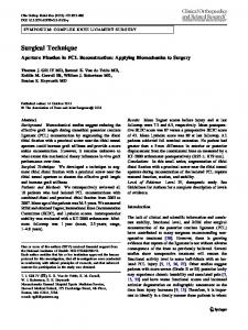
long axis along the frequency encoding direction, flipping frequency and phase encoding directions, and the use of turbo or fast spin-echo pulse sequences [4, 5, 6]. Recently, a new metal artifact reduction sequence (MARS) has been designed that reduces metal susceptibility artifacts (Fig. 1) [7]. To quantify its effect on signal intensity artifacts, the authors compared conventional and MARS modified spin-echo images of titanium and stainless steel hip prostheses in water.
399
Fig. 1 MARS pulse sequence. The modified sequence pulse diagram is identical to a conventional spin-echo sequence except for the addition of a new gradient, which is represented as the shaded areas. This gradient has the same direction and amplitude as the slice select gradient and is applied simultaneously with the frequency encoding gradient
Materials and methods Two phantoms were used, each consisting of a metal femoral hip joint replacement prosthesis (titanium/chromium-cobalt – Zimmer 6032-09; stainless steel – Thackray SD 548-13) immersed in a cylindrical glass container of tap water. Spin-echo coronal images, both with and without the MARS modification, were obtained for each phantom (Fig. 2) using a standard head coil in a 1.5 T GE Signa MR scanner (General Electric Medical Systems, Milwaukee, Wis.). Most imaging parameters for the T1-weighted conventional and MARS enhanced spin-echo sequences were identical for the two phantoms (TR 500 ms, TE 20 ms, FOV 30 cm, 3 mm slices, no gap, 256×256 matrix, 1 excitation, 2 acquisitions and frequency direction SI). The MARS sequence employed view angle tilting [1] with a tilt angle of 44°. The read-out bandwidth for the MARS sequence was ±62.5 kHz and ±15.625 kHz for the conventional spinecho sequence. The slice select gradient for the MARS sequence was increased by 20% relative to that used by the conventional spin-echo sequence. A commensurate decrease in slice select pulse width was applied. In all examinations, the long axis of the metal prosthesis was parallel to the frequency encoding direction. Analysis Quantitative measurement of signal intensity artifact on each axial image was performed using a Sun Sparc 10 Workstation and PV Wave software (Visual Numerics). In each image, it was possible to distinguish some signal from water which appeared not to be artifacted by the presence of the metal prosthesis. A global average of this normal signal was obtained for each data set. Volumetric analysis was then performed using a region of interest drawn around the entire phantom, including as much signal from the phantom as possible but excluding air outside the phantom. For each slice, a histogram of signal intensities was produced and the volumes of image corresponding to given ranges of signal intensity were calculated. Specifically, signal intensities ranging from 0 to a lower cutoff threshold were categorized as low signal artifact and signal intensities ranging from an upper cut-off threshold to the highest pixel intensity were categorized as high signal artifact. The lower cutoff threshold was varied from 50% to 80% of the normal signal mean and the upper cut-off threshold was varied from 120% to 150% of the normal signal mean. Using a water displacement technique, the volumes of the titanium and the stainless steel prostheses were measured to be about 35 and 30 ml respectively.
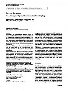
Fig. 2A, B Phantom images. A Coronal conventional spin-echo image and B coronal MARS modified spin-echo image of the stainless steel prosthesis immersed in water. The small dark spots are bands used to hold the prosthesis in position
Results For all thresholds, the MARS spin-echo sequence dramatically reduced the volume of artifact produced in both phantoms (Fig. 3). While the signal artifact volumes varied rapidly with lower and upper threshold cutoff intensity values, the ratio of artifact volumes with and without the MARS sequence was relatively independent of threshold. On images of the titanium prosthesis, the conventional spin-echo sequence demonstrated 50±16% (mean±standard deviation) more volume of low signal artifact and 202±8% more high signal artifact than the MARS modified sequence. Similarly, the conventional spin-echo sequence showed about 53±26% more low signal artifact and 162±9% more high signal artifact than the MARS sequence when imaging the stainless steel prosthesis. The signal-to-noise ratio of the MARS images was about 42% less than for the conventional images.
Discussion MRI requires a static, homogeneous magnetic field (Bo) and applied linear magnetic field gradients in order to produce planar slices which are free from geometric distortion. Metal substances disrupt the magnetic field homogeneity and consequently field gradients are no lon-
400
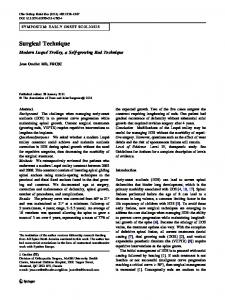
Fig. 3 Threshold analysis. Volumetric analysis comparing conventional and MARS modified T1-weighted images of both metal prostheses was implemented using various cut-off thresholds for low and high signal artifact. For all threshold levels, the reduction in artifact achieved using the MARS protocol was virtually the same
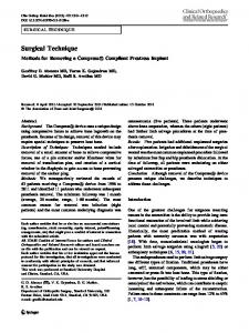
Fig. 4A, B Clinical examination of total hip arthroplasty. A T1-weighted conventional spin-echo MR image (TR 600, TE 15) and B MARS modified T1-weighted image of a 51year-old patient who had undergone total hip reconstruction 3 years previously. Note that the ballooning artifact around the superior aspect of the neck and head of the prosthesis seen on the conventional image is considerably reduced using MARS
ger linear. The overall effect is frequency mis-mapping along both the z- and x-axes which cause the typical image distortion and intensity artifacts commonly associated with metal. In 1988, Cho et al. [1] described a technique designed to correct field inhomogeneity and chemical shift artifacts. This technique involves the application of a second “compensation gradient”, of the same amplitude as the slice select gradient (Gz) and in the direction of Gz, concurrently with the read-out gradient (Gx). This effectively tilts the view angle according to the equation: (θ)=arctan(Gz/Gx) and corrects for the frequency shift along the x-axis. The effect of tilting the view angle can be appre-
ciated simply by realizing that all spins in the slice, at the time of slice selection, possess the same narrow frequency band determined by the frequency profile of the radiofrequency pulse [7]. When the slice select gradient is applied later during data acquisition, the spins will, neglecting frequency encoding and in the absence of motion, possess the same narrow frequency band. Therefore, the resulting image is devoid of in-plane distortion due to inhomogeneous magnetic fields. While image distortion is eliminated by view angle tilting, two other problems occur in the presence of inhomogeneous fields. First, the slice profile is altered in both thickness and curvature by field gradients near the
401
metal object. This effect can be reduced by using a stronger slice select gradient. Also, the slice rotation produced by view angle tilting causes significant image blurring which occurs over the entire image and not just the region near the metal object. This blurring may be reduced by increasing the strength of the frequency gradient. Both these corrections were included in the MARS pulse sequence. The results indicate that MARS was more effective in reducing high signal artifact than low signal artifact; however, the low signal artifact values were artificially increased by the void introduced by the prostheses themselves. Surprisingly, for titanium the low signal artifact volume at a 50% cut-off threshold was actually smaller than the prosthesis volume. This is likely due to slice distortions and partial volume effects. A larger metal artifact volume was produced by the stainless steel prosthesis than the titanium prosthesis. A key determinant of artifact size is the difference in magnetic susceptibility between the metal and the medium.
The major drawback of this new MR protocol is the decrease in the signal-to-noise ratio. However, images from clinical cases (Fig. 4) demonstrated that the reduction in signal did not compromise image interpretability [8, 9, 10]. The decrease in artifact would appear to potentially improve diagnostic ability. A qualitative clinical study [7] comparing image quality of MARS images versus conventional images of patients with spinal hardware found that MARS improved image quality over conventional T1-weighted spin-echo sequences. In conclusion, the new MARS sequence significantly reduces metal artifact on MR images of stainless steel and titanium femoral hip prostheses compared with conventional spin-echo sequences. This reduction in metal susceptibility artifact should increase the level of diagnostic information available. This improvement is attained without any increase in imaging time and is easily integrated with existing spin-echo and fast spin-echo sequences.
References 1. Cho ZH, Kim DJ, Kim YK. Total inhomogeneity correction including chemical shifts and susceptibility by view angle tilting. Med Phys 1988; 15:7–11. 2. Eustace S, Hernan J, Goldberg R, et al. A comparison of conventional spinecho and turbo spin-echo imaging of soft tissues adjacent to orthopedic hardware. AJR 1998; 170:455–458. 3. Eustace S, Goldberg R, Williamson D, et al. MR imaging of soft tissues adjacent to orthopaedic hardware: techniques to minimize susceptibility artefact. Clin Radiol 1997; 52:589–594.
4. Petersilge CA, Lewin JS, Duerk JL, Yoo JU, Ghaneyem AJ. Optimizing imaging parameters for MR evaluation of the spine with titanium pedicle screws. AJR 1996; 166:1213–1218. 5. Tartaglino LM, Flanders AE, Vinitski S, Friedman DP. Metallic artifacts on MR images of the postoperative spine: reduction with fast spin-echo techniques. Radiology 1994; 190:565–569. 6. Czervionke LF, Daniels DL, Wehrli FW, et al. Magnetic susceptibility artifacts in gradient-recalled echo MR imaging. Am J Neuroradiol 1988; 9:1149–1155. 7. McGowan AJ, MacKay AL, Xiang QS, Connell DG, Janzen DL, Munk PL. Reduction of image distortion in the presence of metal (abstract). Proceedings of the International Society for Magnetic Resonance in Medicine 5th Scientific Meeting and Exhibition, 1997:3.
8. Chang SD, Janzen DL, Sallomi DF, Munk PL. MRI of spinal hardware: comparison of conventional T1weighted sequence with a new metal artifact reduction sequence (abstract). Radiology 1998; 209(P):614. 9. Olsen RV, Munk PL, Lee MJ, Janzen DLMAL. Metal artifact reduction sequence: early clinical applications. Radiographics 2000; 20:699–712. 10. Olsen RV, Janzen DL, Sallomi DF, Munk PL, MacKay AL, Xiang Q-S. Clinical application of a new method of metal artifact suppression on MR imaging (abstract). Radiology 1998; 209(P):612.