775
The Journal of Toxicological Sciences (J. Toxicol. Sci.) Vol.41, No.6, 775-781, 2016
Letter
Quinone-mediated induction of cytochrome P450 1A1 in HepG2 cells through increased interaction of aryl hydrocarbon receptor with aryl hydrocarbon receptor nuclear translocator Yumi Abiko1, Fang-Yu Lin2, Hsinyu Lee3, Alvaro Puga4 and Yoshito Kumagai1 Faculty of Medicine, University of Tsukuba, Tsukuba, Ibaraki, Japan 2Winnoz Technology Inc., Taipei, Taiwan 3Department of Life Science, National Taiwan University, Taipei, Taiwan 4Center for Environmental Genetics, Department of Environmental Health, University of Cincinnati College of Medicine, Cincinnati, Ohio, USA 1
(Received July 14, 2016; Accepted September 21, 2016)
ABSTRACT — While it has long been believed that benzenes and naphthalenes are unable to activate the aryl hydrocarbon receptor (AhR) because they are poor ligands, we recently reported that these quinoid metabolites upregulated cytochrome P450 1A1 (CYP1A1) in Hepa1c1c7 cells (Abiko et al., 2015). In the current study, AhR activation, measured with a bioluminescence-based cell free assay, was induced by 1,2-naphthoquinone (1,2-NQ), a metabolite of naphthalene. Consistent with this, 1,4-benzoquinone (1,4-BQ), tert-butyl-1,4-BQ, and 1,4-NQ, as well as 1,2-NQ, all electrophilic mono- and bi-cyclic quinones, upregulated CYP1A1 mRNA and protein in HepG2 cells, whereas their parent aromatic hydrocarbons had little effect. Furthermore, immunofluorescence analysis confirmed that these quinones enhanced translocation of AhR to the nucleus. Key words: Electrophile, Aryl hydrocarbon receptor, Quinone, CYP1A1
INTRODUCTION A variety of mono- and bi-cyclic quinones, including 1,4-benzoquinone (1,4-BQ), 1,2-naphthoquinone (1,2NQ) and 1,4-NQ are found in diesel engine exhaust, burning coal, cigarette smoke (Eiguren-Fernandez et al., 2010; Batterman et al., 2012; Wallace, 1989) and as metabolites during exposure of experimental animals and cultured cells to their parent compounds (Wilson et al., 1996). Butylated hydroxyanisol (BHA) and tert-butyl-1,4-hydroquinone (TBHQ), used as food and cosmetic additives to inhibit oxidation of oils, are metabolized to tert-butyl1,4-benzoquinone (TBQ) (Verhagen et al., 1989; Snyder and Hedli, 1996). Quinones readily cause modification of proteins, often changing protein function and resulting in cytoprotective responses or toxicity (Kumagai et al., 2012). The aryl hydrocarbon receptor (AhR), known to regulate primarily phase-I xenobiotic metabolizing enzymes
such as cytochrome P450 1A1 (CYP1A1) and CYP1A2 (Nebert and Jones, 1989; Nebert et al., 1993), is a sensor for polycyclic aromatic hydrocarbons. These include 2,3,7,8-tetrachlorodibenzo-p-dioxin, biphenyls, polyhalogenated dibenzo-p-dioxins and dibenzofurans (Poland et al., 1976; Waller and McKinney, 1995). However, it was reported that non-classical AhR ligands, such as kynurenine, carbaryl, oltipraz and curcumin, could also induce CYP1A1 through AhR activation (Opitz et al., 2011; Quattrochi and Tukey, 1993; Ciolino et al., 1998; Denison et al., 1998; Miao et al., 2003). In addition, aromatic hydrocarbons with ring numbers of one or two, such as benzenes and naphthalenes, are thought to be poor AhR ligands, except for halogenated naphthalene, which showed ligand activity for AhR (Waller and McKinney, 1995). Surprisingly, our recent study showed that monoand bi-cyclic quinoid metabolites of benzenes and napthalenes could activate AhR, thereby upregulating Cyp1a1 mRNA expression in Hepa1c1c7 cells and in liver from
Correspondence: Yoshito Kumagai (E-mail:
[email protected]) Vol. 41 No. 6
776 Y. Abiko et al.
C57BL6 mice (Abiko et al., 2015). However, the contributions of quinones to AhR activation in human hepatic cell lines is still unclear. To address this issue, we examined AhR activation in human hepatoma HepG2 cells exposed to 1,4-BQ, TBQ, 1,2-NQ or 1,4-NQ as examples of electrophilic mono- and bi-cyclic quinones. MATERIALS AND METHODS Materials The electrophilic mono- and bi-cyclic quinones, TBQ (98% purity determined by GC), 1,4-BQ (98% purity determined by iodometric titration), 1,2-NQ (97.4% purity determined by HPLC) and 1,4-NQ (98% purity determined by GC), and TBHQ (98% purity determined by GC) were from Tokyo Chemical Industry (Tokyo, Japan). BHA (98% purity determined by GC) was from Nacalai (Kyoto, Japan). The AhR antagonist, 1-methyl-N-[2-methyl-4-[2-(2-methylphenyl)diazenyl]phenyl-1H-pyrazole5-carboxamide (CH223191), was from ChemBridge (San Diego, CA, USA). Anti-ARNT and anti-CYP1A1 (H-70) antibodies were from Santa Cruz Biotechnology (Santa Cruz, CA, USA). Anti-AhR and anti-actin (20-33) antibodies were from Enzo Life Sciences Inc. (Framingdale, NY, USA) and Sigma-Aldrich (St. Louis, MO, USA), respectively. All other reagents were of the highest grade available. Cell free assay for AhR activation The bioluminescence-based bioassay was performed as previously described (Wang et al., 2013) with modifications. Briefly, the coding region sequences of AhR (NM_001621), ARNT (NM_001668) and HSP90 (NM_007355) were amplified using cDNA derived from HEK293 cells. The NaNoLuc luciferase template was from pNL1.1 (Promega, Madison, WI, USA). PCR was performed using Phusion High Fidelity DNA Polymerase (ThermoFisher Scientific Inc., Waltham, MA, USA). AhR-NanoLuc was ligated into pcDNA5/TO, between the Kpn I and Xba I restriction enzyme sites, to generate pcDNA5/TO-AhR-NL. Hsp90 and ARNT were ligated to pBudCE4.1 (Invitrogen, Carlsbad, CA, USA) to generate pBudCE4.1-Hsp90-His and pBudCE4.1-ARNT-His. Lentivector pLKO_AS2.puro was purchased from the National RNAi Core Facility Platform, Academia Sinica (Taipei, Taiwan). His-tagged Hsp90 was inserted into pLKO_AS2. puro, between the Nhe I and Eco RI restriction enzyme sites, to generate pLKO_AS2.puro-Hsp90-His. His-tagged ARNT was inserted into pLKO_AS2.puro, between the Nhe I and Age I restriction enzyme sites, to generate pLKO_AS2-Arnt-His. T-REx 293 cells were transfectVol. 41 No. 6
ed with the pcDNA5/TO-AhR-NL and the pLKO_AS2. puro-Hsp90-His or with pLKO_AS2-Arnt-His using the Lipofectamine 2000 transfection reagent according to the manufacturer’s protocol. The cells were cultured in Dulbecco's Modified Eagle's Medium (DMEM, Invitrogen) supplemented with 10% fetal bovine serum (FBS), 5 mg/mL blasticidin, 200 mg/mL hygromycin B and 2 μg/mL puromycin to generate ANH cells or 10% FBS and 2 μg/mL puromycin to generate ARNT-His cells. The cells were washed with phosphate buffered saline (PBS), collected by trypsin-EDTA, and centrifuged 2000 rpm for 5 min. The pellet was washed with PBS twice and lysed into lysis buffer (1% Nonidet P-40 (NP-40), 10% glycerol, 200 mM Tris-HCl, pH 8.0 and 100 mM NaCl). After centrifugation at 21,920 × g for 15 min, supernatants were collected into new tubes, protease inhibitor cocktail (Merck, Darmstadt, Germany) was added and samples were stored at -80°C. Dynabeads His Tag Isolation and Pulldown (2 μL, Thermo Fisher Scientific Inc.) were washed with 1 mL lysis buffer and incubated with ARNT-His lysate (500 μL) and lysis buffer (500 μL) on a shaker for 10 min at 4°C. Then, the ARNT-His bound magnetic beads were washed with lysis buffer and resuspended in 1 mL lysis buffer. A 50 μL aliquot of the magnetic bead suspension was incubated with a mixture of 10-fold diluted ANH cells, which co-expressed nano-luminescence tagged-hAhR with his tagged-hHSP90, lysate (90 μL) and 1,2-NQ solution (10 μL) in a 96-well white plate on a shaker for 30 min at room temperature. The beads were washed with PBS after removing unbound Nano-luminescence taggedhAhR. Nano-luminescence was measured using the NanoGlo Luciferase Assay System (Promega) according to the manufacturer’s protocol. Cells and cell culture Human hepatoma HepG2 (RIKEN Cell Bank, Tsukuba, Japan) cells were cultivated in DMEM (Wako, Osaka, Japan) containing 10% FBS, antibiotics (100 U/mL penicillin and 100 μg/mL streptomycin, Wako) and 2 mM L-alanyl-L-glutamine (Invitrogen) at 37°C with 5% CO2. The cells were cultured in serum-free medium for 24 hr and then exposed to each of the compounds as described for individual experiments. The cells were exposed to lower concentrations of bi-cyclic compounds (1,2-NQ, 1,4-NQ, and naphthalene) than of mono-cyclic compounds (1,4-BQ, TBQ, benzene, and tert-buthyl-1,4-BQ) because bi-cyclic quinones showed stronger CYP1A1 induction than mono-cyclic compounds (Abiko et al., 2015).
777 Quinones activate aryl hydrocarbon receptor in HepG2 cells
Real-time reverse transcription polymerase chain reaction (RT-PCR) Total RNA was extracted with ISOGEN (Wako) according to the manufacturer’s protocols, and complementary DNAs (cDNAs) were synthesized from 2 μg total RNA using the High Capacity RNA-to-cDNATM Kit (Applied Biosystems, Grand Island, NY, USA). The resulting cDNA products were dissolved in a final volume of 200 μL and a 5 μL aliquot was used as a template for subsequent quantification by RT-PCR. PCR was conducted in duplicate in a total volume of 20 μL using SYBR Green PCR Master Mix (Applied Biosystems) and 0.1 μM primer. Primers were: CYP1A1 (5'-CCGGGACATCACAGACACCC-3' and 5'-CCAGCTCCAAAGAGGTCCAACA-3') and GAPDH (5'-GGGCTGCTTTTAACTCTGGTAA-3' and 5'-ATGGGTGGAATCATATTGGAAC-3'). Amplification was performed in an ABI 7500 Real Time PCR (Applied Biosystems); thermal cycling parameters were 50°C for 2 min, 95°C for 10 min, 40 cycles of 95°C for 15 sec and 60°C for 1 min. Relative differences among samples were determined by the ∆∆Ct method using each threshold cycle (Ct) values. The Ct values were normalized to values for GAPDH to determine ∆Ct values. Western blot analysis The cells were exposed to the chemicals, as indicated for each experiment, washed with ice-cold PBS, then collected by scraping into 2% sodium dodecyl sulfate (SDS) and heated at 95°C for 20 min. Protein concentrations of the resulting extracts were determined using the BCA Protein Assay (Pierce, Rockford, IL, USA). Each sample was mixed with a half-volume of SDS-polyacrylamide gel electrophoresis (SDS-PAGE) loading buffer (62.5 mM Tris-HCl, pH 6.8, 6% SDS, 24% glycerol, 15% 2-mercaptoethanol and 0.015% bromophenol blue) and incubated at 95°C for 5 min. The cellular proteins were then separated by SDS-PAGE and transferred to polyvinylidene difluoride membranes (Bio-Rad Laboratories, Hercules, CA, USA). The membranes were blocked with 5% skim milk in Tris-buffered saline with Tween 20 (TTBS, 20 mM Tris-HCl, pH 7.5, 150 mM NaCl and 0.1% Tween 20) and then incubated with anti-AhR, antiARNT, anti-CYP1A1 or anti-actin antibody as the primary antibody. To detect immunoreactive proteins, we used a horseradish peroxidase (HRP) conjugated secondary antibody and an enhanced chemiluminescence system (Chemi-Lumi One; Nacalai). Representative blots are shown from three independent experiments. Immunofluorescence Cells were seeded on 10 mm glass coverslips, fixed
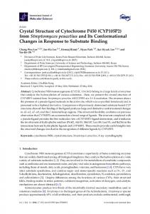
with 4% paraformaldehyde in PBS for 20 min, permeabilized with 0.1% Triton X-100 for 20 min, blocked with 5% bovine serum albumin for 30 min and incubated with anti-AhR antibody at 4°C overnight. To detect immunoreactive proteins, we used an Alexa 488 labeled secondary antibody (ThermoFisher Scientific Inc.) and affixed coverslips with mounting medium containing 4',6-diamino-2-phenylindole (DAPI). Micrographs were taken using a Zeiss Axio microscope (Carl Zeiss Microscopy, Thornwood, NY, USA). Immunoprecipitation Cells were washed twice with PBS and lysed for 30 min on ice with 100 μL radio-immunoprecipitation assay (RIPA) buffer (50 mM Tris-HCl, pH 8.0, 150 mM NaCl, 0.1% SDS, 0.5% deoxycholic acid and 1% NP-40) containing 1% protease inhibitor cocktail (SigmaAldrich). Samples were then centrifuged at 13,000 × g for 10 min to obtain cell lysates. The cell lysates (400 μg) were incubated with 2 mg Protein A Sepharose CL-4B (GE Healthcare, Little Chalfont, England) crosslinked with 2 μg anti-AhR or anti-ARNT antibody, prepared according to the manufacturer’s instructions. The beads were centrifuged at 13,000 × g for 1 min, washed 4 times with 1 mL ice-cold RIPA buffer and then boiled in SDSPAGE loading buffer. Proteins were detected by western blot analysis as described above. Statistical analysis Statistical significance was assessed by one-way ANOVA followed by Holm-Sidak’s multiple comparisons post hoc test using Prism (GraphPad software, San Diego, CA, USA) and p < 0.05 was considered significant. RESULTS AND DISCUSSION To assess AhR activation by 1,2-NQ, we used a cell free assay developed by Wang et al. (2013) to detect dioxin and dioxin-like compounds, also AhR activators. Exposure to 3-methylcholanthrene (1 nM) caused an approximately 1.3-fold activation, compared with control, of the luminescence signal (data not shown), suggesting that the assay could sense low concentrations of AhR activator. As shown in Fig. 1, 1,2-NQ significantly enhanced the luminescence signal (at 0.1 nM, an approximately 1.38-fold increase), indicating that 1,2-NQ was clearly able to increase the interaction between AhR and ARNT, which is involved in substantial upregulation of the CYP1A1 gene. Consistent with this concept, TBQ, 1,4-BQ, 1,2-NQ and 1,4NQ — but not BHA, TBHQ, naphthalene, benzene, and Vol. 41 No. 6
778 Y. Abiko et al.
1,4-hydroquinone — induced CYP1A1 gene expression (Figs. 2A and B). These observations were also confirmed by CYP1A1/2 protein levels detected by western blotting with antibody against CYP1A1 (Figs. 2C-G). Pretreatment with the AhR inhibitor CH223191 markedly blocked 1,2-NQ- and TBQ-mediated upregulation of CYP1A1 (Fig. 3), suggesting participation of AhR in the induction of CYP1A1. Once ligands bind to AhR, the transcriptional factor translocates into the nucleus and interacts with ARNT to bind XRE (Henry and Gasiewicz, 2003; Harper et al., 1991; Fujisawa-Sehara et al., 1987; Soshilov and Denison, 2008). As expected, treatments with TBQ, 1,4-BQ, 1,4-NQ and 1,2-NQ caused translocation of AhR into the nucleus in HepG2 cells as detected by immunostaining (Fig. 4A). Immunoprecipitation analysis with anti-AhR or ARNT antibody and western blotting with these antibodies showed that TBQ enhanced the interaction of AhR with ARNT (Fig. 4B). Our previous study demonstrated that 1,2-NQ enhanced the interaction of AhR with ARNT in Hepa1c1c7 cells (Abiko et al., 2015). 1,2-NQ was a stronger AhR activator than TBQ. If an interaction between AhR and ARNT occurred dur-
Fig. 1.
1,2-NQ-mediated AhR activation detected by a cellfree assay. A lysate from ARNT-His overexpressing cells was incubated with magnetic beads for 10 min at 4°C. The ARNT-His bound magnetic beads were washed and resuspended in 1 mL buffer, then the beads were incubated for 30 min at room temperature with a mixture of lysate from ANH cells and the indicated concentrations of 1,2-NQ. After the reaction, the beads were wash with PBS, then nano-luminescence was measured using the Nano-Glo Luciferase Assay System. Each value is the mean ± S.E. of three replicates. *p < 0.05 and **p < 0.01 compared with 0 nM.
Fig. 2.
Increased CYP1A1 mRNA and protein expression in HepG2 cells exposed to quinones. HepG2 cells were treated with (A) TBQ, 1,4-BQ, 1,2-NQ or 1,4-NQ; or (B) BHA, TBHQ, naphthalene (NP), benzene, 1,4-hydroquinone (HQ) or 3-methylcholanthrene (MC) for 4 hr. The total RNA was converted to cDNA, and quantitative PCR was performed using a CYP1A1 primer. Each value is the mean ± S.E. of three replicates. The cells were treated with 1,2-NQ (C), 1,4-NQ (D), 1,4-BQ (E) and TBQ (F) for 8 hr. (G) HepG2 cells were treated with TBQ, TBHQ, BHA, 1,2-NQ, NP or MC for 8 hr and cell lysates were analyzed by western blotting with the indicated antibodies.
Vol. 41 No. 6
779 Quinones activate aryl hydrocarbon receptor in HepG2 cells
Fig. 3.
Quinone mediated increases in CYP1A1 levels in the presence of an AhR antagonist. HepG2 cells were treated with 10 μM CH223191 for 1 hr and then with 50 μM TBQ, 20 μM 1,2-NQ or 10 nM 2,3,7,8-tetrachlorodibenzo-p-dioxin (TCDD) for 4 hr. The total RNA was converted to cDNA and quantitative PCR was performed using a CYP1A1 primer. Each value is the mean ± S.E. of three replicates.
Fig. 4.
Quinone mediated translocation of AhR and enhancement of the interaction between AhR and ARNT. (A) HepG2 cells were treated with quinones for 60 min and immunofluorescence staining was performed with an anti-AhR antibody and DAPI. (B) HepG2 cells were pretreated with 10 μM MG132 for 3 hr to inhibit protein degradation before TBQ or 3-methylcholanthrene (MC) treatment for 30 min. The association of AhR with ARNT in whole cell lysates was detected by immunoprecipitation using the indicated antibodies.
ing exposure of cells to TBQ, 1,2-NQ would also enhance the interaction. We noted that the luminescence intensity, corresponding to AhR activation as evaluated in the cell-free assay, was increased at 0.1 nM but then decreased at 100 nM 1,2NQ (Fig. 1). This suggested that quinones such as 1,2-NQ appear to have dual effects on AhR activation. Consistent with this, while CYP1A protein levels were upregulated at 50 μM TBQ at both timepoints (8 hr and 24 hr) and interaction of AhR with ARNT was enhanced at 25 and 50 μM TBQ, this induction was suppressed and the interaction was dissociated at 100 μM TBQ (Figs. 4 and 5). Similar results were described previously in Hepa1c1c7 cells (Abiko et al., 2015). We reported that activation of the Akt/CREB signaling pathway in response to MeHg, which is an environmental electrophile and easily bind to thiol groups on the cellular proteins, in SH-SY5Y cells followed a bell-shaped curve (Unoki et al., 2016). This was attributed to Akt activation through S-mercuration of its negative regulator, PTEN, at lower MeHg concentrations and suppression of Akt/CREB signal transduction through S-mercuration of CREB at higher concentrations (Unoki et al., 2016). Considering these data together, we speculate that quinones similarly have a bell-shaped effect on AhR/XRE signaling. Further study will be required to elucidate the contributions of covalent modification of AhR related proteins to this non-linear response of the receptor to quinones. In contrast, TBQ exposure caused a concentration dependent HO-1 induction, detectable at even 100 μM, through activation of the Nrf2/antioxidant responsive element (ARE) pathway (Fig. 5). This process regulates phase-II drug metabolizing enzymes and phaseIII transporters to detoxify and excrete electrophiles (Itoh et al., 1999a, 1999b; Maher et al., 2007). This suggested that different thresholds exist for the AhR/XRE and Nrf2/ ARE pathways. Our study indicated that quinones are activators for
Fig. 5.
TBQ mediated induction of CYP1A1 and HO-1. HepG2 cells were treated with TBQ and 3-methylcholanthrene (MC) for 8 or 24 hr and western blotting was performed with the indicated antibodies.
Vol. 41 No. 6
780 Y. Abiko et al.
AhR, inducing upregulation of CYP1A1 in HepG2 cells as well as in Hepa1c1c7 cells (Abiko et al., 2015). Of interest, quinones themselves, formed from benzenes and naphthalenes by phase-I xenobiotic metabolizing enzymes, were able to activate not only CYP1A1 and phase-II xenobiotic metabolizing enzymes but also phaseIII transporters responsible for detoxification and excretion of quinones and regulated by Nrf2/ARE pathways. This observation implied that there are multiple adaptive responses to reactive chemicals such as quinones, through activation of various signal transduction pathways. ACKOWLEDGMENTS We thank Chan Yayun, Master’s Program, National Taiwan University, for her help to perform the cell free assay detecting AhR activation by 1,2-NQ. This work was supported by a Grant-in-Aid (#JP25220103 to Y.K. and #JP15K18906 to Y.A.) for scientific research from the Ministry of Education, Culture, Sports, Science, and Technology of Japan. Work done at the University of Cincinnati was supported by NIH grants R01 ES006273 and R01 ES010807. Conflict of interest---- The authors declare that there is no conflict of interest. REFERENCES Abiko, Y., Puga, A. and Kumagai, Y. (2015): Covalent binding of quinones activates the Ah receptor in Hepa1c1c7 cells. J. Toxicol. Sci., 40, 873-886. Batterman, S., Chin, J.Y., Jia, C., Godwin, C., Parker, E., Robins, T., Max, P. and Lewis, T. (2012): Sources, concentrations, and risks of naphthalene in indoor and outdoor air. Indoor Air, 22, 266-278. Ciolino, H.P., Daschner, P.J., Wang, T.T. and Yeh, G.C. (1998): Effect of curcumin on the aryl hydrocarbon receptor and cytochrome P450 1A1 in MCF-7 human breast carcinoma cells. Biochem. Pharmacol., 56, 197-206. Denison, M.S., Phelan, D., Winter, G.M. and Ziccardi, M.H. (1998): Carbaryl, a carbamate insecticide, is a ligand for the hepatic Ah (dioxin) receptor. Toxicol. Appl. Pharmacol., 152, 406-414. Eiguren-Fernandez, A., Shinyashiki, M., Schmitz, D.A., DiStefano, E., Hinds, W., Kumagai, Y., Cho, A.K. and Froines, J.R. (2010): Redox and electrophilic properties of vapor- and particle-phase components of ambient aerosols. Environ. Res., 110, 207-212. Fujisawa-Sehara, A., Sogawa, K., Yamane, M. and Fujii-Kuriyama, Y. (1987): Characterization of xenobiotic responsive elements upstream from the drug-metabolizing cytochrome P-450c gene: a similarity to glucocorticoid regulatory elements. Nucl. Acids Res., 15, 4179-4191. Harper, P.A., Prokipcak, R.D., Bush, L.E., Golas, C.L. and Okey, A.B. (1991): Detection and characterization of the Ah receptor for 2,3,7,8-tetrachlorodibenzo-p-dioxin in the human colon adenocarcinoma cell line LS180. Arch. Biochem. Biophys., 290, Vol. 41 No. 6
27-36. Henry, E.C. and Gasiewicz, T.A. (2003): Agonist but not antagonist ligands induce conformational change in the mouse aryl hydrocarbon receptor as detected by partial proteolysis. Mol. Pharmacol., 63, 392-400. Itoh, K., Ishii, T., Wakabayashi, N. and Yamamoto, M. (1999a): Regulatory mechanisms of cellular response to oxidative stress. Free Radic. Res., 31, 319-324. Itoh, K., Wakabayashi, N., Katoh, Y., Ishii, T., Igarashi, K., Engel, J.D. and Yamamoto, M. (1999b): Keap1 represses nuclear activation of antioxidant responsive elements by Nrf2 through binding to the amino-terminal Neh2 domain. Genes Dev., 13, 76-86. Kumagai, Y., Shinkai, Y., Miura, T. and Cho, A.K. (2012): The chemical biology of naphthoquinones and its environmental implications. Annu. Rev. Pharmacol. Toxicol., 52, 221-247. Maher, J.M., Dieter, M.Z., Aleksunes, L.M., Slitt, A.L., Guo, G., Tanaka, Y., Scheffer, G.L., Chan, J.Y., Manautou, J.E., Chen, Y., Dalton, T.P., Yamamoto, M. and Klaassen, C.D. (2007): Oxidative and electrophilic stress induces multidrug resistance-associated protein transporters via the nuclear factor-E2-related factor-2 transcriptional pathway. Hepatology, 46, 1597-1610. Miao, W., Hu, L., Kandouz, M. and Batist, G. (2003): Oltipraz is a bifunctional inducer activating both phase I and phase II drugmetabolizing enzymes via the xenobiotic responsive element. Mol. Pharmacol., 64, 346-354. Nebert, D.W. and Jones, J.E. (1989): Regulation of the mammalian cytochrome P1-450 (CYP1A1) gene. Int. J. Biochem., 21, 243252. Nebert, D.W., Puga, A. and Vasiliou, V. (1993): Role of the Ah receptor and the dioxin-inducible [Ah] gene battery in toxicity, cancer, and signal transduction. Ann. N. Y. Acad. Sci., 685, 624640. Opitz, C.A., Litzenburger, U.M., Sahm, F., Ott, M., Tritschler, I., Trump, S., Schumacher, T., Jestaedt, L., Schrenk, D., Weller, M., Jugold, M., Guillemin, G.J., Miller, C.L., Lutz, C., Radlwimmer, B., Lehmann, I., von Deimling, A., Wick, W. and Platten, M. (2011): An endogenous tumour-promoting ligand of the human aryl hydrocarbon receptor. Nature, 478, 197-203. Poland, A., Glover, E. and Kende, A.S. (1976): Stereospecific, high affinity binding of 2,3,7,8-tetrachlorodibenzo-p-dioxin by hepatic cytosol. Evidence that the binding species is receptor for induction of aryl hydrocarbon hydroxylase. J. Biol. Chem., 251, 4936-4946. Quattrochi, L.C. and Tukey, R.H. (1993): Nuclear uptake of the Ah (dioxin) receptor in response to omeprazole: transcriptional activation of the human CYP1A1 gene. Mol. Pharmacol., 43, 504508. Snyder, R. and Hedli, C.C. (1996): An overview of benzene metabolism. Environ. Health Perspect., 104 Suppl 6, 1165-1171. Soshilov, A. and Denison, M.S. (2008): Role of the Per/Arnt/Sim domains in ligand-dependent transformation of the aryl hydrocarbon receptor. J. Biol. Chem., 283, 32995-33005. Unoki, T., Abiko, Y., Toyama, T., Uehara, T., Tsuboi, K., Nishida, M., Kaji, T. and Kumagai, Y. (2016): Methylmercury, an environmental electrophile capable of activation and disruption of the Akt/CREB/Bcl-2 signal transduction pathway in SH-SY5Y cells. Sci. Rep., 6, 28944. Verhagen, H., Furnee, C., Schutte, B., Hermans, R.J., Bosman, F.T., Blijham, G.H., ten Hoor, F., Henderson, P.T. and Kleinjans, J.C. (1989): Butylated hydroxyanisole-induced alterations in cell kinetic parameters in rat forestomach in relation to its oxidative cytochrome P-450-mediated metabolism. Carcinogenesis, 10,
781 Quinones activate aryl hydrocarbon receptor in HepG2 cells 1947-1951. Wallace, L.A. (1989): The exposure of the general population to benzene. Cell Biol. Toxicol., 5, 297-314. Waller, C.L. and McKinney, J.D. (1995): Three-dimensional quantitative structure-activity relationships of dioxins and dioxin-like compounds: model validation and Ah receptor characterization. Chem. Res. Toxicol., 8, 847-858.
Wang, B.J., Wu, P.Y., Lu, Y.C., Chang, C.H., Lin, Y.C., Tsai, T.C., Hsu, M.C. and Lee, H. (2013): Establishment of a cell-free bioassay for detecting dioxin-like compounds. Toxicol. Mech. Methods, 23, 464-470. Wilson, A.S., Davis, C.D., Williams, D.P., Buckpitt, A.R., Pirmohamed, M. and Park, B.K. (1996): Characterisation of the toxic metabolite(s) of naphthalene. Toxicology, 114, 233-242.
Vol. 41 No. 6