®
Genes, Genomes and Genomics ©2008 Global Science Books
Rapid and Efficient Method for the Extraction of Fungal and Oomycetes Genomic DNA Ajay Kumar Mishra • Kamal Sharma • Raj Shekhar Misra* Central Tuber Crops Research Institute, Thiruvananthapuram, Kerala, 695017 India Corresponding author: *
[email protected]
ABSTRACT An improved protocol for the isolation of high-quality DNA from fungi is described. The method involves inactivating proteins by SDS/proteinase K and precipitating polysaccharides in the presence of high salt. Further purification is based on differential solubility of DNA and high molecular weight polysaccharides in aqueous media. The procedure does not use the toxic and potentially hazardous chemical such as phenol and as many as 50 samples can be processed per day. The purity of isolated genomic DNA was confirmed by means of spectrophotometer analysis (A260/280 ratio of 1.80-1.96), indicated a minimal presence of contaminating metabolites. The method yielded 0.55-0.92 µg DNA mg-1 freeze dried mycelia, when tested on three fungal species Fusarium solani, Colletotrichum capsici, and Rhizoctonia solani and two oomycetes Phytophthora colocasiae and Pythium aphanidermatum. The DNA was completely digested with EcoRI and HindIII. PCR-based technique such as random amplification of polymorphic DNA (RAPD), showed the DNA’s compatibility with downstream applications.
_____________________________________________________________________________________________________________ Keywords: DNA extraction, fungi, RAPD, restriction enzyme digestion
INTRODUCTION The development of PCR-based molecular marker techniques have become the method of choice for plant pathologists to characterize pathogens to understand or elucidate the principles or factors underlying molecular evolution, population genetics, plant fungus interactions, or pathogen evolution at molecular level (Leung et al. 1993; Milgroom and Fry 1997). A prerequisite for taking advantage of this method is the ability to isolate genomic DNA of superior quality and quantity for analyzing through PCR, restriction enzyme digestion and subsequent Southern blot hybridization. The lack of an easy DNA extraction method suited to conditions commonly encountered in many laboratories, which are not equipped to handle toxic organic substances such as phenol, commonly used during DNA extraction (Graham et al. 1994; Zhang et al. 1996). Thus the method must limit the use of toxic chemicals as much as possible and should potentially remove inhibitory materials, i.e. polysaccharides, proteins, etc., which limit the sensitivity of different reactions in which isolated DNA is applied (Sambrook et al. 1989). The major challenge for isolation of DNA of good quality from fungi lies in breaking the rigid cell walls, as they are often resistant to traditional DNA extraction procedures (Fredricks et al. 2005). Fungal nucleases and high polysaccharide contents add to the difficulties in isolating DNA from filamentous fungi (Zhang et al. 1996; Muller et al. 1998). The methods include use of SDS, CTAB or proteinase K (Wilson 1990), SDS lysis (Syn and Swarup 2000), lysozyme or SDS (Flamm et al. 1984), highspeed cell disruption (Muller et al. 1998) and bead-vortexing or SDS lysis (Sambrook and Russel 2001). Additionally, some give poor yields of DNA, as cell walls or capsule are difficult to lyse (Muller et al. 1998). The method that we developed for extracting DNA from filamentous fungi does not use organic reagents such as phenol, chloroform or isoamyl alcohol but yields DNA of high quality and purity suitable for restriction digestion and PCR-based analysis. The procedure involves pulverReceived: 1 July, 2008. Accepted: 28 July, 2008.
izing the mycelia by vortexing with glass beads as this reduces the shearing of DNA (Hollick et al. 2004), inactivating proteins by SDS or proteinase K and precipitating polysaccharides and proteins in the presence of high salt (Kim et al. 1990). The removal of polysaccharides and other contaminating hydrates is based on the differential solubility of DNA versus the high-molecular weight polysaccharides in aqueous media (Rozman and Komel 1994). MATERIALS AND METHODS Biological materials This study included three fungal species (Fusarium solani, Colletotrichum capsici and Rhizactonia solani) and two oomycetes (Phytophthora colocasiae and Pythium aphanidermatum). Mycelium was grown on potato dextrose agar medium (PDA; 250 g L-1 potato, 20 g L-1 dextrose and 20 g L-1 agar) at 25°C for 1 week. Erlenmeyer flasks (250 mL) containing 100 mL of potato dextrose broth were inoculated with two 1-cm discs removed from actively growing cultures. The cultures were placed on a rotary shaker (100 rpm) and incubated at 27°C for 4-5 d. Mycelia were harvested by filtration through cheesecloth, blotted dry with sterile paper towels and used immediately for DNA extraction.
Extraction of genomic DNA 50 mg of mycelia was ground to a fine powder in 2 ml of extraction buffer (Tris-HCl, 100 mM; EDTA, 10 mM; NaCl, 1M; SDS, 1%; proteinase K, 0.05 mg ml-1; pH 8.0) and 10% (v/v) glass beads. Samples were vortexed and incubated at 65°C for 30 min. After incubation, samples were centrifuged at 10,000 × g for 15 min and supernatant was transferred to a fresh tube. To the supernatant, 150 µl of 3 M guanidine hydrochloride was added and incubated at 20°C for 10 min. Samples were centrifuged at 10,000 × g for 10 min. After centrifugation supernatant was transferred to a fresh tube, and an equal volume of isopropanol was added. Samples were incubated at -20°C for 1 h. The samples were then centrifuged for 10 min at 10,000 × g and 70% ethanol was added and
Techniques Paper
Genes, Genomes and Genomics 2 (1), 57-59 ©2008 Global Science Books
centrifuged once more for 10 min at 10,000 × g. The pellet obtained were air dried and dissolved in 50 µl of TE buffer (Tris-HCl, 10 mM, pH 8; EDTA, 1 mM). The nucleic acid dissolved in TE buffer was treated with 3 µl of RNase A (10 mg ml-1), incubated at 37°C, and stored at -20°C until use.
Measurement of amount and purity of DNA DNA was quantified by spectrophotometric measurement of UV absorption at 260 nm (Shimadzu UV-260). DNA was also quantified by means of 0.8% agarose gel electrophoresis followed by ethidium bromide visualization using 1-kbp DNA ladder (Fermentas) as DNA size marker. DNA purity was determined by calculating the absorbance ratio A260/280. DNA purity was further confirmed by digestion with EcoRI and HindIII, incubating reaction mixture at 37°C for 3 h and followed by 0.8% agarose gel electrophoresis.
RAPD analysis of DNA Suitability of the isolated DNA for downstream analysis was assessed by RAPD. PCR reactions for RAPD analysis were performed in a 25 µl volume containing 200 ng of genomic DNA, 2.5 µl of 10 x reaction buffers, 4 µl of 25 mM MgCl2, 2 µl of 2.5 mM dNTPs, 200 ng screened random decamer oligonucleotide primer OPA-1 (5′-CAGGCCCTTC-3′). Amplification was performed in a thermal cycler (Techne progene). The reactions were heated in an initial step of 94°C for 2 min and subjected to 4 cycles of the following program 94°C for 30 s, 37°C for 1 min, 72°C for 2 min and further to 40 cycles of same program. After the last cycle, the temperature was maintained at 72°C for 8 min. Amplified DNA was electrophoresed in a 1.4% agarose gel containing 0.5 mg ml-1 ethidium bromide and photographed on UV transilluminator.
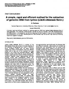
Fig. 1 Agarose gel electrophoresis of extracted genomic DNA. Lanes 1, 2, 3, 4, 5: DNA from C. capsici, P. colocasiae, F. solani, P. aphanidermatum and R. solani, respectively. Lane M: molecular weight marker (1 kb).
RESULTS AND DISCUSSION The present extraction method yielded good quantity and quality of pure, high molecular weight DNA (Table 1, Fig. 1). The present extraction method yielded 0.55-0.92 µg DNA mg-1 freeze dried mycelium (Table 1). The A260/280 ratio ranged from 1.80-1.96, showing that DNA was of high purity (Table 1). The purity of DNA was further confirmed by means of digestion by two restriction enzymes EcoRI and HindIII and monitoring the banding profile of digested DNA (Fig. 2). This indicated that isolated DNA was amenable for further downstream applications. Successful and reproducible amplification was obtained by using screened random decamer primer OPA-1 (5′-CAGGCCCTTC-3′) in RAPD analysis for all the samples (Fig. 3). The purpose of this study was to improve and simplify the currently available DNA extraction method for filamentous fungi (van Burik et al. 1998; Haugland et al. 1999; Alsamarrari and Schmidt 2000) for use by researchers in developing countries. The DNA extraction protocol described here is rapid and technically easy for preparing high molecular weight DNA without any ultra centrifugation or column purification steps. The key step of modified protocol is the use of guanidine hydrochloride and proteinase K to remove protein completely. This step precludes the use of organic solvent (e.g., phenol, chloroform) that is normally used during DNA isolation in other methods, making it suitable for use in areas where facilities for handling such chemical do not exist. These simple modifications make the method faster and provide another effective and rapid nucTable 1 DNA yield by following present DNA extraction protocol. Biological species Mean yield of DNA (µg per mg A260/280 mycelium) ± SD by presented DNA extraction protocol C. capsici 0.55 ± 0.05 1.82 ± 0.03 P. colocasiae 0.76 ± 0.05 1.87 ± 0.06 F. solani 0.90 ± 0.2 1.89 ± 0.05 P. aphanidermatum 0.72 ± 0.1 1.90 ± 0.02 R. solani 0.79 ± 0.03 1.88 ± 0.03
Fig. 2 Restriction digestion analysis of DNA prepared by using present method. Lanes 1, 2, 3, 4, 5: double digest with EcoRI and HindIII of DNA products from C. capsici, P. colocasiae, F. solani, P. aphanidermatum and R. solani, respectively. Lane M: molecular weight marker (1 kb).
Fig. 3 RAPD assay conducted with the DNA prepared by means of the present method. Lanes 1, 2, 3, 4, 5: amplification product from C. capsici, P. colocasiae, F. solani, P. aphanidermatum and R. solani respectively. Lane M: molecular weight marker (1 kb).
Fungal and oomycete genomic DNA extraction. Mishra et al.
leic acid extraction procedure. The protocol was successfully extended to recover DNA from Sclerotium rolfsii and Trichoderma, where the genomic DNA isolation is difficult due to high mucilage and protein content (Cassago et al. 2002) and thus we believe that it can easily be adapted to other filamentous fungi. The amount and quality of the DNA obtained by this method were suitable for PCR amplification, restriction digestion and further downstream analysis. This DNA extraction method has several advantages: a) the number of DNA extraction steps is minimal and thus large number of samples can be processed in parallel without any contamination risk and loss of DNA, b) it is efficient and cost effective because as little as 50 mg of mycelium gives good DNA yields with small amount of chemicals and little equipment. The method described here is rapid, reliable and use of this procedure resulting in extraction of DNA sufficiently pure for PCR and other molecular assay. ACKNOWLEDGEMENTS The funding provided for conducting the research work by the Indian Council of Agricultural Research, New Delhi, is gratefully acknowledged. The authors thank Director, Central Tuber Crops Research Institute, Thiruvananthapuram, for providing the infrastructure facilities.
REFERENCES Al-Samarrai TH, Schmid J (2000) A simple method for extraction of fungal genomic DNA. Letters in Applied Microbiology 30, 53-57 Cassago A, Panepucci RA, Baião A-M T, Henrique-Silva F (2002) Cellophane based mini-prep method for DNA extraction from the filamentous fungus Trichoderma reesei. BMC Microbiology 2, 14 Flamm RK, Hinrichs DK, Thomashow MF (1984) Introduction of pAM beta1 into Listeria monocytogenes by congugation and homology between native L. monocytogegenes plasmids. Journal of Bacteriology 159, 214-221 Fredricks DN, Smith C, Meier (2005) A Comparison of six DNA extraction methods for recovery of Fungal DNA as assessed by quantitative PCR. Jour-
nal of Clinical Microbiology 43, 5122-5128 Graham GC, Mayer P, Henry RJ (1994) A simplified method for preparation of fungal genomic DNA for PCR and RAPD analysis. Biotechniques 16, 4850 Haugland RA, Heckman JL, Wymer LJ (1999) Evaluation of different methods for the extraction of DNA from fungal conidia by quantitative competitive PCR analysis. Journal of Microbiological Methods 37, 165-168 Hollick PS, Taylor RJ, Mccomb JA, Dixon KW, Krauss SL (2004) Optimisation of DNA extraction for AFLP analysis of mycorrhizal fungi of terrestrial orchids Caladeniinae and Drakaeinae. Plant Molecular Biology Reporter 22, 307a-307h Kim WK, Mauthe W, Hausner G, Klassen GR (1990) Isolation of high molecular weight DNA and double-stranded RNAs from fungi. Canadian Journal of Botany 68, 1898-1902 Leung H, Nelson RJ, Leach JE (1993) Population structure of plant pathogenic fungi and bacteria. Advances in Plant Pathology 10, 157-205 Milgroom MG, Fry WE (1997) Contributions of population genetics to plant disease epidemiology and management. Advances in Botanical Research 24, 1-30 Muller FM, Werner KE, Kasai M, Francesconi (1998) A Rapid extraction of genomic DNA from medically important yeasts and filamentous fungi by high-speed cell disruption. Journal of Clinical Microbiology 36, 1625-1629 Rozman D, Komel R (1994) Isolation of genomic DNA from filamentous fungi with high glucan level. Biotechniques 16, 382-384 Sambrook J, Fritsch EF, Maniatis TA (1989) Molecular Cloning: A Laboratory Manual (2nd Edn), Cold Spring Harbor Laboratory, Cold Spring Harbor, New York, pp 745 Sambrook J, Russel DW (2001) Rapid isolation of yeast DNA. In: Sambrook J, Russell DW (Eds) Molecular Cloning: A Laboratory Manual (2nd Edn), Cold Spring Harbor Laboratory, New York, pp 631-632 Syn CK, Swarup S (2000) A scalable protocol for isolation of large-sized genomic DNA within an hour from several bacteria. Annals of Biochemistry 278, 86-90 van Burik JA, Schreckhise RW, White TC, Bowden RA, Myerson D (1998) Comparison of six extraction techniques for isolation of DNA from filamentous fungi. Medical Mycology 36, 299-303 Wilson K (1990) Preparation of genomic DNA from bacteria. In: Ausubel FM, Brent R (Eds) Current Protocols in Molecular Biology, Greene Publishing Association and Wiley Interscience, New York, pp 241-245 Zhang D, Yang Y, Castelbury LA, Cerniglia CE (1996) A method for large scale isolation of high transformation efficiency genomic DNA. FEMS Microbiology Letters 145, 261-265