probe sequence segments, i.e., POL, MAC, P-M, and pan-16S, used in this study are also indicated. kindly provided) by Gary Olsen of the University of Illinois.
APPLIED AND ENVIRONMENTAL MICROBIOLOGY, Aug. 1992, p. 2571-2578
Vol. 58, No. 8
0099-2240/92/082571-08$02.00/0 Copyright C 1992, American Society for Microbiology
Rapid In Situ Hybridization Technique Using 16S rRNA Segments for Detecting and Differentiating the Closely Related Gram-Positive Organisms Bacillus polymyxa and Bacillus macerans REBECCA J. JURTSHUK,l* MARK BLICK,1 JOEL BRESSER,l GEORGE E. FOX,2 AND PETER JURTSHUK, JR.3 Molecular Analysis Inc., 8000 El Rio, Houston, Texas 77054, 1 and Department of Biochemical and Biophysical Sciences2 and Department of Biology, 3 University of Houston, Houston, Texas 77204 Received 7 October 1991/Accepted 1 May 1992
A rapid, sensitive, inexpensive in situ hybridization technique, using 30-mer 16S rRNA probes,
can
specifically differentiate two closely related Bacillus spp., B. polymyxa and B. macerans. The 16S rRNA probes were labeled with a rhodamine derivative (Texas Red), and quantitative fluorescence measurements were made on individual bacterial cells. The microscopic fields analyzed were selected by phase-contrast microscopy, and the fluorescence imaging analyses were performed on 16 to 67 individual cells. The labeled 16S rRNA probe, POL, whose sequence was a 100%o match with B. polymyxa 16S rRNA but only a 60%v match with B. macerans 16S rRNA, gave quantitative fluorescence ratio measurements that were 34.8-fold higher for B. polymyxa cells than for B. macerans cells. Conversely, the labeled probe, MAC, which matched B. polymyxa 16S rRNA in 86.6% of its positions and B. macerans 16S rRNA in 100% of its positions, gave quantitative fluorescence measurements that were 59.3-fold higher in B. macerans cells than in B. polymyxa cells. Control probes, whose 16S rRNA sequence segment (P-M) was present in both B. polymyxa and B. macerans as well as a panprokaryotic probe (16S), having a 100o match with all known bacteria, hybridized equally well with both
organisms. These latter hybridizations generated very high fluorescence signals, but their comparative fluorescence ratios (the differences between two organisms) were low. The control paneukaryotic probe (28S), which had less than 30% identity for both B. macerans and B. polymyxa, did not hybridize with either organism. Traditional laboratory methods for identifying bacteria have relied heavily on the use of either simple or differential microbiological tests such as the Gram stain, the growth on common (or exotic) diverse substrates, and subsequently the specific use of biochemical tests. In recent years a strong trend in microbial systematics for the use of direct molecular characterizations, e.g., DNA base composition, DNA-DNA hybridization, gene sequencing, etc., to define taxa has emerged. These methods have revolutionized studies in bacterial evolution but, initially at least, did comparatively little for the practitioner who simply needed to identify a particular organism in a given environment or specimen. Thus, in the face of important breakthroughs, the arduous processes of isolating pure cultures and conducting the traditional procedures for culture identification have continued largely unabated. Over the last 10 years, there has been a considerable expenditure of effort to develop methodologies for rapid identification of microorganisms by using a variety of novel procedures. One of the most promising of these has been the use of DNA probes that are specific for an organism's 16S rRNA (5, 8, 9, 12). Indeed, well-received tests with this approach are already on the market. The advantages of using the 16S rRNA methodology are several, not the least of which is the huge data base of sequence information that is currently available. Almost 1,000 16S rRNA sequences have already been documented for bacterial phylogenetic studies, and this data base includes a large variety of bacteria, including pathogens. From a technical *
perspective the 16S rRNA sequence segments can serve as molecular probes, which will allow for species identification of any bacterium. In other instances, 16S rRNA probe hybridizations can give a more general grouping identity (at the genus or family level), depending on the application required. Probably the most important applications of the rapid in situ (RIS) hybridization technique would be for its potential (i) to identify pathogenic bacteria, in blood, stool, or sputum or from other tissue samples; (ii) to identify bacteria directly in environmental applications, even in field studies, by using a portable scanning fluorometer; and (iii) to monitor microbial changes in samples that would occur during a bioremediation procedure to determine which organisms are still present (or absent). The following study seeks to demonstrate that an alternative detection technique, i.e., RIS hybridization, can be used for the specific identification of bacteria, and the improved RIS technique described herein makes for a faster and more convenient assay that can be routinely used in a laboratory setting. In this study, a particular 16S rRNA probe assay which allows for the highly specific detection and differentiation of two closely related Bacillus species, B. polymyxa and B. macerans, is described. As noted in Table 1, these two Bacillus strains are closely related species which are difficult to distinguish by traditional microbiological methods. The 16S rRNA probes used for selectively identifying both B. polymyxa and B. macerans were designed from sequence information obtained from an ongoing study on the 16S rRNA phylogenetic characterization of bacteria of the genus Bacillus (2, 5a, 15, 17). The RIS hybridization approach offers several advan-
Corresponding author. 2571
2572
APPL. ENvIRON. MICROBIOL.
JURTSHUK ET AL.
TABLE 1. Phenotypic characteristics of B. polymyxa and B. maceransa Value, description, or presence
Characteristic
Common Spore morphology (group II)
B. polymyxa
Oval, swollen cells, central
B. macerans
Oval, swollen cells, subtermi-
terminal
Nitrogen fixation Facultative anaerobe Growth on: Glucose Biotin
Differentiating G+C (for type strain) (mol% [range]) Oxidase reaction Voges-Proskauer reaction (2,3-butanediol formation) Dihydroxyacetone formation Growth at 45°C Casein hydrolysis Growth on thiamine
+b
nal terminal
+
+b +
Acid/gasb
Acid/gasb
+
+
45.6 (44.0-48.0)
53.2 (50.0-54.0)
-
+
+ +
-
+
+
+ a A very close relationship exists among B. polymyxa, B. macerans, and B. circulans. (These species form a closely related cluster analogous to the B. subtilis group which includes B. subtilis, B. lichenformnis, and B. pumulus [4, 7, 10, 12, 16]). b Identifying biochemical trait found for both B. polymyxa and B. macerans relative to other Bacillus species.
tages. First, the detection of a specific segment of the 16S rRNA sequence, within intact individual cells, will allow the identification of a specific bacterial cell, even within a mixed heterogeneous population. Second, in situ hybridization inherently exhibits a greater sensitivity for the detection of specific bacterial cells than the other nucleic acid detection methods currently in use. Third, this 16S rRNA probe procedure does not require extraction of nucleic acids from isolated bacterial cells that are to be tested. Fourth, the RIS hybridization technique allows for the use of multiple probes, with different labels. Multiple probes of this type will permit the simultaneous detection of several target sites even within an individual bacterial cell. For example, in a mixed population of bacterial cells, one can in theory identify an Escherichia coli cell with one specific fluorescent probe; with a different tag on another probe, one might identify the drug resistance nucleotide segment, and with yet a third tag and different probe, one might identify a toxinproducing gene. Finally, because RIS hybridization analysis can be performed on single cells, minimal sample sizes of bacteria (104 cells) can be used, and only microquantities of reagents are normally required. The specific implementation described herein is faster than the protocol described previ-
ously (1).
MATERUILS AND METHODS rRNA probe design and synthesis. A data base of 16S rRNA sequences was assembled from the literature on a VaxServer 3300 (Digital Equipment Corporation) running the VMS operating system. Sequence alignment and editing were facilitated by the use of a specialized editor, SEQEDT, designed for rRNA studies. This editor was developed (and
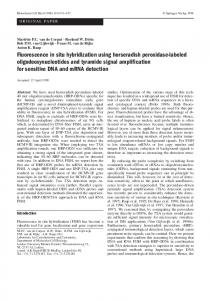
FIG. 1. Schematic diagram of the Bacillus subtilis 16S rRNA. The molecule is drawn so as to indicate the usual secondary structure elements (8). The locations of the four 30-mer 16S rRNA probe sequence segments, i.e., POL, MAC, P-M, and pan-16S, used in this study are also indicated.
kindly provided) by Gary Olsen of the University of Illinois in Urbana-Champaign, Urbana-Champaign. Assembly of the data base was facilitated (i) by the collection of small subunit rRNA sequences which is published semiannually (13) and (ii) by electronic access to the recently established rRNA data base project at the University of Illinois (14). The selection of specific Bacillus 16S rRNA probes was especially facilitated by recently published sequences from B. polymyxa and B. macerans (15) as well as from a data base collection of 16S rRNA sequences gathered from a variety of specific Bacillus species (2, 5a, 15, 17). A total of four 16S rRNA probes were designed by examination of the above data base (Fig. 1). Two of these probes (POL and MAC) were specifically designed to selectively distinguish B. macerans from B. polymyxa cells. Probe POL provided a 100% sequence match with B. polymyxa 16S rRNA but had a reduced affinity (60% match) for B. macerans, while probe MAC was 100% specific for a sequence match with B. macerans but exhibited a reduced affinity (86.6%) for B. polymyxa. These probes were selected from two different regions of the 16S rRNA molecule. In addition, two other positive control bacterial 16S rRNA probes were constructed. One of these probes, designated P-M, possessed a 16S rRNA segment that had a 100% sequence match with both B. polymyxa and B. macerans, while the other control probe, designated 16S, was designed from a highly conserved region, and thus its sequence segment would presumably interact with any bacterial 16S rRNA. The 16S rRNA sequence segments for the four probes used are shown in Table 2, with their corresponding
VOL. 58, 1992
RIS HYBRIDIZATION USING 16S rRNA SEGMENTS
2573
TABLE 2. Sequences of the rRNA probes used for the in situ hybridization studies monitoring fluorescence incorporation in B. polymyxa and B. maceransa Probe designation
% Homology
Sequence B.
POL MAC P-M 16S 28S
ATACQCGATAT
TCTGCATQGGA
TACAACGGGAAGCGAAGTAGTGATATGGAG CCAGGGAAGAACGTCTTCTAGAGTAACTGC
AAACTCCTACGGGAGGCAGCAGTAAGGAAT CGCCOGGTGAAATACCACTACTCTGATCGTT
polymyxa 100 86.6 100 100