CASE STUDY
Rapidly progressive intracranial artery stenosis in primary antiphospholipid syndrome € rmel2, Achim Berthele1, Sascha Prothmann3 & Christian L. Seifert1, Markus C. Kowarik1, Klaus Thu 1 Silke Wunderlich €nchen, Munich, Germany Neurologische Klinik und Poliklinik, Klinikum rechts der Isar der Technischen Universit€ at Mu €r Rheumatologie, Klinikum rechts der Isar der Technischen Universit€ €nchen, Munich, Germany Abteilung fu at Mu 3 €r Neuroradiologie, Klinikum rechts der Isar der Technischen Universit€ €nchen, Munich, Germany Abteilung fu at Mu 1 2
Correspondence Christian L. Seifert, Neurologische Klinik und Poliklinik, Klinikum rechts der Isar der €nchen, Ismaninger Technische Universit€at Mu €nchen, Germany. Str. 22, 81675 Mu Tel: +49 89 4140 4601; Fax: +49 89 4140 4675; E-mail:
[email protected] Funding Information There was no funding for the conduction of this case study.
Abstract Antiphospholipid antibody syndrome (APS) is usually a disease of young adults. In elderly stroke patients APS was not associated with progressive intracerebral stenosis in the past. Here, we report a 65-year-old patient who presented with recurrent ischemic strokes associated with progressive stenosis of the right middle cerebral artery. Antiphospholipid antibodies were detected and treatment with plasma exchange, tapered steroids, and anticoagulants was successful. This case demonstrates that APS should be considered also in elderly stroke patients. This is of particular relevance since APS confers a significant risk to angioplasty and stenting procedures which therefore should be avoided in APS.
Received: 2 March 2015; Revised: 14 April 2015; Accepted: 23 April 2015 Annals of Clinical and Translational Neurology 2015; 2(7): 780–782 doi: 10.1002/acn3.215
Introduction Cerebral infarcts associated with antiphospholipid antibody syndrome (APS) predominantly occur in younger adults. In a recent retrospective study the mean age of patients with APS and stroke was 37.6 years with a predominance of females (80%); usually multiple infarcts in subcortical and cortical areas with an asymmetric distribution occurred.1 In elderly patients APS is rarely diagnosed. In a subgroup of subjects with severe carotid stenosis, antiphospholipid antibodies (aPL) were found to be elevated.2 One larger series of 23 patients with aPL could show various abnormalities like vasculitis-like findings and extracranial as well as intracranial stem branch occlusions or stenoses.3 However, APS in association with intracerebral arterial stenosis have rarely been reported.4
Case Study Here, we report the case of a 65-year-old male Caucasian with repeated ischemic strokes and APS. The patient 780
gave written informed consent to publish his medical history. On his first admission, the patient presented with a leftsided hemiparesis. Cerebral MRI (Fig. 1A) disclosed subcortical, periventricular and cortical infarcts in the right middle cerebral artery (MCA) territory. Ultrasound revealed a stenosis of the right MCA (maximal flow velocity 370 cm/ sec), and bilateral plaques in the internal carotid arteries. Treatment with acetylsalicylic acid (ACA), clopidogrel and atorvastatin was started.5 Transthoracic echocardiography and holter-ECG were without pathological findings. However, 3 days after admission, the patient suffered from a myocardial ischemia, which made stenting of the left anterior descending coronary artery necessary. Thus, the MCA stenosis was considered to be atherosclerotic as well. Treatment with ACA, clopidogrel, and atorvastatin was continued and the patient transferred to a rehabilitation unit. Four weeks later the patient had recovered well except slight deficits in fine motor skills of the left hand. Ultrasound of the right MCA showed a decrease in the maximal flow velocity (200 cm/sec).
ª 2015 The Authors. Annals of Clinical and Translational Neurology published by Wiley Periodicals, Inc on behalf of American Neurological Association. This is an open access article under the terms of the Creative Commons Attribution-NonCommercial-NoDerivs License, which permits use and distribution in any medium, provided the original work is properly cited, the use is non-commercial and no modifications or adaptations are made.
C. L. Seifert et al.
Intracranial Artery Stenosis
valve endocarditis in transesophageal echocardiography, which is frequently reported in APS.7 Dermatological screening was unremarkable, the patient showed no livedo reticularis. Not fulfilling the American College of Rheumatology criteria for systemic lupus, the patient was finally diagnosed with a primary APS. Slightly elevated creatinine of 1.7 mg/dL without significant proteinuria was compatible with this disease.8 Ultrasound of the kidneys was unremarkable with normal perfusion parameters. To rule out a solid tumor, abdominal sonography as well as X-ray of the chest were carried out and were unremarkable. Treatment with phenprocoumon 3–4.5 mg per day was initiated and heparin was stopped as soon as the patient reached therapeutic INR (international normalized ratio) of 2–3, in addition to the platelet inhibitors (which were as well necessary for the coronary stent), and the steroid was slowly tapered. No further cardiovascular events occurred in the following 4 months, and the intracranial MCA stenosis remained stable.
Conclusion
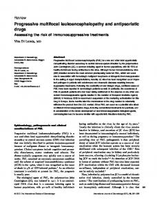
Figure 1. (A) Cerebral MR-angiography and diffusion-weighted MRI showing a stenosis of the right middle cerebral artery; on the left at first presentation, on the right 6 weeks later (second hospitalization). (B) Corresponding to (A) infarcts in diffusion-weighted (DWI)- MRI scans. (C) Antiphospholipid antibody titer in the serum before and after plasma exchange (PE): aCL, anticardiolipin antibodies; Antib2GBI, beta-2 glycoprotein antibodies.
Ten days later the patient relapsed with a left-sided hemiparesis and slurred speech. Transcranial ultrasound revealed deterioration of the MCA stenosis. Flow velocity in the stenosis could not be determined, and the poststenotic flow was significantly reduced. The corresponding MRI scan (Fig. 1B) also showed a progress of the stenosis of the right MCA and new ischemic infarcts in the MCA territory. Because of this rapid progression additional laboratory tests were ordered, and revealed a very high aPL titer (Fig. 1C), positive lupus anticoagulant, positive ANA (antinuclear antibodies)-titers of 1:240 (without ENA [extractable nuclear antigens] and without double-stranded DNA antibodies) and slightly reduced C3 complement (87 mg/ dL). The patient showed no thrombocytopenia or hemolytic anemia. APS was suspected and plasma exchange (PE) as well as concomitant glucocorticoids (prednisolone 60 mg) were initiated according to data on catastrophic APS (CAPS)6 to reduce aPL titers (Fig. 1C) and to prevent occlusion of the MCA. In addition, treatment with intravenous heparin at a dose of 900 units/h was started. The further diagnostic workup revealed signs of an aseptic mitral
To the best of our knowledge, this is the first case report of an elderly male patient (>65 years) presenting with a rapidly progressive intracranial stenosis in the context of highly elevated aPL. The patient responded favorably to PE and high-dose glucocorticoids. The case seems of particular relevance, since the clinical course with a rapid progression of an intracranial artery stenosis within 6 weeks despite therapy with ACA and clopidogrel is unusual. Although intracranial stenting is a possible escape strategy in refractory cases of intracranial stenosis, the complication of a rapid in-stent thrombosis has to be considered under these circumstances: It is well known that stenting procedures in APS are associated with a higher periinterventional risk for neurological complications9 including in-stent thrombosis. Knowledge on cerebral manifestations in elderly patients with APS is scarce, even in the context of its worst manifestation, the CAPS. CAPS is a rare complication encountered in a subset of patients with APS, and characterized by the development of multiple blood clots in multiple organs leading to rapid general organ failure. Interestingly, in the large group of patients (n = 282) described in the CAPS-registry cohort (http://www.med.ub.es/MIMMUN/FORUM/CAPS.HTM), 15 patients were aged ≥65 years, and only five of them presented with cerebral infarcts, whereof four are described as microinfarcts in the registry.10 The occurrence of cerebral stenosis is not exactly reported in this cohort, it is not clear whether inflammatory or preexisting atherosclerotic processes or even an acute cerebrovascular event could trigger the development of such a stenosis in APS.11 One
ª 2015 The Authors. Annals of Clinical and Translational Neurology published by Wiley Periodicals, Inc on behalf of American Neurological Association.
781
Intracranial Artery Stenosis
could also argue that repetitive embolic events might have been causal for the progression in the stenosis of our patient, but this is unlikely because no ischemia in other cerebral vascular beds had been observed in this patient. Moreover, after immunomodulatory treatment of his APS, he did clinically well, and the intracranial stenosis improved. Further studies are warranted to clarify whether APS could play a major role in rapid progressive intracranial stenosis in elderly patients. This seems of particular clinical relevance since stenting should be avoided in these patients as far as possible.
Conflict of Interest None declared. References 1. Zhu DS, Fu J, Zhang Y, et al. Neurological antiphospholipid syndrome: clinical, neuroimaging, and pathological characteristics. J Neurol Sci 2014;346:138–144. 2. Marcucci R, Sofi F, Fedi S, et al. Thrombophilic risk factors in patients with severe carotid atherosclerosis. J Thromb Haemost 2005;3:502–507. 3. Provenzale JM, Barboriak DP, Allen NB, et al. Antiphospholipid antibodies: findings on arteriography. AJNR Am J Neuroradiol 1998;19:611–616.
782
C. L. Seifert et al.
4. Wong M, Sangle S, Jan W, et al. Intracerebral arterial stenosis with neurological events associated with antiphospholipid syndrome. Rheumatology 2005;44:948–949. 5. Chimowitz MI, Lynn MJ, Derdeyn CP, et al. Stenting versus aggressive medical therapy for intracranial arterial stenosis. N Engl J Med 2011;365:993–1003. 6. Espinosa G, Cervera R. Antiphospholipid syndrome: frequency, main causes and risk factors of mortality. Nat Rev Rheumatol 2010;6:296–300. 7. Turiel M, Sarzi-Puttini P, Peretti R, et al. Five-year follow-up by transesophageal echocardiographic studies in primary antiphospholipid syndrome. Am J Cardiol 2005;96:574–579. 8. Gigante A, Gasperini ML, Cianci R, et al. Antiphospholipid antibodies and renal involvement. Am J Nephrol 2009;30:405–412. 9. Steiner S, Sadushi R, Bartok A, et al. Association of periprocedural neurological deficit in carotid stenting with increased anticardiolipin antibodies. Thromb Res 2009;123:827–831. 10. Cervera R. CAPS registry. Lupus 2012;21: 755–757. 11. Sherer Y, Shoenfeld Y. Antiphospholipid antibodies: are they pro-atherogenic or an epiphenomenon of atherosclerosis? Immunobiology 2003;207: 13–16.
ª 2015 The Authors. Annals of Clinical and Translational Neurology published by Wiley Periodicals, Inc on behalf of American Neurological Association.