RESEARCH ARTICLE 1461
Development 137, 1461-1471 (2010) doi:10.1242/dev.042911 © 2010. Published by The Company of Biologists Ltd
RBPjk-dependent Notch signaling regulates mesenchymal progenitor cell proliferation and differentiation during skeletal development Yufeng Dong1,*, Alana M. Jesse1,*, Anat Kohn1, Lea M. Gunnell1, Tasuku Honjo2, Michael J. Zuscik1, Regis J. O’Keefe1 and Matthew J. Hilton1,†
SUMMARY The Notch pathway has recently been implicated in mesenchymal progenitor cell (MPC) differentiation from bone marrow-derived progenitors. However, whether Notch regulates MPC differentiation in an RBPjk-dependent manner, specifies a particular MPC cell fate, regulates MPC proliferation and differentiation during early skeletal development or controls specific Notch target genes to regulate these processes remains unclear. To determine the exact role and mode of action for the Notch pathway in MPCs during skeletal development, we analyzed tissue-specific loss-of-function (Prx1Cre; Rbpjkf/f), gain-of-function (Prx1Cre; Rosa-NICDf/+) and RBPjk-independent Notch gain-of-function (Prx1Cre; Rosa-NICDf/+; Rbpjkf/f) mice for defects in MPC proliferation and differentiation. These data demonstrate for the first time that the RBPjk-dependent Notch signaling pathway is a crucial regulator of MPC proliferation and differentiation during skeletal development. Our study also implicates the Notch pathway as a general suppressor of MPC differentiation that does not bias lineage allocation. Finally, Hes1 was identified as an RBPjk-dependent Notch target gene important for MPC maintenance and the suppression of in vitro chondrogenesis.
INTRODUCTION The Notch pathway is an evolutionarily conserved signaling system that regulates cell proliferation, differentiation, cell fate determination and stem/progenitor cell self-renewal in both embryonic and adult organs (Artavanis-Tsakonas et al., 1999; Chiba, 2006; Lai, 2004). In mammals, Notch signaling is initiated when a ligand (jagged 1, jagged 2, delta-like 1 or delta-like 4) binds to a single-pass transmembrane cell surface Notch receptor (Notch1-4) on the neighboring cell. Ligand-receptor interactions ultimately induce cleavage of the Notch receptors via the gamma-secretase complex, releasing the Notch intracellular domain (NICD) to activate both canonical and non-canonical Notch signaling mechanisms. During canonical Notch signaling, the NICD translocates to the nucleus and binds the transcriptional repressor, RBPjk, converting it into an activator and inducing the expression of downstream target genes (Kopan and Ilagan, 2009; Lai, 2002). Some of the most well-defined canonical, or RBPjk-dependent, Notch target genes include specific members of the Hes/Hey family of basic helix-loop-helix transcription factors: Hes1, Hes5, Hes7, Hey1, Hey2 and HeyL, which are thought to mediate much of Notch function (Iso et al., 2003; Iso et al., 2001). The NICD also has the ability to function independently of RBPjk via non-canonical interactions with proteins and complexes of other signaling pathways (Martinez Arias et al., 2002). RBPjk1
Department of Orthopaedics and Rehabilitation, Center for Musculoskeletal Research, University of Rochester School of Medicine and Dentistry, Rochester, NY 14642, USA. 2Immunology and Genomic Medicine, Kyoto University Graduate School of Medicine, Kyoto 606-8501, Japan. *These authors contributed equally to this work Author for correspondence (
[email protected])
†
Accepted 25 February 2010
independent Notch signaling was originally identified in Drosophila when specific Notch alleles were shown to induce Deltex-dependent Notch signaling events and the repression of neural cell fate (Matsuno et al., 1997; Ramain et al., 2001). Mammalian NICDs also bind to many other signaling molecules and transcriptional regulators such as SMADs 1, 5 and 8 (Dahlqvist et al., 2003; Itoh et al., 2004), SMADs 2 and 3 (Blokzijl et al., 2003), Lef1 (Ross and Kadesch, 2001), b-catenin (Hayward et al., 2005), dishevelled (Dvl) (Axelrod et al., 1996), IkB kinase a subunit (IKKa) (Vacca et al., 2006; Vilimas et al., 2007), the p65/p50 subunits of the NF-kB complex (Wang et al., 2001) and deltex (Axelrod et al., 1996; Diederich et al., 1994; Hayward et al., 2005; Ramain et al., 2001; Vacca et al., 2006; Vilimas et al., 2007), potentially regulating these pathways in an RBPjk-independent manner. Recently, compelling data for RBPjk-independent Notch signaling in the skin was demonstrated by comparing conditional genetic mouse models removing core components of the gamma-secretase complex (presinilin 1/2; PS1/PS2), the Notch receptors (Notch1/2; N1/N2) or the RBPjk transcriptional repressor (Demehri et al., 2009). These reports highlight the complexity of RBPjk-dependent and -independent Notch signaling in various cell systems and the reasons why detailed genetic studies are required to identify the mechanisms by which Notch regulates context-dependent cell proliferation and differentiation. The Notch pathway is important in regulating stem/progenitor cell self-renewal, proliferation and differentiation from various tissues including: hematopoietic (Hadland et al., 2004; Kunisato et al., 2003; Robert-Moreno et al., 2005; Stier et al., 2002; VarnumFinney et al., 2000), neural (de la Pompa et al., 1997; Hatakeyama et al., 2004; Hitoshi et al., 2002; Ohtsuka et al., 1999), pancreatic (Apelqvist et al., 1999; Jensen et al., 2000) and intestinal (Fre et al., 2005). However, genetic investigations into Notch regulation of
DEVELOPMENT
KEY WORDS: RBPjk, Notch, Hes1, Mesenchymal progenitor cell, Limb development, Mouse
1462 RESEARCH ARTICLE
MATERIALS AND METHODS Mouse strains
All mouse strains including Rosa-NICD, Rbpjk and Prx1Cre are as previously described (Han et al., 2002; Logan et al., 2002; Murtaugh et al., 2003). Prx1Cre mice were obtained from the Jackson Laboratory (Bar Harbor, ME, USA), whereas Rosa-NICD and Rbpjk floxed mice were generous gifts from Dr Douglas Melton (Harvard University, MA, USA) and Dr Tasuku Honjo (Kyoto Graduate School of Medicine, Japan), respectively. Analyses of mouse embryos
Embryonic tissues were harvested at E11.0-E12.5 in PBS, fixed in 10% neutral buffered formalin, then processed and embedded in paraffin prior to sectioning at 4 m. Standard Alcian Blue/Orange G (AB/OG) staining was performed in order to analyze tissue architecture and cartilage composition of the limb-buds. In situ hybridization was performed as described previously (Hilton et al., 2005; Hilton et al., 2007; Hilton et al., 2008), using 35 S-labeled riboprobes. Unpublished riboprobes were generated from the following cDNA clones: Sox9 (4165469), Agc1 (5345931), Hes1 (10469606), Hey1 (9792713), jagged 1 (Jag1) (10699187), delta-like 1(Dll1; 10698888), and delta-like 4 (Dll4; 7492828), available from Open Biosystems or ATCC. The Gfp probe was generated by cloning the enhanced Gfp coding sequence into the pGEM-T Easy vector. Notch1, Notch2, Notch3, Fgf8 and Fgf10 cDNAs and riboprobes are as described (Bellusci et al., 1997; Crossley and Martin, 1995; Mitsiadis et al., 1995). For BrdU immunostaining analyses, pregnant females were injected with BrdU at 0.1 mg/g body weight 2 hours prior to harvest. BrdU detection was performed on paraffin sections using a kit from Invitrogen as per manufacturer’s instructions. Analyses of apoptotic MPCs were performed using both anticleaved caspase 3 immunostaining (Cell Signaling) and TUNEL staining (Roche Cell Death In Situ Kit) on limb-bud sections according to the manufacturers’ instructions. Wholemount skeletal staining of embryos was performed as previously described (Hilton et al., 2005; McLeod, 1980). Limb-bud MPC and C3H10T1/2 cell culture
Limb-bud derived MPCs were isolated from E11.5 CD1 mouse embryos as previously described (Zhang et al., 2004). For chondrogenic differentiation, MPCs were seeded in micromass (1 ⫻ 105 cells in 10 l) for 1.5 hours before adding standard media, media containing the gamma secretase and Notch inhibitor N-(3,5-difluorophenylacetyl-L-alanyl)]-S-phenylglycine t-butyl ester (DAPT; 1 M; Calbiochem), or media containing Hes1/control shRNA lentivirus. Cells were cultured for a time-course of 6 hours, at 3, 5 and 7 days prior to harvest for cartilage staining (ABH/OG) or total RNA isolations. C3H10T1/2 micromass chondrogenic cultures were treated and harvested in a similar manner (Denker et al., 1999; Haas and Tuan, 1999). Limb-budderived MPCs were also cultured in monolayer for 21 days and treated with either osteogenic (10 nM dexamethasone; 50 M ascorbic acid; 10 mM bglycerolphosphate) or adipogenic medium (Millipore) in the presence and absence of DAPT. Fixed MPCs were stained for osteoblastic differentiation using an Alkaline Phosphatase stain (nitro blue tetrazolium chloride/5bromo-4-chloro-3-indolyhosphate P-toluidine salt) or adipogenic differentiation using an Oil Red-O staining solution (Millipore). Total RNA was isolated from monolayer cultures at day 21 for use in real-time RT-PCR analyses. Real-time RT-PCR
Real-time RT-PCR was performed on RNA extracted from both embryonic limb-bud tissues or micromass cultures as previously described (Dong et al., 2005). Primer sequences are available upon request. Mouse-specific PCR primers were developed for: Sox9, Runx2, Col2a1, Agc1, Col1a1, alkaline phosphatase (Ap), Oc, Pparg, jagged 1, jagged 2, delta-like 1, delta-like 3, delta-like 4, Notch1, Notch2, Notch3, Notch4, Hes1, Hes3, Hes5, Hes7, Hey1, Hey2, HeyL and cyclin D1. Gene expression was normalized to bactin expression levels and then normalized to control samples. Western blot analyses
Total protein was isolated from either whole mouse limb-bud tissue or cultured limb-bud-derived MPCs in the presence and absence of DAPT (1 m). Protein samples (~100 g) from each isolation were subsequently separated on 10% SDS-polyacrylamide and subjected to standard western
DEVELOPMENT
mesenchymal stem/progenitor cells (MPCs) have gone largely unexplored. This has probably been owing to the fact that many mutant mice for crucial components of the Notch pathway are embryonic-lethal prior to MPC differentiation and overt skeletogenesis (Donoviel et al., 1999; Hamada et al., 1999; Hrabe de Angelis et al., 1997; Oka et al., 1995; Swiatek et al., 1994; Xue et al., 1999). Recently, work by Hilton et al. (Hilton et al., 2008) used conditional genetic approaches to demonstrate that ‘upstream’ components of the Notch pathway (PS1/PS2 and N1/N2) were crucial in regulating osteoblastic differentiation of bone marrowderived MPCs in mice. In vitro studies using human bone marrowderived MPCs (hMPCs) have also implicated Notch signaling and jagged 1 in the induction of hMPC proliferation and a contextdependent regulation of differentiation (Oldershaw et al., 2008; Vujovic et al., 2007). Collectively, these data suggest that various Notch signaling components might play crucial and differential roles in MPC self-renewal, proliferation and differentiation. MPCs are not only multi-potent cells found in various adult tissues including bone, cartilage, muscle and fat, but are also prominent cells populating the embryonic limb-bud, which are required for chondrogenic and osteogenic differentiation during skeletal development. During limb development, various cell types migrate into the limb field, a great many of which include MPCs of the lateral plate mesoderm, which begin rapid proliferation driving growth of the limb. During early phases of limb development, the apical ectodermal ridge (AER) maintains an apical zone of cells in an undifferentiated state primarily via fibroblast growth factor (Fgf) signals (Niswander et al., 1994). As MPCs withdraw from control of the AER, many of the cells are recruited into mesenchymal condensations that undergo chondrogenic and osteogenic differentiation, giving rise to the skeletogenic elements. The MPC cell fate decision to undergo chondrogenesis versus osteogenesis is dictated by a balance between both the expression and activity of Sox9 and Runx2 (Akiyama et al., 2005). Sox9 is expressed in all MPCs, maintaining their ability to undergo both chondrogenesis and osteogenesis (Akiyama et al., 2002; Bi et al., 1999). MPCs localized near the center of condensations strongly upregulate Sox9 expression and activity, inducing important downstream targets such as type II collagen (Col2a1) and aggrecan (Agc1; Acan – Mouse Genome Informatics), and therefore undergo the process of chondrogenesis. Meanwhile, Sox9 ‘low’-expressing cells around the borders of the condensations upregulate Runx2 expression and activity, thereby inducing osteogenic molecules such as type I collagen (Col1a1), osterix (Osx) and osteocalcin (Oc), and suppressing the chondrogenic fate (Akiyama et al., 2005; Drissi et al., 2000). Although Sox9 and Runx2 are crucial regulators of MPC differentiation, very few additional molecules have been identified to control MPC differentiation at or upstream of Sox9 and/or Runx2. To determine the exact role and mode of action for the Notch pathway in MPCs we analyzed tissue-specific loss-of-function (Prx1Cre; Rbpjkf/f), gain-of-function (Prx1Cre; Rosa-NICDf/+) and RBPjk-independent Notch gain-of-function (Prx1Cre; RosaNICDf/+; Rbpjkf/f) mice for defects in MPC proliferation and differentiation during early limb development. Additionally, we used limb-bud tissue sections, in vitro limb-bud-derived MPC cultures and C3H10T1/2 mesenchymal cell cultures to identify the expression of specific Notch pathway components, to determine if the Notch pathway was a generic regulator of MPC differentiation and to identify potential Notch target gene(s) regulating MPC differentiation.
Development 137 (9)
Notch signaling maintains and expands MPCs
RESEARCH ARTICLE 1463
blotting procedures. NICD1 and NICD2 cleaved proteins were detected using the bTAN 20 (Notch1) and C651.6DdHN (Notch2) primary antibodies (0.4 g/ml) and then further probed with appropriate secondary antibody (1:3000). Anti-b-actin antibody (Sigma) was used as a control for equal protein loading.
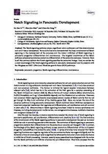
Fig. 1. Notch pathway component expression during MPC differentiation and chondrogenesis both in vitro and in vivo. (A-C)Real-time RT-PCR gene expression analyses of the Notch ligands (A), Notch receptors (B) and the Hes/Hey family of RBPjk-dependent Notch target genes (C). (Da-j) In situ hybridization for the indicated Notch pathway components at E11.5 (Da-Dh) and for N2 (Di) and Hes1 (Dj) at E12.0. Insets show high magnification of N1- and Dll4-associated endothelial cells surrounding vascular canals on alternative sections. (E)Western blot for cleaved Notch2 protein (NICD2) isolated from limbbud-derived MPCs (LB-MPCs) cultured in the presence and absence of DAPT or from whole limb-bud (WLB). The y-axis of graphs is the relative gene expression normalized to b-actin represented in arbitrary units. d, days; hr, hours.
overlapping regions where Jag1 expression is concentrated (Fig. 1Da,g). By E12.0-E12.5, most of the Notch pathway components are difficult to detect via in situ hybridization. Interestingly, only Notch2 and Hes1 expression were maintained in limb-bud MPCs surrounding chondrogenic condensations, but showed significant downregulation within the condensations themselves (Fig. 1Di,j, black and white contours), whereas components like Hey1 maintained a more ubiquitous expression pattern (data not shown). To determine which Notch receptor is active in the limb-bud mesenchyme, we isolated total protein from cultured MPCs in the presence and absence of the gamma-secretase and Notch inhibitor, DAPT. We also directly isolated protein from wild-type E11.5 whole limb-bud tissue and performed western blot analyses for each sample using Notch1 and Notch2 antibodies to detect the active (NICD) form of the receptor. Western blot analyses revealed that Notch2 was the prominent receptor activated in E11.5 limb-bud MPCs, and that DAPT treatment of cultured MPCs can reduce the abundance of cleaved Notch2 (NICD2; Fig. 1E). Notch1 (NICD1) was nearly undetectable at total protein concentrations up to 100 g (data not shown) and, unfortunately, a Notch3 antibody that can
DEVELOPMENT
RESULTS Expression of Notch pathway components during MPC differentiation in vitro and in vivo We performed real-time RT-PCR to identify the exact temporal expression of the five murine Notch ligands (Jag1,2 and Dll1,3,4), the four Notch receptors (Notch1-4), and the six canonical Notch target genes of the Hes/Hey family (Hes1, Hes5, Hes7, Hey1, Hey2 and HeyL) during limb-bud MPC differentiation and in vitro chondrogenesis. Limb-bud MPCs were isolated from E11.5 mouse embryos and cultured for 6 hours, 3 days and 7 days in micromass. Of the five possible Notch ligands, only Jag1, Dll1 and Dll4 were detected at significant levels, with Jag1 showing the highest level of expression at all time-points (Fig. 1A). Only 3 of the 4 Notch receptors (N1, N2 and N3) were detected during limb-bud MPC differentiation, with Notch2 displaying dramatically higher levels of expression at each time-point as compared with the other Notch receptors (Fig. 1B). To begin understanding which ‘downstream’ components of the Notch signaling pathway are important during limb-bud MPC differentiation and chondrogenesis, we examined the expression of classical RBPjk-dependent Notch target genes. Of the six possible targets within the Hes/Hey family, only Hes1, Hey1 and HeyL were identified. Hey1 and HeyL were the most abundant Notch target genes, showing similar levels of expression at each time-point that increased during MPC differentiation in vitro (Fig. 1C). Whereas Hes1 displayed a lower level of expression as compared with Hey1 and/or HeyL, Hes1 expression was most pronounced in early limb-bud MPCs, with declining expression levels during MPC differentiation, indicating a potential role in regulating the earliest stages of MPC commitment to the chondrocyte lineage (Fig. 1C). We also performed in situ hybridization analyses on E11.5 and E12.0 limb-bud sections to identify the exact in vivo spatial expression pattern for the Notch signaling molecules identified in our real-time RT-PCR analyses. These data demonstrated that Notch ligands Jag1, Dll1 and Dll4 all had very different expression profiles. At E11.5, Jag1 was expressed moderately throughout much of the limb-bud mesenchyme but was highly expressed in a concentrated region of the distal medial mesenchyme adjacent to the apical zone (Fig. 1Da). Of the other two Notch ligands, Dll1 was sporadically expressed throughout the limb-bud mesenchyme (Fig. 1Db), while Dll4 demonstrated a more concentrated expression pattern around vascular structures (Fig. 1Dc, high magnification insert) at E11.5. Dll4 is a well-known regulator of angiogenesis, which, along with Notch1, have been documented previously as crucial factors expressed in the vascular endothelium (Hellstrom et al., 2007; Shutter et al., 2000). The Notch receptor, Notch1, was also primarily expressed in regions of vascular tissues (Fig. 1Dd, high magnification insert) and the early ectoderm in E11.5 limb-buds, with lower levels of expression observed throughout some of the mesenchyme. Notch2 was expressed ubiquitously throughout most of the limb-bud MPCs at the same stage (Fig. 1De). Notch3 was expressed sporadically in the limb-bud mesenchyme, with higher concentrations in the proximal and peripheral MPCs. The Notch target genes, Hes1 and Hey1, each had expression patterns similar to that of Notch2 at E11.5 (Fig. 1De,g,h), although a slight elevation of Hes1 expression could be observed in the distal medial MPCs
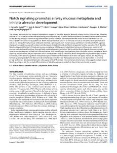
Fig. 2. DAPT-mediated Notch inhibition enhances limb-bud MPC differentiation without biasing lineage determination. (A) Alcian Blue/Orange G (AB/OG) staining of limb-bud MPC micromass cartilage nodules and real-time RT-PCR for Sox9, Col2a1 and Agc1 at 3, 5 and 7 days. (B)Alkaline Phosphatase staining of limb-bud MPC osteogenic cultures and real-time RT-PCR for Col1a1, AP and Oc at 21 days. (C)Oil Red-O staining of limb-bud MPC adipogenic cultures and real-time RTPCR for Pparg at 21 days. The y-axis of graphs is the relative gene expression normalized to b-actin and to the control. *, P