due to a Cu2+-catalyzed oxidation reaction which resulted in the formation of a .... H+ are released/mol ATP cleaved, in agreement with the value of. Alberty et al. ..... end of the experiment, cysteine (25 PM) was added to calibrate the spectral .... the release, phase A 23187 was added to abolish any remaining Ca2+ gradient ...
Vol. 264, No. 36, Issue of December 25, pp. 21725-21736.1989 Printed in U.S .A.
THEJOURNAL OF BIOLOGICAL CHEMISTRY 0 1989 by The American Society for Biochemistry and Molecular Biology, Inc.
Reactive Disulfides TriggerCa2+Release from Sarcoplasmic Reticulum via an Oxidation Reaction* (Received for publication, November 23, 1988)
Nikhat F. Zaidi$**, Carl F. LagenaursQ, Jonathan J. Abramsonsll, Isaac PessahlJ, and Guy Salama**$$ From the Departmentsof **Physiology, and §§Neurobiology,Anatomy, and Cell Science, School of Medicine, University of Pittsburgh, Pittsburgh, Pennsylvania 15261, the !$Physics Department, Portland State Uniuersity, Portland,Oregon 97207, and the 1I Department of Pharmacology and Toxicology, School of Veterinary Medicine, Universityof California, Davis, California 95616
Reactive disulfidecompounds (RDSs) with a pyridyl ring adjacent to theS-S bond such as 2,2’-dithiodipyridine (2,2’-DTDP), 4,4’-dithiodipyridine, and N-succinimidyl 3(2-pyridyldithio)propionate (SPDP)trigger Ca2+release from sarcoplasmic reticulum (SR) vesicles. They are known to specifically oxidize free SH sites via a thiol-disulfide exchange reaction with the stoichiometric production of thiopyridone. Thus, the formation of a mixed S-S bond between an accessible SH site on an SR protein and a RDS causes large increases in SR Ca2+permeability. Reducing agents, glutathione (GSH) or dithiothreitol reverse the effect of RDSs and permit rapid re-uptake of Ca2+by the Ca2+,Mg2+-ATPase.The RDSs, 2,2’-DTDP, 4,4‘-dithiodipyridineandSPDP displaced [‘Hlryanodine binding to the Ca2+-receptor complex at IC60 values of 7.5 f 0.2, 1.5 f 0.1, and 15.4 f 0.1 pM, respectively. RDSs did not alter the rapid in.itia1 phase of Ca2+ uptake by the pump, stimulated ATPase activity, and induced release from passively loaded vesicles with nonactivated pumps; thus they act at a Ca2+ release channel andnot at theCa2+,Mg2+-ATPase.Efflux rates increased in 0.25-1.0 mM [Mg2+]frW then decreased in 2-5 mM [Mg2+]free. Adenine nucleotides inhibited the oxidation of SHs on SR protein by RDSs and thus reduced Ca2+efflux rates. However, once RDSs oxidized these SH sites and opened the Ca2+release pathway, subsequent additions of nucleotides stimulated Ca2+ efflux. In skinned fibers, 2,2‘-dithiodipyridine elicited rapid twitches which were blocked by ruthenium red. These results indicate that RDSs trigger Ca2+ release from SRby oxidizing a critical SH group,and the prothus provide a method tocovalentlylabel tein&)involvedincausingthesechangesin Ca2+ permeability.
* This work was supported in part by American Heart Association Grant 87-1065 and Western Pennsylvania Heart Affiliate (to G. S.) and by American Heart Association Grant 87 915 and the Oregon Affiliate of the American Heart Association (to J. J. A.), and a Biomedical Support Grant (to I. P.) The costs of publication of this article were defrayed in part by the payment of page charges. This article must therefore be hereby marked “aduertisement” in accordance with 18 U.S.C. Section 1734 solely to indicate this fact. $ Recipient of aMuscularDystrophy Association Postdoctoral Fellowship. ll Established investigator 84130 of the American Heart Association. $$ Recipient of Research Career Development Award 5 KO4 NS00909 from the National Institutes of Health. To whom correspondence should be addressed.
The propagation of an action potential along transversetubular membranes in skeletal muscle triggers the release of Ca2+from the sarcoplasmicreticulum (SR)’ networkand thereby initiates a contraction (1-3). The exact mechanism responsible for the coupling of electrical impulses to SR Ca2+ release remains unknown (4). Current theories have implicated the dihydropyridinereceptor as the t-tubule voltage sensor ( 5 ) .The L-type Ca2+ channels a t theot-tubulesmay be linked toSR Ca2+ releaseacross a 100-200-A gap through the influx of Ca2+(4,6), the synthesis and diffusion of a chemical messenger inositol 1,4,5-triphosphate across the gap (7,8), or by direct physical connections via “feet” spanning thegap (9, 10). The SR Ca2+release channel was recentlyidentified by several laboratories as a M, 400,000 protein through its high affinity binding sitefor ryanodine, a plant alkaloid known to interferewiththe releaseprocess (11-16). Theryanodine receptor complex was purifiedfromdetergent-solubilized junctional or heavy SR proteins using radiolabeled ryanodine (17) or immunoaffinity chromatography(18) and found tobe similar to the native SR Ca2+ release channel. Incorporation of the receptor complex in planar bilayers revealed a Ca2+permeable pore with a large unitary conductance which was blocked by micromolar ruthenium red and millimolar M e andactivatedintheopenstate by micromolarCa2+ and millimolar ATP (17, 19). The criteria strongly suggest that the 400-450 kDa receptor is the “physiological” SR release channel since similar properties areobserved for Ca2+ release from skinned fibers (6, 20), heavy SR vesicles (21, 22), or “native” SR Ca2+channels incorporatedby fusing SR vesicles with planarbilayers (23-25). Eecently, we have demonstrated an alternative method to activate SR Ca2+releaseusingsulfhydrylreagents.Heavy metals like H e , Ag+, Cu2+, Cd2+, and Zn2+ were found to induce Ca2+ release from SR vesicles by binding to anaccessible free sulfhydryl group on a protein (26). The concentrations of heavy metals used to trigger rapid Ca2+ releasewere too low to affect the Ca2’,Me-ATPase and their potency was similar to their relative binding affinity to SH groups. Moredetailed experiments revealed that Ag+ interacts directly with theCa2+ release mechanism from SR and not with Ca2+,Mg2+-ATPase (27). Several lines of evidence support this The abbreviations used are: SR, sarcoplasmic reticulum; HEPES, 4-(2-hydroxyethyl)-l-piperazineethanesulfonic acid; MOPS, morpholinopropanesulfonic acid; AP 111, antipyrylazo 111; RR, ruthenium red; EGTA, [ethylenebis(oxyethylenenitrilo)Jtetraaceticacid; 2,2’DTDP, 2,2’-dithiodipyridine; 4,4’-DTDP, 4,4’-dithiodipyridine; SPDP, N-succinimidyl 3(2-pyridyldithio)propionate; 2-TP, 2-thiopyridone; 4-TP, 4-thiopyridone; DTT, dithiothreitol; AMP-PCP, B,ymethyleneadenosine 5”triphosphate; RDS, reactive disulfide; DIFP, diisopropyl fluorophosphate; Me2S0, dimethyl sulfoxide.
21725
2 1726
SH Oxidation-reduction Opens and Closes SR Ca2+Channels
interpretation. (a) Ag+ (0.1-10 p M ) was found to be the most mM AP 111,20mM Tris-HC1, at pH 6.8, and room temperature. The potent of theagents known to triggerCa2+release from cuvette was placed in the spectrophotometer and two sequential isolated triads and terminal cisternae (28). ( b ) It had little aliquots of CaZ+(25 ,UM) were manually but rapidly added to the cuvette to calibrate the API11 signal. Active Ca2+ uptakewas initiated effect on light SR vesicles which are thought to have few by adding an ATP-regenerating system consisting of creatine kinase release channels but arerich in Ca2+ pumps.(c) Itseffect was (5-10 units) and creatine phosphate (5 mM), followed by equimolar modulated by the known blockers and stimulators of excita- concentrations of Mg-ATP. In a typical experiment, 50 ,UMMg-ATP tion-contraction coupling (27, 28). ( d ) Ag+ caused the rapid was sufficient to obtain optimal rates of Ca2+accumulation. Another dissociation of [H3]ryanodinefrom its Ca2+-receptorcomplex ATP-regenerating system consisting of pyruvate kinase and phosinterfered with release induced by RDSs and thus (29). Besides heavy metals, mercaptans like cysteine caused phoenolpyruvate could not be used for these experiments. Upon completion of Ca2+ Ca2+release from heavy SR vesicles, in the presence of Cu2+ uptake, when the dye signal leveled off, efflux was elicited by a single (1-2 p M ) (30). TheCu2+ plusmercaptan effect appeared tobe addition of a sulfhydryl reagent. Efflux was kinetically and quantidue to a Cu2+-catalyzed oxidation reaction which resulted in tatively measured; once the efflux phase settled, the Ca” ionophore the formation of a mixed disulfide bond between the exoge- A 23187 (2 pg/ml) was added to determine the total releasable Ca2+ nous mercaptan and asulfhydryl on an SR protein. Con- stored in the lumen of the vesicles. A t low concentrations of RDSs, versely, reduction of the disulfide bond with D T T reversed the onset of efflux was delayed by seconds to minutes after the addition of these reagents. Thus, the rate of release could not be the effect and resulted in Ca2+re-uptake by the SR (30). Other practically determined from the initial slope of the traces. Instead, reagents such as sulfhydryl reactive dyes(31), phthalocyanine the kinetics of release were expressed as the time taken for 50% dyes (32),andanthraquinones (e.g. doxorubicin) (33), all release. In several experiments, DIFP (1 mM) was added tothe cause SR Ca2+ release most likely by acting at a free SH reactionmixture and did not alter active Ca2’ uptake or release induced by RDSs. Spectrophotometric measurements of Ca2+transgroup. port were confirmed with a calcium ion selective electrode (Cal 1, In view of mounting evidence for the existence of a “critical” WPI, New Haven, CT), under identical conditions, but without AP S H group at or near therelease channel (34),we searched for 111. The response of the electrode was calibrated in the reaction a method tocovalently link a label to this SH site in order tomedium by adding aliquots of Ca” (25 p ~ )as, well as by calibrating identify and purify the protein(s)involved and examine their the electrode with buffered Ca-EGTA solutionsat variouspcavalues. Efflux from Passively Loaded SR Vesicles-In some experiments, possible participation in theCa2+ releaseprocess. This paper shows that a class of compounds called “reac- SR vesicles were passively loaded with Ca2+,to measure rates and extent of efflux in the absence of functional Ca2+-pumps.SR vesicles tive”disulfides such as 2,2’-DTDP, 4,4’-DTDP, or SPDP (18-22 mg protein/ml) were incubated for 90 min in 100 mM KCl, 20 caused Ca2+ release from actively loaded SR vesicles. These mM Tris-HC1, 4 “C at pH 6.8, and 2 mM CaC12. Passive efflux from agents are known to be absolutely specific to free sulfhydryls, vesicles loaded with 2 mM Caz+was monitored spectrophotometrically which they oxidize with the concommittant release of thio- by adding an aliquot of stock SR solution to a cuvette containing 2 pyridone in the medium (35). As in the case for other sulfhy- ml of 100 mM KC1, 20 mM Tris-HC1, pH 6.8, 0.1 mM AP 111, at 23 “C, dryl-oxidizing agents, they appear to specifically react with and various agents that modulate release (i.e. M e , RDSs, or adenine nucleotides). The addition of Ca2+-loadedvesicles resulted in a sudden the Ca2+release channel but not the ATPase. signal change due to theopacity of the vesicles and theextravesicular In the following paper, we describe strategies for the syn- Ca2+,followed by a slower exponential change in differential absorpthesis of biotin-conjugated RDSs which covalently linked a tion of AP 111. The latter represented a passive release of Ca2+from biotin moiety to a 106,000 M , SR protein. This protein was the lumen of the SR which could be accelerated by additions of purified by biotin-avidin chromatography and upon reconsti- sulfhydryl reagents. After the release phase, A 23187 was added to tution in planar bilayers revealed the existence of Ca2+ release obtain complete loss of the Ca2+gradient across the vesicles. This followed by the addition of aliquots of Caz+ (5 ,UM) to calibrate channels. We present evidence thatthisprotein is not a was the dye signals. Rates of release induced by 2,2’-DTDP were normalproteolytic fragment of the 400 kDa highmolecular mass ized with respect to control rates of passive efflux. ryanodine receptor complex and is not the Ca2+,Mg2+-ATPATPase Actiuity-Kinetic measurements of Ca2+transport by SR ase. Preliminary reportsof these findings have been presented and ATPase activity were carried out in parallel experiments under identical conditions. Ca2+transport was measured using AP I11 as in abstract form (36, 37). EXPERIMENTALPROCEDURES
Preparation of SR Vesicles-SR vesicles were prepared from rabbit white skeletal muscle as previously described (38). After the last centrifugation step, the vesicles were suspended at 10-12 mg protein/ ml, in a medium containing 0.9 M sucrose, 10 mM HEPES, pH 7.0, and stored in liquid nitrogen, until use. Protein concentration was determined by the method of Lowry et al. (39). In some experiments, junctional SR vesicles were prepared as previously described (21), in the presence of DIFP (1mM) and EGTA (2 mM) to prevent proteolytic breakdown of 400-kDa “foot” proteins. Measurement of Ca2+Transport-Caz+ uptake and release from SR vesicles were measured spectrophotometrically through the differential absorption changes of the metallochromic indicator, antipyrylazo I11 (AP 111), an indicator of extravesicular free Ca2+. Differential absorption was measured at 720-790 nm with a time sharing dual wavelength spectrophotometer (SDB-3A, University of Pennsylvania, Biomedical Instrumentation, Philadelphia, PA). The application of AP I11 for similar SR experiments, standard controls, and its specificity as anindicator of free [Ca’+] have been previously demonstrated (40). The RDSs, DTT, and GSH at the concentrations used in this study produced negligible changes in the absorption of AP I11 and did not interfere with measurements of free Caz+ in the medium. DTT (up to 5 mM) and GSH (up to 2 mM) did not alter the rate of active Ca2+ uptake by SR vesicles. Unless otherwise stated, measurements of Ca2+ uptake and release were made in a2-mlassay medium containing 0.5 mg/ml SR vesicles, 100 mMKC1, 0.5 mMMgC12, 0.1
described above, then ATPase activity was monitored in a magnetically stirred cuvette by measuring the acidification of the medium due to ATP hydrolysis through the differential absorption changes of phenol red at 540-507 mm. For these experiments, the buffer contained 2 mM Tris-HC1 at pH 6.8; under these conditions, 0.5 mol of H+ are released/mol ATP cleaved, in agreement with the value of Alberty et al. (41). Spectral Measurements-The spectral characteristics of RDSs, e.g. 2,2’-DTDP, 4,4’-DTDP, have been extensively studied and shown to undergo large spectral changes following an oxidation reaction initiated by the addition of a mercaptan (42,43). Thelarge shifts inpeak absorption in the UV region are associated with a loss of the parent compound and theproduction of the corresponding thiopyridone. The oxidation of cysteine by 2,2’-DTDP, 4,4’-DTDP, or SPDP results in the stoichiometric production of 2 molecules of 2-TP, 4-TP, or 2-TP, respectively. Measurements of RDSs spectra were repeated with our standard buffer, with or without SR vesicles to determine whether or not SH groups on the SRwere involved in a thiol-disulfide exchange reaction, with the release of thiopyridone in themedium. Cysteine was used as the control mercaptan to initiate oxidation reactions and to calibrate the optical detection of thiopyridone production. Absorption spectra in the UV region were measured with a double beam, single wavelength spectrophotometer, with microprocessors to digitally record, store, add, or/and subtract spectral data (HITACHI Ltd., model 557, Tokyo, Japan). RDS reagents were incubated in a reaction mixture containing (in millimolars): 100 KC1, 5 creatine phosphate, creatine kinase (5 units), 20 Tris-HC1, pH 6.8, at 23 “C and spectra were
SH Oxidation-reduction Opens and recorded in the range of 200 to 400 nm. Additions of cysteine or SR vesicles were then made to measure spectral changes of the RDSs. Kinetics of Thiopyridone Production during Ca2+Efflux from SROxidation of SH groups on SR proteins was monitored quantitatively through the production of thiopyridones. The time course of thiopyridone production was compared with that of Caz+ release from the SR by performing parallel experiments to measure production of 2T P or 4-TP, respectively, at 340-310 and 324-350 nm and extravesicular [Ca"] by AP 111 at 720-790 nm, respectively. SR vesicles (0.5 mg/ml) were suspended in 2 ml of 100 mM KCl, 20 mM Tris-HC1, pH 6.8,0.5 mM MgC12,5 mM creatine phosphate, and 10 units of creatine phosphokinase. ATP (0.1 mM) was added to initiate Caz+ uptakeand 2,2'-DTDP (10 p ~ to) elicit efflux. Differential absorption at 340310 nm measured thiopyridone production, and cysteine was added to calibrate the signals. Caz+transport was measured under identical conditions, except that AP 111 (0.1 mM) was added to the reaction mixture and thedifferential absorption was measured at 720-790 nm. Skinned Fiber Experiments-Chemically skinned fibers were prepared from psoas muscles of rabbits as described by Wood et al. (44). In brief, bundles of several hundred fibers were removed and skinned in a solution consisting of (in millimolars): 170 potassium gluconate, 5 magnesium gluconate, 2.5 NaZATP, 5 EGTA, 5 imidazole, at pH 6.8 and 4 "C. After 24 h at 4 "C, the bundles were transferred to a solution of the same ionic composition made up in 50% glycerol for storage at -20 "C, until used. Isometric force was measured from 2 to 4 fibers separatedfrom the psoas muscles mounted between a fixed post and the head stage of a tension transducer (model 400A, Cambridge Technology, Inc. Watertown, MA). The distance between the fixed post and transducer was adjusted by a micromanipulator to stretch thefibers to 120% of their resting length. Fibers were bathed in a magnetically stirred chamber in relaxing solutions containing (in millimolars): 170 potassium gluconate, 10 MOPS, 2.5 NazATP, 1 MgS04,pH 6.75,23 "C. The solutions and stocks were made from double distilled, deionized water, then they were passed through a Chelex 100 (Bio-Rad) ion-exchange column (30 X 1 cm) to reduce calcium contamination from the water and added chemicals. Free M$+ and Caz+ levels in the relaxing solution were determined from computercalculations(45) to be approximately 100 p M and 150 nM (pCa 6.8), respectively. Free Ca2+ levels were confirmed with a Ca2+ selective electrode and aqueorin luminescence. Fibers were bathed in solution at pCa 6.5 to load the SR network with Ca2+, thenwashed in pCa 6.8. Aliquots of Ca2+(1 p~ total equivalent to 50 nM free) were added to elicit Ca2+induced Ca2+release twitches, followed by caffeine (2 mM) to determine the maximum releasable Ca2+from the SR. Fibers were washed, the SR reloaded with Caz+, thenRDSs reagents were added in the presence or absence of ruthenium red. A second addition of caffeine was made to ensure that no significant fiber run-down had occurred, and saturating concentrations of Ca2+were added to determine the maximum force that could be generated by the fibers. Ryanodine Binding to the CaZ+-ReceptorComplex-Heavy SR fractions were centrifuged through a six-step discontinuous sucrose gradient as described by Inui et al. (16). Vesicles enriched in junctional markers were isolated at the 36-45% sucrose interface, diluted in buffer A (in millimolars): 115 KCl, 15 NaCl, 40 Tris maleate, pH 7.1, and repelleted. Pellets were resuspended in 2 mg protein/ml in buffer A and rapidly frozen in 0.5-ml aliquots in liquid nitrogen, until needed. One mlof250 nM [3H]ryanodine and 50 p1 of 0.1 M CaC12 was added to buffer A (100 ml of final volume) to obtain a stock concentration of 2.5 mM radioligand and approximately 50 p~ free Ca2+. Aliquots (1 ml) were divided among 100 test tubes and 10 pl of each RDS reagent (from 100 X stock in dimethyl sulfoxide, MezSO) was added to yield final concentrations of 1 nM to 0.1 mM. Control experiments were done in duplicate tubes where MezSO (10 pl) was added to control and nonspecific (with cold 10 p~ ryanodine) samples. Junctional SR membranes (50 pg protein/ml) were added lastly to initiate thereaction and incubated for 90 min at 37 "C. The reactions were quenched by rapid filtrationthrough WhatmanGF/B glass filters, rinsed twice with 5 ml of ice-cold deionized buffer. Experiments were repeated at least three timesondifferentmembrane preparations,ondifferent days. The concentrations of eachRDS compound which reduced specific [3H]ryanodine binding by 50% of control value (ICso)were calculated from linear regressions of Hill plots using the pooled data from two experiments, each in duplicate. Materials-All reagents were of analytical grade. Reactive disulfides, Na,-ATP, Mg-ATP, creatine phosphate, creatine phosphokinase, Tris-HC1, ruthenium red, Tris, and HEPES were purchased
Closes SR Ca2+Channels
21727
from Sigma. Antipyrylazo I11 was from ICN Pharmaceuticals (Plainview, NY). A 23187 was purchased from Behring Diagnostics and dissolved in ethanol stock solutions at 1 mg/ml. RESULTS
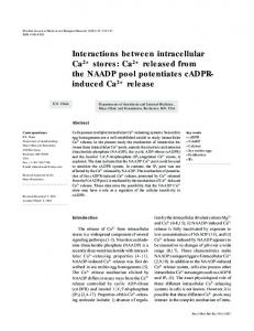
Reactive Disulfides Induce SR Ca2+Release-RDS reagents are known to be absolutely specific to free SH groups and to readily oxidize S H groups at low substrate concentrations and high reaction rates (35). These features make them particularly attractive to test the theory that the SR contains a critical SH site which can be oxidized (via formation of a disulfide bond) then reduced to open andclose a Ca2+ release channel (30, 34). The basic phenomenon is demonstrated in Fig. 1. SR vesicles are suspended in a KC1 buffer and AP I11 to measure changes in extravesicular free [Ca"]. After additions of Ca2+ with an ATP-regeneratingsystem then Mg-ATP, thevesicles actively pumped Ca2+. The addition of 2,2'-DTDP (10 PM) caused the release of accumulated Ca2+and GSH at0.5 or 1.0 mM resulted in the complete re-uptake of the released Ca2+. The final addition of A 23187 released similar amounts of Ca2+. Thus,2,2'-DTDP causes thetranslocation of Ca2+ stored in the lumenof the SR anddoes not merely dissociate membrane bound Ca2+(Fig. lA). In Fig. lB, SR vesicles were Ca2+ loadedas in Fig. lA and release was induced by various concentrations of 2,2'-DTDP (2.5-100 WM). At the endof the release phases, DTT (1mM) like GSH reversed the chemical reaction between the RDS and the SR resulting in active reuptake. Oxidized glutathione, GSSGdid not reverseCa2+ efflux caused byRDSs nordoes it stimulate Ca2+ release when A
1 2.2DTDP
c
_
2 mln
FIG. 1. SR CaZ+release and re-uptake by sulfhydryl oxidation then reduction with 2,2'-DTDP and a reducing agent, respectively. A , SR (0.5 mg/ml) were suspended in 2 mlof (mM) 100 KC1,20 Tris-HC1, 0.1 AP 111,0.5MgCl,, at 23 "C, pH 6.8. Two aliquots of Ca2+were added to calibrate the AP I11 response, then ATP (100 p ~ along ) with an ATP-regenerating system to initiate Ca2+ uptake. Ca2+efflux was induced by an addition of 2,2'-DTDP (10 p ~ )After . completion of the release phase, the addition of reduced glutathione (GSH)(0.5-1 mM) resulted in complete re-uptake of Ca2+. B, SR vesicles were Ca2+loaded as in A and release was induced by various concentrations of 2,2'-DTDP. After the completion of release, the addition of DTT (1 mM) caused the active re-uptake of Ca2+.The same results were obtained in the absence or presence of DIFP (1 mM) in the reaction mixture.
Closes SR Ca2+ Channels
SH Oxidation-reduction Opens and
21728
added following completion of the Ca2+uptake phase. In some SR preparations, the onset of Ca2+ release following the addition of a RDS was delayed, and lag phases were particularly pronounced at low [RDS] (compare Fig. 1,A and B, from two different preparations). These differences in the onset and rates of release were attributed todifferent ratios of heavy to light SR in these preparations since rates of release in heavy SR were -4 times faster than in light SR (data not shown). The chemical reaction responsible for this reversible Ca2+ efflux was examined further by testing various disulfide compounds. From previous studies, disulfides like cystine and cystamine did not cause SR Ca2+release (30). As shown in Table I, the lowest concentrations of RDSs that could elicit calcium release was2.5 ~ L Mof 2,2'- or 4,4'-DTDP which released 50% of total releasable Ca2+.Disulfides with a pyridyl ring immediately adjacent to the S-S bond (e.g. 2,2'-DTDP, 4,4'-DTDP, and SPDP) elicited release. However, when a structure contained a methyl between the pyridyl ring and the disulfide (e.g. pyrithioxin) or lacked a pyridyl ring (i.e. dithiobis(succinimidy1propionate)), therewas no stimulation of Ca2+efflux at up to 100 ~ L M In Fig. . 2, % Ca2+efflux and
the time for 50% efflux is plottedasa function of RDS concentration. The data shows that the compound with a disulfide bond at the 2 position on the pyridyl ring (2,2'DTDP) produces the highest % Ca2+efflux, 8 5 4 8 % of total releasable Ca2+.When the disulfide bond is at the 4 position (4,4'-DTDP), the ratesof efflux were maximum. Compounds with two pyridyl rings adjacent to the disulfides (2,2') and 4,4'-DTDP) are considerably more effective at inducing release compared to a compound with a single pyridyl (SPDP). The succinimidyl propionate moiety (i.e. dithiobis(succinimidyl propionate)) did not cause release. SR Ca2+Release and Thiopyridone Production-RDSs discriminate so effectively in favor of thiol groups with low pKa, and for practical purposes, their specificity for free sulfhydryls may be considered absolute (35). The most likely thiol-disulfide exchange reactions between a free SH on an SR protein and the RDSs are shown in Fig. 3. These reactions result in the formation of a covalent disulfide bond between the free sulfhydryl on the SR and either a pyridyl ring (Fig. 3, a and b) or a succinimidyl propionate (Fig. 3c). In all three cases, 1 mol of 2-TP (Fig. 3, a and c), or 4-TP (Fig. 3b) is produced/ mol of oxidized SH sites on an SR protein. These reactions
TABLE I Structure-function relationship of various disulfide compounds and SR Ca2+release Compound
Ca2+
Minimum effective conc. for release
Half-time of release at minimum effective conc.
2.5 p M
8.7 min
2.5 p M
5.1 min
-
1.
2,2'-Dithiodipyridine
2. 4,4'-Dithiodipyridine
25 p M
3.
N-Succinimidyl 3(2-pyridyldithio) propionate 4.
H HO
CHZ-OH
&q \
qH
3
I"CH.-S-S-CHz
\
OH
No effect
CHZ-OH Pyrithioxin 3,3-(dithiodimethy1ene)bis(5hydroxy-6-methyl-4-pyridine methanol)
No effect
5.
Dithiohis(succinimidy1propionate)
15 min
SH Oxidation-reduction Opens and
loor 80
I
A
T
1 //
I
Q 2.2'DTDP
e
4,4 DTDP
A
SPDP
Closes SR Ca2+Channels
21729
are effectively unidirectional because of the thiopyridone production, even under conditionswith equimolar concentrations of free SHs and RDSs (35). The absorption spectra of RDSs (50 PM) were measured in the same reaction medium used to study SR Ca2+transport (Fig. 4A)and 2 min after the addition of an excess of cysteine (100 PM) (Fig. 4B). The presence of cysteine activated the thiol-disulfide exchange reaction resulting in spectralchanges due to theproduction of thiopyridones. The absorption peaks for the various RDSs and their respective thiopyridones were identical to previous reports (42,43). SPDP and2,2'-DTDP have absorption peaks at 232 and 280 nm, while 4,4'-DTDP has a major peak at 247 nm. Reduction of these RDSs by cysteine decreased the absorption peaks of the parentcompounds and produced new absorption peaks at 270 and 340 nm for 2-TP and at230 and 324 nm for 4-TP, respectively. The addition of SR vesicles to a medium containing 2,2'DTDP (50 PM) resulted in a rapid (within 2 min, the time needed to measure a spectrum) increase of 270 and 340 nm absorption (i.e. peaks for 2-TP) with an equivalent reduction in the absorption of 2,2'-DTDP at 280 nm (Fig. 5). At the end of the experiment, cysteine (25 PM) was added to calibrate the spectral measurement with a known concentration of free SHs. In Fig. 6, Ca2+release and thiopyridone production were measured in parallel runs, from the same preparation of Ca2+loaded SR. In the top truce, extravesicular free Ca2+ was monitored with AP 111; after additions ofCa", uptake was initiated with an ATP-regenerating system, release was in-
moo 25
50
8
75
2b0
Concentration of reactive disulfide (pM)
18
e
2.2'DTDP 4.4'DTDP
I
A
.G 14 E 16[
SPDP
f
DL 10 l2I
//
I
1 25
50
75
1;o
2b0
Concentration of reactive disulfide (pM)
?*
FIG. 2. Rate and extent of Ca" release induced by RDSs. SR vesicles were Ca2+loaded using an ATP-regenerating system, as described for Fig. 1. After completion of uptake, various concentrations of RDSs were added to induce Ca2+release. DTT reversed the effect of the RDSs, and A 23187 was added to determine the maximum [Ca"] that could be released by the vesicles. A , the % Ca2+efflux was normalized with respect to the maximum releasable Ca2+ measured in thepresence of A 23187 and plotted as a function of [RDS]. B, the rate of Ca2+release was analyzed as the time for 50% Ca2+release (min) andplotted versus [RDS].
Wavelength (nm) THIOL-DISULFIDE EXCHANGE REACTION
20
i o H
0
200
+ U
I
FIG. 3. Thiol-disulfide exchange reaction between free SH sites on SR and RDSs. The free SH sites on SR proteins attack the RDSs to form mixed disulfides and the production of thiopyridones.
400
Waved?gth (nm) FIG. 4. AbsorptionspectraofRDSsand their respective thiopyridones. A , RDS (50 p M ) was added to a 2-ml cuvette containing (in mM) 100 KCl, 20 Tris-HC1, 0.5 MgC12, at pH 6.8. The RDSs 2,2'-DTDP and SPDP have peaks at 232 and 280 nm, and 4,4'-DTDP has a main peak at 247 nm. B, an excess of cysteine (200 nmol) caused the complete reduction of the RDSs, resulting in the production of their respective thiopyridones. The spectra of 2,2'DTDP ( a ) and SPDP (b) shift to maxima at 270 and 340 nm, due to the production of the orthoisomer of thiopyridone (z.e. 2-TP). As expected, the molar production of 2-TP is twice as great for 2,2'DTDP compared with SPDP. In the presence of cysteine, 4,4'-DTDP (c) produces the paraisomer (i.e. 4-TP) which has a higher molecular extinction coefficient, and peaks at 230 and 324 nm. The vertical scale is in units of optical density.
SH Oxidation-reduction Opens and Closes SR Ca2’ Channels
2 1730 as
2.2’OTOP
A
/ A
50 pM
3 4 0 nm T h i o p y r l d o n e Ab..
r
t
\ GSH
Warei.ngth(nm)
FIG. 5. RDSs oxidize SH groups on SR. SR vesicles (0.5 mg/ ml) were suspended in a 2-ml cuvette containing (in mM) 100 KC1, 20 Tris-HC1, 0.5 MgC12, pH 6.8. The UV absorption spectrum of the suspension was recorded, stored in memory, and subtracted from subsequent spectra. An addition of 50 /IM 2,2’-DTDP (100 nmol) produced a mixed spectrum of the parent compound 2,2’-DTDP and 2-TP. The main peak for 2,2’-DTDP at 280 nm shifted toward 271 nm, and a shoulder appeared at 340 nm which was used to measure 2-TP production in the reaction mixture.After 30 min, the OD change leveled off and was related to moles of T P produced. The oxidation of cysteine (50 nmol) results in the formation of cystine (25 nmol) and 2-TP (50 nmol); the latter resulted in a 0.22 increase in OD at 340 nm. Thus, the reaction between 2,2’-DTDP and SR vesicles produced 5.6 nmol of 2-TP (OD = 0.025) within2min, and a maximum of 25 nmol after 30 min. A similar spectral analysis with 4,4’-DTDP in the presence of SR vesicles indicated the production of about 5 nmol of 4-TP/mg SR, at a peak absorption of 324 nm, thus confirming the results obtained with 2,2’-DTDP.
duced with 2,2’-DTDP, re-uptakeoccurred upon the addition of GSH, and finally complete release with A 23187. In the bottom trace, thiopyridone production was monitored a t 340310 nm, under identical conditions, except for the absenceof AP 111. The additionof 2,2’-DTDP elicited a fast phase of 2TP production which preceded slightly Ca2+efflux, followed by a slower phase of 2-TP generation. The addition of a large concentration of GSH reduced sulfhydryls on SR proteins,as well as the remaining 2,2’-DTDP. The latter effect resulted in a large thiopyridone absorption signal. The maximum number of SH sites on SR vesicles oxidized by 2,2’-DTDP was 4 nmol/mg SR protein. From similar experiments with 4,4’-DTDP and measurements of 4-TP production at 324 nm, 4-5 nmol of SH sites on SR vesicles were oxidized/mg SR protein. Forboth compounds, there was a fast phase of thiopyridone production of -2 nmol/mg SR associated with the fast initial phase of Caz+efflux, followed by a slow phase. Site of Action of RDSs-The effect of RDSs was examined in the presence of agents that modulate SR Ca2+ release to elucidate their site of action. For instance, ionized Mg2+ is known to inhibit (a)Ca2+releaseinduced by Ca2+ (6), (b) binding of ryanodine to itsreceptor site (11, 15), and(c) Ca2+ release caused by sulfhydryl reagents like A$+ or cysteine plus Cu2+(27,30). Tomeasure the effect of [M$+]freeon RDSinduced Ca2+ release, SR vesicles were actively loaded using 100 h~ Mg-ATP and an ATP-regenerating system in the presence of varying [Mg2+]f,,; once the phase of Ca2+uptake leveled off, 2,2’-DTDP (10 WM) was added to measure release. Fig. 7 shows that ratesof Ca2+release induced by 2,2’-DTDP are stimulated by increasing concentrations of free [M$+] from 0.25 to 1.0 mM, then decreasedfrom 2.0 to 5.0 mM [ M e ] . The lower rates of Ca2+ release at low [M$+] were not caused by diminished Ca2+gradients across the SR since total Ca’+ uptake was the same (150 nmol Ca2+/mg SR) at
t
10
! pM
lox
FIG. 6. Kinetics of Caz+ release and thiopyridone production. SR vesicles were Ca2+loaded, release of Ca2+was initiated by the addition of 2,2’-DTDP (10 WM), GSH (1mM) reversed Ca2’ efflux, and anaddition of A 23187 was used to determine the totalreleasable Ca2+ from the vesicles. Top trace, Ca2+ transport was measured through the differential absorption of AP I11 (0.1 mM) at 720-790 nm. Bottom trace, in a parallel experiment under identical conditions but without AP I11 in the reaction mixture, the production of 2-TP release was monitored a t 340-310 nm. The release of Ca2+ induced by 2,2’-DTDP (20 nmol) was associated with the concomitant production of 4 nmol of 2-TP. The gain on the instrument was reduced by five times and the addition of GSH reacted with the remaining 2,2’-DTDP to produce 16 nmol of 2-TP. Four similar measurements were repeated on differentSR preparations with 2,2’-DTDPand 4,4’DTDP where thiopyridone production was measured at 340-310 and 320-350 nm, respectively. Thiopyridone production with both RDSs was in the range of 4-6 nmol of TP/mg SR protein.
1.0
5.0
2.0
[Me2+] in m M
FIG. 7. Effect of ionized [ M 2 + ] on Caz+release induced by RDSs. SR (0.5 mg/ml) were suspended in 2 ml ofKC1 buffer containing 0.1 mM AP I11 and various [ M p ] . Extravesicular Ca2+ was measured at 720-790 nm, and Ca2‘ uptake was initiatedas described for Fig. 1. Caz+efflux was initiated by adding 2,2’-DTDP (10 WM), and upon completion of release, A 23187was added to determine thetotal releasable Ca2+ from the vesicles. Foreach [ M e ] , the % Ca2+efflux (0)was calculated from the amount of Ca2+ released by 2,2’-DTDP divided by thetotal releasable Ca2+ and plotted uersus [MgZ+]. Rates of Ca2+ efflux (0)were expressed as nmoles of Ca2+/mg protein/min.
low and high [M$+], in thepresence of the ATP-regenerating system. Further tests revealed that ionized M e modified Ca2+ releaseby interacting with the oxidized SR Ca2+ release channel protein. First, changes in[ M e ] from 0 to 2 mM did
SH Oxidation-reduction Opens and Closes SR Ca2+Channels not alter the rates or total amountsof thiopyridone production which indicated that it neither stimulated nor inhibited the oxidation of SH sites on SR proteins by RDSs (not shown). Thus, changes in [Mg"] altered release from the oxidized or open channel. Second, in SR vesicles loaded passively with Ca", in the complete absence of adenine nucleotides, stimulation of Ca2+release by RDSs becomes independent of the activity of the Ca2+ pumps regardless of the [Mg2+] in the reaction. This is shown in Fig. 8 for which SR vesicles were passively loaded with 2 mM Ca2+,0 [Mg2+]free and 0 adenine nucleotides. The SR vesicles were then reacted with 0, 25, or 50 FM 2,2'-DTDP and transferred to a Ca2+ efflux medium with various [M$+]. In [MPlf,,, = 0.2 mM, the rateof passive Ca2+efflux was stimulated (-2-fold) by 2,2'-DTDP (Fig. 8, left truces). At 1 mM [MgP+Ifree, the rate of passive Ca2+efflux was considerably slower and even after stimulation of release by 25 and 50 PM 2,2'-DTDP, rates of release were still slower compared toefflux at lower [M?] (Fig. 8, right truces). Thus, the inhibitionof oxidation-dependent Ca2+release by M$+ is similar to the effects ofMg2+ on Ca2+-induced Ca2+ release. The rate and extent of Ca2+ release induced by 2,2'-DTDP increased with pH from 6.0 to 6.8 and thendecreased for more alkalinepH (Fig. 9). Thedifferentrates of release as a function of pH were not. due to differences in Ca2+ gradients across the SR membranes since similar levels of active Caz+ accumulation and subsequent passive Ca2+ leakage were observed in this range of pH values. Measurements a t higher p H (i.e. pH 8 ) are not shown since the diminished rates of release were a reflection of reduced active Ca2+ uptake. Adenine nucleotides enhance Ca2+-induced Ca2+release in SR vesicles (21, 22), SRCa2+ releasein skinned fibers (6, 20), and the probability of Ca2+ channel openingin proteins incorporated in planar bilayers (24, 25). The effect of adenine nucleotides on Ca2+release induced by RDSs was examined in detail since it is a salient feature of physiological Ca2+ release. As shown in Fig. 10, ATP ( A ) and ADP ( B ) in the range of 50-500 FM gradually reducedthe ratesof Caz+release induced by 2,2'-DTDP and blocked release at 1 mM. On the other hand, increasing concentrations (0.1-1 mM)of cAMP (Fig. lOC) and (2 mM) AMP-PCP (Fig. 1OD) stimulated Caz+
21731
- 100
A
x
-80
2
c Q
-60
+
mrn
V
6.0
6.5
7.0
8
-
40
-
20
7.5
PH
FIG. 9. pH dependence of RDS induced Ca2+ release. Ca'+ transport atvarious pH values was monitored with a calcium selective electrode (WPI, Cal 1, New Haven, CT). Vesicles were suspended in 2 ml of KC1 buffer of different pH. Ca2' uptake was initiated with a ATP-regenerating system and release with 2,2'-DTDP (10 PM). Rates (A) and % Ca" efflux (A)were calculated as described for Fig. 7 and plotted versus pH.
release induced by 2,2'-DTDP, while higher concentrations inhibited release. Several experiments indicated that adenine nucleotides inhibit Ca2+efflux induced by RDSs by interferring with the oxidation of S H sites on the SR rather than a blockade of efflux from reacted channels. First, thepresence of increasing [ATP] from 0 to 1 mM gradually inhibited the oxidation rate of S H sites on SR vesicles by 2,2'-DTDP, as monitored through thiopyridine production (data not shown). This implied that adenine nucleotides inhibit the reaction between the RDS and the SR protein when the nucleotide was added beforethe RDS. Second, when the RDS was added before the adenine nucleotide, the presence of nucleotides caused a further stimulation of Ca2+efflux beyond that induced by the RDSalone. As demonstrated inFig. 11,the rate of Ca2+efflux from passively loaded SR was stimulated by a factor of 3.7 in 2,2'-DTDP (50 FM) (compare traces 0 and 3) and was further stimulatedby another factorof 1.4 when 2,2'DTDP was added first (for 10 min) before adding cAMP (2 mM). Conversely, when cAMP is added first followed by 2,2'DTDP, the RDS is ineffective at further stimulation of the Ca" efflux rate(compare truces 1 and 2, Fig. 11). Third, whereas ATP added before 2,2'-DTDP blocked Ca2+release and resulted in the maintenanceof a Ca2+gradient across the SR vesicles (Fig. 10);the reverse additions of 2,2'-DTDP (for 5-10 min) before the addition of ATP caused the openingof release channels, uncoupled the SR, and prevented active Ca2+ uptake(Fig. 13). Fourth,high ATP (ie. 1mM) inhibited Ca2+ release due to 2,2'-DTDP most likely by blocking the SR 1 6 oxidation reaction rather thanby a stimulation of active Ca2+ 1 6 mg SR loaded wllh loaded wllh uptake since the ATP-regenerating systemwith 100 PM ATP 2 mM Ca2' 2 mM Ca" FIG. 8. RDSs inducedCa2+ release from passively loaded already produced maximum rates of Ca2+ uptake. In addition, were obtained with other nucleotides SR vesicles. SR vesicles (20 mg/ml) were passively loaded by incu- similarinhibitions (ADP, CAMP, and AMP-PCP) which are not substrates for bation in 2 mM CaCI2 in 100 mM KC1, 20 mM Tris-HCI, pH 6.8, a t room temperature for 90 min. Ca2+efflux was measured from pas- the Ca2+,M$+-ATPase (Fig. 10). sively loaded SR vesicles, spectrophotometrically. An aliquot (80 11) Ryanodine has been extensively studied as an agent that of loaded vesicles equivalent to 1.6mg of SR protein was rapidly binds to thephysiological site of SR Ca2+ release and stimutransferred to a cuvette containing 2 ml of 100 mM KC1,20 mM TrisHCI, pH 6.8, 0.1 mM AP 111 a t room temperature, with 0.2 mM ( A ) lates or inhibits release depending on its concentration (14). The effect of ryanodine on Caz+release induced by RDSs was or 1.0 mM ( B ) of ionized M P . Addition of SR vesicles causeda examined by incubating vesicles in various concentrations of sudden change in the differential absorption of AP 111, due to the opacity of vesicles, and an increase in external Ca2+ concentration, ryanodine (10 nM to 20 PM) for 1-2 h a t 37 "C, then attempting followed by a slow changeinabsorption, which representedthe to releaseCa2+with an addition of a RDS reagent. Prior passive release ofCa" from loaded SR vesicles. Prior additions of incubation of the vesicles with ryanodine (100 nM) slightly 2,2'-DTDP in the loading medium accelerated the rate of passive CaZ+release a t both 0.2 and 1.0 mM Mg2+. After the completion of reduced (20-30%) the rates of Ca2+efflux triggered by RDS, of release (data not shown). The the release, phase A 23187 was added to abolish any remaining Ca2+ with no change in the extent gradient across the SR vesicles. At the end of each run, analiquot of converse experiment was to incubate SR proteins with radioCaz+(5 FM) was added to calibrate the API11 signals. labeled['Hlryanodine (2.5 PM) andtomeasureryanodine
FIG. 10. Effect of adenine nucleotides on RDS induced Ca2+release. SR vesicles were Ca2+loaded and uptake was measured as in Fig. 1 but in the presence of various concentrations of ATP ( A ) ,ADP ( B ) ,CAMP (C), or AMP-PCP (D). After uptake, 2,2’-DTDP was added to induce Ca” release.
binding to the Ca2+release receptor complex in the presence of various concentrations of a RDS reagent. As shown in Fig. 12, 2,2’- and 4,4’-DTDP altered ryanodine binding in a biphasic manner, i.e. enhancing then blocking ryanodhe binding with increasing concentrations. Low concentrations of 2,2’- and 4,4’-DTDP caused a significant stimulation of radioligand binding to SR proteins whereas higher concentrations dramatically inhibited ryanodine binding. SPDP revealed a monophasic response and only inhibited ryanodine binding at concentrations greater than 5 X M. The possibility that RDSs might actatthe level of Ca2+,Mg2+-ATPasewas examined by measuring Ca2+uptake and ATPase activity of SR vesicles with or without a prior incubation with a RDS followed by an addition of ATP since ATP inhibits the reaction between the SR release mechanism and RDSs. Ca2+uptake and ATPase activity were measured
in parallel experiments under identical conditions through the differential absorption changes of A P I11 (Fig. 13, top truces) and phenol red (Fig. 13, bottom traces), respectively. Phenol red (100 PM) was used to measure one H+ produced per two ATP hydrolyzed at pH6.8. Control measurements of Ca2+uptake (top) and ATPase activity (bottom) are shown in the traces: “0,” in the absence of a RDS. When the vesicles are incubated in 25 PM 2,2’-DTDP for 10 min before the addition of 1 mM ATP, there is a rapid and partial uptakeof Ca2+followed by a release (top, truce I ) and a stimulation of ATPase activity (bottom, truce 1).After the release phase (top, truce l ) ,A 23187 was added to show that a small fraction of the SR vesicles did not contain release channels that react with 2,2’-DTDP. With 50 PM 2,2’-DTDP, there was a further reduction of Ca2+accumulation (top, truce 2) and stimulation of ATPase activity (bottom, truce 2). Although, 2,2’-DTDP
2 1733
SH Oxidation-reduction Opens and Closes SR Ca2+Channels 1 mM Mg A T P
pFg A 231 87
li
\
\\ '
+ V 4 4 rng SR loaded w t h 2 mM CaZ'
FIG. 11. Ca2+efflux from passively loaded SR caused by 2,2'-DTDP before and after additions of CAMP.A stock solution of passively loaded vesicles was prepared by incubating 22 mg SR/ml in 100 mMKC1,20mM Tris-HC1, pH 6.8, 4 "C, 2 mM CaC12 plus various agents thatmodify release as described below. An aliquot (20 pl) of Ca*+-loaded SR stocksolution (0.44mg SR) was then rapidly added to 2 ml of 100 mM KC1, 20 mM Tris-HC1, pH 6.8, 0.1 mM AP 111, at 23 "C. A: trace 0, control; trace I , cAMP (2 mM) was added to the stock solution of Ca2+-loadedS R truce 2, cAMP (2 mM) was added first followed by 2,2'-DTDP (50 p ~ ) trace ; 3, 2,2'-DTDP (50 p ~ ) trace ; 4, 2,2'-DTDP (50 p ~ was ) added first, followed by cAMP ( 2 mM) 2 min later.
liI
AA=O!0061
I
Abslnc.
720 - 790 nm
\
i I
25 )IM
Ca2*
t " (
100 pM H'
2 min
Abs. Inc.
540 - 507 nm
1 mM Mg ATP
LoS
Aryldisulfide, M
FIG. 12. Effect of RDSs on [3H]ryanodineequilibrium binding to SR proteins. The presence of 2,2'- and 4,4'-DTDP produce biphasic (stimulatory and inhibitory) effects on radioligand binding tot be Ca2+-receptorcomplex. Both isomers show significant stimulation of [3H]ryanodine bindingabove 3 X lo-' M, but the2,2'-isomer is stimulatory over a broader range of concentration. SPDP does not stimulate andonly inhibits [3H]ryanodine binding. Inhibitory protencies (ICsvs),and theirrespective standard errors aresummarized. [3H] Ryanodine binding in control samples lackingRDSs (i.e. corresponding to 100% bound) averaged 1.5 & 0.1 nmol/mg SR protein. This value for ryanodine binding to junctional SR at subsaturating concentrations of radioligand is in excellent agreement with previous reports (13, 16, 33).
uncoupled the SR and as expected stimulated ATPase activity, the RDScould still be causing a partial inhibition of the Ca2+,Mg2+-ATPase since the maximum rate of ATPase activity (bottom, truce 4 ) measured in vesicles uncoupled with A 23187 was greaterthan for vesicles uncoupled with 2,2'DTDP (bottom, truce 2). However, the lower rate of ATPase activity in thepresence of 50 PM 2,2'-DTDP (bottom, truce 2 ) compared with that measured with A23817 was primarily due to a partial uncoupling of the vesicles rather than an inhibition of ATPase activity by the RDS. This was confirmed by comparing the ATPase activityof vesicles treated with up to 50 PM 2,2'-DTDP plus A 23187 (bottom, trace 3) or with A 23187 alone (bottom, trace 4 ) . Initial rates of ATPase activity in traces 3 and 4 were similar within experimental error and in many vesicle preparations these traces superimposed precisely. Another possibility was that RDSs could act on the Ca2+, Mg2+-ATPaseby uncoupling active Ca2+ translocation into the lumenof the SR without inhibiting ATP hydrolysis. This was addressed by measuring the initial rate of active CaZ+ uptake initiated with 50 F M ATP and an ATP-regenerating
FIG. 13. Effect of 2,2'-DTDP on ATPase activity. Ca2+uptake (top traces) and ATPase activity (bottom truces) were measured in parallel experiments, through the differential absorption changes of AP Ill (at 720-790 nm) and phenol red (at 540-507 nm), respectively. Assay media were identical except that they contained 100 pM AP I11 for the top traces and 100 p~ phenol red for bottom traces. The reaction mixture contained 0.5 mg SR/ml, 100 mM KC1, 20 mM Tris-HC1, 0.5 mM MgCl, in traces 0; truce 1, 25 p M 2,2'-DTDP; truce 2, + 50 pM 2,2'-DTDP; truce 3, + 50 p M 2,2'-DTDP and 2 pg of A 23187; and trace 4, + 2 pg of A23187. Experiments were carried out in 2-ml cuvettes with two sequential additions of 25 p M CaCI,, then 1 mM MgATP to initiate CaZf uptake and ATPase activity. In the absence of 2,2'-DTDP and A23187, the ATPase activity represents the rateof ATP hydrolysis coupled to Ca2+ translocation(truces 0). With A 23187 with or without 2,2'-DTDP, Ca2+-dependentATPase activity is maximally stimulated due to the uncoupling of SR vesicles (truces 3 and 4 ) indicating that 2,2'-DTDP did not inhibit ATP hydrolysis by Ca2+ pumps.
+
system, in the absence or presenceof a low concentration of RDS. Fig. 14 shows that the initial rapid phase of Ca2+ uptake is not altered by prior incubation in 5 PM 2,2'-DTDP for 515minanduptakeismaintained,untilCa2+ efflux rates dominate uptake. Taken together, these results show that the site of action of RDS reagents is a Ca2+ releasepathway and not the Ca2+,M$+-ATPase. Ca2+Release from SkinnedFiber-Data obtained with fragmented SR indicate that RDSs oxidize accessible free sulfhydryls on SR proteinsinduce to Ca2+release. A pivotalquestion is whether or not this sulfhydryl site isnormally accessible in native SR networks linked to transverse tubulesor becomes accessible as a result of SR vesicle isolation procedures. To address thisissue, the effect of RDSs on the SR network was examined on chemically skinned rabbitpsoas fibers (see "Experimental Procedures"). Release of Caz+ from the SR was indirectly monitored through the force generated by skinned fibers. In a typicalexperiment,skinnedfibers were Ca2+ loaded at pCa6.75, then washed with pCa 6.8 solution. Additions of Ca2+ (1 FM total equivalent to 0.05 p~ free) were made until a Ca2+-induced CaZ+release contraction was obtained, then thefibers were washed with pCa 6.8 solution. In Fig. 15 (top truce),two additions of 2,2'-DTDP elicited rapid
S H Oxidation-reduction Opens and Closes SR Ca2+Channels
21734 50pM ATP
1 dA:0.0056
1
I
Ca2+Etflux AP Ill Abs.lncrease 720-790 nm
2,Z"DTDP-induced contractions were effectively blocked by 1pM ruthenium red (RR), a specific blocker of the ryanodinereceptor complex (Fig. 15, lower truce). In contrast, RR did not effectively block Ca2+ release induced by RDSs in SR vesicle experiments. DISCUSSION
The main results are that reactive disulfides (i.e. dithiopyridines) induce Ca2+release from Ca2+-loadedSR vesicles by oxidizing free sulfhydryls on SR proteins. Release induced by RDSs was not due to permanent damage to Ca2+ pumps or the SR membrane since the effect was completely reversible through the addition of disulfide-reducing agents like DTT or Control GSH. The chemistry of RDS reagents has been studied in detail and is well understood. A key feature of RDSs is that their interaction is absolutely specific for free sulfhydryl and that they oxidize free SH groups via a thiol-disulfide exchange reaction. The oxidation reaction results in the production of T thiopyridone in the medium which was monitored spectroFIG. 14. Effect of 2,2'-DTDP on active Ca2' translocation photometrically and demonstrated that SH sites on the SR by Ca2+ pumps. Ca2+ uptake was measured as in Fig. 1 in the were oxidized. The kinetics of SH oxidation reaction slightly presence or absence of 2,2'-DTDP (5 PM). The presence of the RDS preceded Ca2+release. All the RDSs that were tested caused did not alter the fast initial phase of Ca2+ uptake which could be release and their potency was similar to their relative effecprecisely superimposed on a control trace before the rate of release tiveness at oxidizing SH groups. Conversely, nonreactive dioverwhelmed uptake. sulfides had no effect on Ca2+ transport. A representative RDS, 2,2'-DTDP, was examined in detail to identify its site of action. Ca2+release induced by 2,2'-DTDP was inhibited by [M$+jf,,,, RR (in skinned fibers),and adenine nucleotides. The RDSs did not alter initial rates of active Ca2+uptake or ATPase activity but did inhibit binding of radiolabeled ryanodine to its receptor complex. Thus, a most reasonable interpretation of the data is that RDSs act at a sulfhydryl located at or near the Ca2+release channel. Chemistry of RDS Reagents-Our previous studies have shown that heavy metals or mercaptans like cysteine caused Ca2+release at the apparent physiological pathway by binding or forming mixed disulfide bonds with SH groups on SR proteins (26, 27,30). Bothclasses of reagents appearedto act at a site related to physiological Ca2+release. Similarly, agents Lasdlng like phthalocyanine dyes (32) and anthraquinones like doxorubicin (33) induce SR Ca" release most likely through an oxidation reaction of a free sulfhydryl. Even though these reagents appear to act by a common mechanism, they are not known to exclusively interact with thiols nor do they selectively interact with certain classes of SH sites. On the other hand, RDS reagents are of special Loading phase R.R. car importance for several reasons. ( a ) The chemical reactions of FIG. 15. P,a'-DTDP-induced contractions in skinned fibers. RDSs are absolutely specific to free sulfhydryls (35, 36). ( b ) Small bundles of three to four chemically skinned rabbit psoas fibers The oxidation of free SH groups by a RDS results in the were bathed in relaxing solution, the SR network was Ca" loaded at stoichiometric production of thiopyridone which can be monpCa 6.75, then washed a t pCa 6.8. A, at W, fibers are washed at pCa itored spectrophotometrically (42). (c) For stearically hind6.8 and addition of Ca2+ (1 PM total = 50 nM free) did not elicit a contraction, but subsequent additions of 2,2'-DTDP (10 PM) elicited ered SH groups on proteins, the reaction between a RDS and large phasiccontractionapproximately equal to caffeine-induced a thiol is a thiol-disulfide exchange reaction which does not contraction. Total Ca2+ contamination in stocksolutions of 2,2'- proceed beyond the formation of mixed disulfide bonds (46). DTDP was below lo-' M and could not accountfor these contractions. The reaction is initiated by the attackof the mercaptide anion B, prior addition of ruthenium red (1 PM) blocked subsequent addi- on the disulfide, and the reaction rate depends on both the tions of the RDS; such low concentration of RR did not block caffeineinduced Ca2+release indicating that the SRnetwork was functional nature of the disulfide and the nucleophilic properties of the mercaptide anion (35, 46, 47). ( d ) The RDSs tested in this and Ca'+ loaded. study preferentially react with SH groups of low pK, even in twitches which were of comparable magnitude to caffeine- the presence of large concentrations of other types of SH induced contractions and to the maximum force generated by groups, and the exchange reaction proceeds at rapid rates direct addition of Ca2+to themyofibrils. Contractions elicited with low RDS concentrations, at physiological pH and temby 2,2'-DTDP were caused by Ca2+release from the SRsince peratures (35). This means that RDSs can be used to selec( a ) fibers devoid of functional SR after treatment with Triton tively oxidize aparticular population of SH sites and to X-100 (0.1%)did not respond to 2,2'-DTDP, ( b ) such con- covalently tag them to identify the relevant protein(s). It is of critical importance to note that many reagents have tractions required the presence of Ca2+ loaded SR, and ( c )
\
SH Oxidation-reduction Opens and Closes SR Ca2+Channels been used to oxidize thiols in enzymes and other proteins (48), but,with the exception of the RDSs discussed here, none of them combine specificity to thiols, reactivity over a wide range of pH, and thepossibility to easily monitor and quantify the reaction. Sulfhydryls in SR vesicles have been traditionally studied with iodoacetamides and maleimides which often require long reaction times and thepresence of excess reagent (millimolar). Furthermore, their specificity for thiols is not absolute since maleimides also react with amino and imidazole groups (35). Sulfhydryl Oxidation Causes SR Ca2+Release-Present observations show that RDSs cause SR Ca2+release by oxidizing a specific class of SH groups on SR proteins. The postulated reaction is that themercaptide anions located on SR proteins promotes a nucleophilic attack on the exogenously added disulfide compound resulting in theformation of mixed disulfides. This exchange reaction has been extensively studied and found to proceed at rapid rates for disulfide compounds bearing electron withdrawing groups, specially with pyridine rings adjacent to the disulfide (35, 49). The chemical predictions are entirely in agreement with our observations that 2,2’- and 4,4’-DTDP (ortho-and paraisomers)containing two-electron withdrawing groups (pyridine rings) were most effective in causing release while SPDP with one pyridine ring released Ca2+at slower rates and required greater concentrations of SPDP. Thismay bepartly due to thesuccinimidyl moiety which readily reacts with amino groups (lysines), thereby decreasing the effective concentration of SPDP. In addition, disulfide compounds lacking electron withdrawing groups, like cystine, cystamine (30), dithiobis (succinimidyl propionate), and pyrithioxin with a pyridine ring one carbon bond away from the disulfide bond would not attack SHsites of proteins and consequently failed to cause Ca2+efflux. In principle, aliphatic disulfides (i.e. cystine or pyrithioxin) could oxidize SH groups on proteins but thereaction would only be expected to proceed in alkaline media, pH 9.5, and atconsiderably slower rates (50). If the oxidation of SH groups in SR proteins and formation of mixed disulfide bonds (Figs. 1 and 3) is responsible for the opening of a Ca2+efflux pathway, then reduction of the postulated disulfide bond should reverse the effect and close this pathway. The addition of DTT or GSH but not GSSG elicited the active re-uptake of Ca2+by SR vesicles (Fig. 1, A and B ) by reducing the mixed disulfide bond since controls showed that the reducing agents had no direct effects on ATP-driven Ca2+ uptake or ATPase activity. The reaction between RDSs and thiols results in the production of thiopyridones in themedium; 2,2’- and 4,4’-DTDP produce 2- and 4-TP, respectively (Fig. 3). The ultraviolet absorption spectra of thiopyridones are different from their parent compounds which possess resonating pyridine rings (Fig. 4). Thus, the reaction of DTDPs with thiolcan be quantitatively monitored through the production of pyridones at the appropriate wavelength. As anticipated, Ca2+release induced by DTDP was associated with the production of thiopyridones (Fig. 5 ) . The kinetics of Ca2+release and thiopyridone production were similar, and the number of SH groups oxidized during this process was in the range of 2-5 nmol of SH groups/mg SR, according to 2-thiopyridone production (Fig. 6). However, SR proteins contain 185 nmol of accessible SH groups/mg SR (27) which implied that a very small subpopulation of SH sites are involved in this release process (2-3%). The selective interaction of RDSs with this particular subclass of SH groups in SR proteins is entirely in line with the chemistry of RDSs (35,46). site of Action of RDSs and Other SH Reagents-RDSs do not oxidize lipids to cause permanent damage to SR mem-
21735
branes and Ca2+leakage from the vesicles since their effect is completely reversible with the addition of GSH or DTT. Similarly, these reagents do not act at Ca2+ pumpssince the initial phases of Ca2+uptake and ATPase activity are not altered by theseagents (Fig. 11). In addition,2,2’-DTDP elicited rapid twitchesin skinned psoas fibers which indicates the lack of irreversible damage to SR membranes and functional Ca2+ pumps (Fig. 12). Thus, RDSs may interact at nonspecific sites not associated with the Ca2+release mechanism or at the physiological Caz+ release channel. We addressed this issue by examining RDS-induced CaZcrelease in the presence of agents that are known to modulate SR Ca2+ release. Ruthenium red and ionized M$+ inhibit Ca2+-induced Ca2+ release from SR in vesicle preparations and skinned fibers (6, 20,28). RReffectively blocked contractions induced by RDSs in skinned rabbit psoas fibers and the blockade by RR could be overcome by raising the concentration of RDS reagent (not shown). The present experiments also show that ionized M P from 0.25 p M to 1 mM stimulate ca2+release induced by RDSs and inhibition of release occurs for M e levels above 1 mM. This biphasic Mg2+dependence is similar to that observed for Ag+-induced Ca2+release (27). Adenine nucleotides are potent stimulators of Ca2+-inducedCa2+ release and enhance the probability of single channel opening when the ryanodine receptor complexis incorporatedin planar bilayers (14, 17). In contrast,adenine nucleotides, ATP, ADP, inhibited Ca2+release induced by RDS reagents whereas CAMP and AMP-PCP stimulate at low and inhibit at high concentrations (Fig. 9). One may surmise that adenine nucleotides stearically hinder access of the RDS to the important free sulfhydryl andthus block the thiol-disulfide exchange reaction. Alternatively, adenine nucleotides may interact with the SHcontaining protein without blocking the oxidation reaction, yet may still cause an allosteric modification of the channel to inhibit Ca2+release induced by RDSs. The latter explanation is less likely since treatment of SR vesicles with 2,Y-DTDP first, followed by ATP, prevented Ca2+accumulation and stimulated ATPase activity (Fig. 13) and since treatment with 2,2’-DTDP followedby CAMPstimulated Ca2+release by the RDSfrom passively loaded SR (Fig. 11). Ryanodine binding to its Ca2+receptor complex in the presence of various concentrations of RDSs provides the strongest evidence in support of a critical SH site on the physiological Caz+release channel. The binding of [3H]ryanodine to itsCa2+receptor complex reaches an equilibrium value in 1-2 h, and theradiolabeled ligand can be slowly dissociated in the presence of a large excess of unlabeled ryanodine (12). The data show that low concentrations of RDSs influenced markedly the binding of [3H]ryanodine to its Ca2+receptor complex (Fig. 12). In viewof the identification of 400-kDa foot proteins as thephysiological Ca2+release channel and its susceptibility to proteolytic breakdown, the effect of RDSs was also examined in junctional SR prepared in the presence of DIFP and EGTA (2 mM) as described by Meissner (21) to protect the ryanodine-receptor complex from degradation. Figs. 1, 6, 7, 8, 12, and 13 were successfully repeated and duplicated using junctional SR (21), indicatingthat thepresent results are independent of the SR preparation technique. The presentstudies do notyetaddress several important questions: (a) which SR proteins are oxidized by RDSs, (b) how many oxidized SH sites are specifically associated with Ca2+release channel proteins uersus other proteins unrelated to Ca2+efflux, (c) what is the number of oxidized SHs/Ca2+ channel protein, and ( d ) how many moles of Ca2+ channel proteins/mg SR. In conclusion, RDSreagents cause SR Ca2+ release by
21736
S H Oxidation-reduction Opens and
Closes SR Ca2+Channels
22. Meissner, G., Darling, E., and Evelett, J . (1986) Biochemistry 2 5 , 236-244 23. ~ Smith, ~ ~ J. S., ~ Coronado, ~ d iR., ~and ~Meissner, G. (1985) Nature 3 1 6 , 446-449 24. Smith, J. S,, Coronado, R,, and Meissner, G. (1986) Biophys, J , 50,921-928 25. Smith, J. S., Coronado, R., and Meissner, G. (1986) J . Gen. Physiol. 88,573-588 26. Abramson, J. J., Trimm, J. L., Weden, L., and Salama, G. (1983) Proc. Natl. Acad. Sci. U. S. A. 80, 1526 27. Salama, G., and Abramson, J . (1984) J . Biol. Chem. 259,1336313369 Acknowledgments-We thank Thomas Brown, Richard Tress, and 28. Palade, p . (1987) J. ~ i o l C. h m . 246, 6149-6154 Benjamin Lurid for their technical support, and Dr. Sumanth Prabhu 29. Pessah, I. N., Stambuk, R. A,, and Casida, J. E. (1987) Mol. formeasurements of passive Ca2+efflux inthe presence of CAMP.Pharmacol. 3 1, 232-238 30. Trimm, J . L., Salama, G., and Abramson, J. J. (1986) J. Biol. Chem. 261,16092-16098 REFERENCES 31. Salama, G., Waggoner, A., and Abramson, J . J. (1985) Biophys. 1. Melzer, W., Rios, E., and Schneider, M. F. (1986) J . Physiol. J. 47,456a 372,261-292 32. Abramson, J. J., Cronin, J . R., and Salama, G. (1988) Arch. 2. Melzer, W., Schneider, M. F., Simon, B. J., and Szucs, G. (1986) Biochem. Biophys. 2 6 3 , 245-255 J. Physiol. 373, 481-511 33. Abramson, J . J., Buck, E., Salama, G., Casida, J. E., and Pessah, 3. Berwe, D., Gottschalk, G., and Luttgau, H.Ch. (1987) J. Physiol. I. N. (1988) J. Biol. Chem. 263,18750-18758 385,693-707 34. Abramson, J. J., and Salama, G. (1987) J. Membr. Sci. 3 3 , 2414. Agnew, W. S. (1988) Nature 334, 299-300 248 5. Rios, E., and Brum, G. (1987) Nature 3 2 5 , 717-720 35. Brocklehurst, K. (1979) Znt. J. Biochem. 1 0 , 259-274 6. Martonosi, A. (1984) Physiol. Reu. 64, 1240-1320 36. Salama, G., Zaidi, N. F., Abramson, J. J., and Lagenaur, C. (1988) 7. Volpe, P., Salviati, G., Di Virgilio, F., and Pozzan, T. (1985) Biophys. J. 5 3 , 420 (abstr.) Nature 316,347-349 37. Zaidi, N. F., Abramson, J. J., Lagenaur, C., and Salama, G. (1988) 8. Vergara, J , Tsien, R. Y., and Delay, M. (1985) Proc. Natl. Acad. Biophys. J. 53, 456 (abstr.). S C ~U. . S. A. 82,6352-6356 38. Salama, G., and Scarpa, A. (1983) Biochem. Pharmacol. 83,34659. Chandler, W. K., Rakowski, R. F., and Schneider, M. F. (1976) 3477 J. Physiol. (London) 254, 285-316 39. Lowry, 0. H., Rosebrough, N. J., Farr, A. L., and Randall, R. J . 10. Ferguson, D. G., Schwartz, H. W., and Franzini-Armstrong, C. (1953) J. Biol. Chem. 103,265-274 (1984) J. Cell Biol. 9 9 , 1735-1742 40. Scarpa, A., Brinley, F. J., Jr., andDubyak, G. (1978) Biochemistry 11. Pessah, I. N., Waterhouse, A. L., and Casida, J. E. (1985) 17,1378 Biochem. Biophys. Res. Commun. 128,449-456 41. Alberty, R. A,, Smith, R. M., and Beck, R. M. (1951) J. Biol. 12. Pessah, I. N., Francini, A. O., Scales, D. J., Waterhouse, A. L., Chem. 193,425-434 and Casida, J. E. (1986) J . Biol. Chem. 2 6 1 , 8643-8648 42. Grassetti, D. R., and Murray, J. F. (1967) Arch Biochem. Biophys. 13. Fleischer, S., Ogunbunmi, E. M., Dixon, M. C., and Fleer, E. A. 119,41-49 M. (1985) Proc. Natl. Acad. Sci. U. S. A . 82, 7256-7259 43. Grassetti, D. R., Murray, J. F., and Ryan, H. T. (1969) Biochem. 14. Meissner, G. (1986) J . Biol. Chem. 2 6 1 , 6300-6306 Pharmacol. 1 8 , 603-611 15. Lattanzio, F. A., Schlatterer, R. G., Nicar, M., Campbell, K. p., 44. Wood, D. S., Zollman, J . R., Reuben, J. P., and Brandt, P. W. and Sutko, J. L. (1987) J. Biol. Chem. 262,2711-2718 (1975). Science 1 8 7 , 1075-1076 16. Inui, M., Saito, A., and Fleischer, s. (1987) J . Bioi. Chem. 262, 45. Fabiato, A., and Fabiato, F. (1979) J. Physiol. (Paris) 75, 4631740-1747 505 17. Lai, F. A., Erickson, H. P., Rousseau, E., Liu, Qi-Yi, and Meis- 46. Torchinsky, Y. M. (1981) in Sulfur in Protein, p. 27, Pergamon sner, G. (1988) Nature 331, 315-319 Press, Inc., Elmsford, NY 18. Campbell, K. P., Knudson, C. M., Imagawa, T., Leung, A. T., 47. Wilson, J. M., Bayer, R. J., and Hupe, D. J. (1977) J. Am. Chem. SOC.9 9 , 7922-7926 Sutko, J . L., Kahl, S. D. Raab, C. R., and Madson, L. (1987) J. Biol. Chem. 262,6460-6463 48. Webb, J. L. (1966) Enzyme and Metabolic Inhibitors, Vol. 2, pp. 19. Smith, J . S., Imagawa, T., Ma, J., Fill, M., Campbell, K. P., and 655-700, Academic Press, New York 49. Jocelyn, P. C. (1972) Biochemistry of the -SH Group, Academic Coronado, R. (1988) J . Gen. Physiol. 9 2 , 1-26 20. Volpe, P., and Stephenson, E. W. (1986) J. Gen. Physiol. 8 7 , Press, New York A., R. Kanarek, L., and Hill, R. L. (1969) J. Biol. 271-288 50. Bradshaw, Chem. 242,3789-3798 21. Meissner, G. (1984) J. Biol. Chem. 259, 2365-2374
oxidizing a critical sulfhydryl group through a thiol-disulfide exchange reaction. The available evidence suggests that the site of action of these R D S ~may be the ~ ~ z + receptor The paper identifies the protein associated with this critical sulfhydryl site by labeling SR proteins via adisulfide link to an easily detectable marker. Furtherexperimentsare needed to elucidate the possible role of sulfhydryl redox reactions during a muscle twitch and long term maintenance of SR Ca2+permeability.