database and a medical call center where the acquired signals. 1 âRemoteâ monitoring .... IVR. Idioventricular rhythm. AFL. Atrial flutter. AFIB. Atrial fibrillation. BII.
Real-Time Classification of ECGs on a PDA. Rodríguez, Goñi and Illarramendi
1
Real-Time Classification of ECGs on a PDA JIMENA RODRÍGUEZ, ALFREDO GOÑI and ARANTZA ILLARRAMENDI. University of the Basque Country. Spain. Donostia - San Sebastián. http://siul02.si.ehu.es/
Abstract-- The new advances in sensor technology, PDAs and wireless communications favor the development of a new type of monitoring systems that can provide patients with assistance anywhere and at any time. Of particular interest are the monitoring systems designed for people that suffer from heart arrhythmias, due to the increasing number of people with cardiovascular diseases. PDAs can play a very important role in these kinds of systems because they are portable devices that can execute more and more complex tasks. The main questions answered in this paper are whether PDAs can perform a complete ECG beat and rhythm classifier, if the classifier has a good accuracy and if they can do it in real time. In order to answer these questions, in this paper we show the steps that we have followed to build the algorithm that classifies beats and rhythms, and the obtained results, which show a competitive accuracy. Moreover we also show the feasibility of incorporating the built algorithm into the PDA. Index Terms--ECG classifier, monitoring systems, ubiquitous computing. I.
INTRODUCTION
P
ATIENTS with heart rhythm irregularities which are not detected on a normal stationary electrocardiogram (ECG) require some type of monitoring. By looking at the different devices and monitoring systems commercially available and some other research proposals we have made a classification, based on the following features: a) systems that record signals and perform classification off-line; b) systems that perform remote real-time classification; and c) systems that provide local real-time classification. For the last ones, we differentiate them taking into account the level of mobility. Among the first group of systems and devices the Holters stand out [1]. The use of a holter consists in placing electrodes (leads) on the patient’s chest; these leads are attached to the holter. After the patient is sent home and goes back to normal life, a tape records a continuous ECG for 24 or 48 hours. One or two days later, the holter is removed and the tape is analyzed. A physician will see each of the patient’s heart beats and if abnormal beats or rhythms occurred during that period, Manuscript received on August 11, 2003. Revised on March 12 and July 28, 2004. This work was mainly supported by the University of the Basque Country, Diputación Foral de Gipuzkoa (cosupported by the European Social Fund), and and Spanish Ministry of Education and Science under grant TIN2004-07999-C02-01. All authors are from the Computer Language and Systems Department at the University of the Basque Country, Donostia, San Sebastián Spain Apdo 649 (20080), e-mail: {jibroarf, alfredo, jipileca}@si.ehu.es}. Digital Object Identifier 10.1109/TITB2004.838369
they would be identified by the physician. Nowadays, there are also other more sophisticated recording devices like Medtronic Reveal® Insertable Loop Recorder [2] that allows for up to 14 months recording of ECG episodes. With that device, in case the user experiments a fainting episode, for example after waking, the user can activate a button in a hand-held device and a physician can analyze the stored information a posteriori and determine whether the fainting episode was caused by an abnormal heart rhythm. Although these solutions (holters and new devices) have the advantage that patients can continue living a normal life, they present a serious drawback: if the patient suffers from a serious rhythm irregularity, only recording is performed and not real-time classification of ECGs: the classification is performed off-line. In order to overcome the previous restriction, there are proposals, which belong to the second group, where remote real-time classification is performed while patients continue living a normal life. Vitaphone [3] commercializes a card that can transmit ECG data by infrared to a mobile phone that automatically transmits the ECG to a service center where the ECG analysis can be made. QRS Diagnostic [4] (which acquired Ventracor's Cardiac e-Health Division in 2003) commercializes the EKGCard, that can convert any computer (PC, laptop or PDA) into an electrocardiograph that allows the visualization and storing of ECG data. They also provide analyzer software of the ECG signal that runs only at PCs or laptops but not at PDAs, although the result of the analysis can be made available at the PDA for reviewing purposes. Cardio Control [5] commercializes a product that allows the visualization and recording of ECG signals in a PDA. Those signals can be transferred to a workstation where they can also be analyzed and printed. Additional features like GSM/GPRS transmission to an analyzing unit are also being developed. Active Corporation [6] commercializes the ActiveECG, a device that can be connected to a PDA, store ECG signals and perform a basic cardiac monitoring (identification of QRS, mainly). Pulse Medical Limited [7] commercializes a product, MeditSense, that is a complete 12 lead ECG system designed for mobile and stationary use where ECG data can be recorded in a Tablet PC (not in a PDA). The MeditSense system also provides interpretation of the ECG which can be used as a guide for diagnosis conclusions. MobHealth project [29] has developed a vital signs monitoring system based on a body area network and a mobile-health service platform that can transmit sensor measurements via UMTS o GPRS to a backend system, where a remote detection of emergencies is
Real-Time Classification of ECGs on a PDA. Rodríguez, Goñi and Illarramendi performed. Although the previous systems allow for a real-time monitoring of the ECG signal, they perform a remote real-time monitoring. Most of them make use of mobile telephones and/or PDAs (Personal Digital Assistant) to capture the ECG signal and send it to a monitoring center where the real-time classification is performed. They continuously send ECGs through a wireless communication network. In spite of the advantages these kinds of systems provide in relation to recording devices, they still present certain limitations related to the fact that the analysis is not performed in the place where the signal is acquired. In fact, there is a loss of efficiency in the use of a wireless network because normal ECGs are also sent (which implies a high cost); and, in case the wireless network is not available at some moment, there might be a loss of ECG signal with the corresponding risk of not detecting some anomalies (unless the signal is recorded in the mobile device and sent when the wireless network is available again). In the third group of systems we consider those that provide real-time classification by using an architecture that includes an intermediary local computer between the sensors and the control center. Those computers perform some local real-time monitoring in order to detect some anomalies and send alarms to a control center or a hospital. Among them are research projects like @Home [8], TeleMediCare [9] or PhMon [10], whose aims are to build platforms for real time remote1 monitoring. These systems include wireless bio-sensors that measure vital parameters such as heart rate, blood pressure, insulin level, etc. The health monitoring system, carried by the patients, controls these sensors and performs some analysis. In the @Home system, the patients are equipped with ambulatory sensors that acquire health care data (ECG among others). These data are transmitted using wireless communication (Bluetooth or DECT2) to a patient’s local PC station. In that PC, there is analyzing software that can trigger an alert if some thresholds defined by physicians are reached. The TeleMediCare system consists of some intelligent bio-sensors (ECG included) that have a wireless communication module (Bluetooth) which communicates with a Local Patient Computer. This computer samples, stores, processes and analyses patient data through pre-defined procedures and can forward alerts to a Control Center. The PhMon system (Personal Health Monitoring System with microsystem sensor technology) allows to measure all the patient's relevant vital parameters either continuously or at determined time intervals without restricting the patients mobility. Among the considered sensors we can find ECG sensors connected via Bluetooth with a base station (a smartphone or a PDA). In that base station an analysis is performed and, in case a critical vital parameter is acknowledged, the patient is informed. Moreover, the base station, via a mobile communication network, keeps in contact with a central electronic patient database and a medical call center where the acquired signals 1 2
“Remote” monitoring from the viewpoint of the hospital. Digital Enhanced Cordless Telecommunications
2
and vital parameters are reviewed by doctors. However, we have not been able to find precise descriptions of the kind of ECG analysis performed in any of these systems. Bai et al. describe another interesting proposal for a portable ECG (and also blood pressure) telemonitoring system [11]. In a previous version of the system [12] there was a PCbased home monitor that was able to acquire, digitize and analyze the ECG signals transmitted by an ECG detector; and also to send alerts to a monitoring center at the hospital via the public telephone network (PSTN) whenever an abnormality of the ECG exceeded an alarm threshold. Aiming at a more costeffective solution, the PC-based home monitor was replaced by a portable device (with the main board of an IBM 486 compatible PC) to ensure that the ECG analyzer algorithm could run. For this proposal there exists information about the ECG analysis performed: their on-line arrhythmia analysis algorithm is an on-line wavelet-based ECG that can recognize the following abnormalities: asystole, missed beats, bradycardia, tachycardia and premature ventricular contraction. In those systems that promote an intermediate level, some local real-time classification is performed and therefore communication costs are reduced, because only anomalous ECG signals are sent but not whole ECG signals3. For the solutions that make use of PCs in order to perform local realtime classification and make use of wireless communications between sensors and those PCs, the mobility area of the patients is not very large: it is almost reduced to their homes. We advocate for a solution where a PDA performs local real-time classification and detects the ECG anomalies “in situ”. This solution allows a real-time classification anywhere and at any time where the PDAs can analyze ECG signals, detect anomalies, and make use of wireless communications like GSM/GPRS/UMTS in order to send those anomalous situations to the control center. Some known restrictions of PDAs like low battery life and small size of memory will require some ad-hoc solutions. Recharging the PDA battery would restrict the mobility area of the patient during that period (but that could be made while the user is at home). Moreover, other proposed solutions have the same problem. For example, solutions where mobile phones are used to transmit continuously the ECG signal. With respect to the limited size of memory, when the PDA memory is full and it is necessary to keep its content, then that content should be sent to another computer using the best type of connection available at that moment (Bluetooth or cable if it is possible or GSM/GPRS/UMTS in other case). After an exhaustive search we did not find any work that builds a complete4 ECG beat and rhythm classifier in a PDA, nor an open source complete classifier that we could try to install and deploy in a PDA. For this reason we have designed 3 Supposing that tariffs based on “data transmitted” are applied (like in GPRS or UMTS), and not tariffs based on “connection time” (like in GSM). 4 By a complete ECG beat and rhythm classifier, we refer to a classifier for all the beat and rhythms types found in the MIT-BIH database.
Real-Time Classification of ECGs on a PDA. Rodríguez, Goñi and Illarramendi
3
Fig. 1. Process to obtain a correct classification
and implemented one. The goal of this paper is twofold: on the one hand, to try to find a good classifier5 for beats and rhythms and, on the other hand, to demonstrate that it is feasible to implement it into a PDA. In order to obtain the most accurate beat and rhythm classifier, we have used several tools and methods, in machine learning area. Among those methods we can mention: decision trees [13], nearest neighbor methods [14], neural networks [15], and boosting methods [14]. In the rest of the paper we describe the steps followed to build the most accurate classifier. Then, we present a comparison with other classification works. Later, we show some details of the implementation and experiments on a PDA. And finally, we present our conclusions and future work. II. SELECTING AN ACCURATE CLASSIFIER OF BEATS In this section we explain the process that we followed in order to select a heart beat classifier that provides competitive results. In figure 1, there is an overview of this process, which consists of building and evaluating several experiments using some available tools, which apply some known methods over a 5
Although the classifier obtained in this paper is good enough to be incorporated into any cardioanalyzer software, we think that a classifier to be deployed in a PDA should concentrate in classifying more accurately the high-risk arrhythmias. That can save some life if they are sent as soon as possible by GPRS/UMTS to a control center.
set of known ECG data source. As it is not possible to evaluate all the possible tools and methods with all the existing ECG data sources, we explain the steps arranged in the next subsections. A. Mediator for ECG data First, we had to select an appropriate ECG source from which the data used in the experiments could be extracted. Moreover, building tools that would permit to manage the data stored at any source in a common way (for this reason we call to the module Mediator for ECG data) was also relevant at this step. To select the ECG source we use PhysioNet [16] that provides a set of databases that group records of one or more digitized ECG signals, as well as a set of their corresponding beat and rhythm annotations. Some of those databases are: 1) LongTerm ST Database. Each record contains ST episodes, rhythm changes, and signal quality changes. 2) European ST-T Database, which is used to test the ST segments and the T waves. 3) MIT-BIH Noise Stress Test Database, which contains typical noises in ambulatory ECG recordings. 4) ANSI/AAMI EC13 Test Waveforms, which is used to test various devices that measure heart rate. 5) MIT-BIH Arrhythmia Database, which is used to study the different types of arrhythmias. As this is the appropriate database for our case we have selected it.
Real-Time Classification of ECGs on a PDA. Rodríguez, Goñi and Illarramendi
4
TABLE I BEAT TYPES F L R j f A a V S
Fusion of ventricular and normal beat Left bundle branch block beat Right bundle branch block beat Nodal (junctional) escape beat Fusion of paced and normal beat Atrial premature beat Aberrated atrial premature beat Premature ventricular contraction Supraventricular premature beat
N E | " ! J e /
Normal beat Ventricular escape beat Isolated QRS-like artifact MISSB Ventricular flutter wave Nodal premature beat Atrial escape beat Paced beat
TABLE II TABLE OF RHYTHM TYPES N PREX SBR NOD P IVR AFIB SVTA
Normal sinus rhythm Pre-excitation (WPW) Sinus bradycardia Nodal (A-V junctional) rhythm Paced rhythm Idioventricular rhythm Atrial fibrillation Supraventricular tachyarrhythmia
VFL AB VT B T AFL BII
Ventricular flutter Atrial bigeminy Ventricular tachycardia Ventricular bigeminy Ventricular trigeminy Atrial flutter II heart block
The MIT-BIH database contains forty-eight 30-minute registers (enumerated from 100-124 and from 200-234 with some missing in between). In the first interval of registers, several typical clinical cases can be found. In the second one, we can find several complex anomalies like nodal rhythms, ventricular and supraventricular. These registers were collected from men and women between the ages of 23 and 89. All registers, were first acquired analogically, and later transformed into digital signals, with a frequency of 360 Hz, using 11 bits and a resolution of approximately 5 mV. Afterwards, the registers were analyzed by two independent cardiologists which classified them using the nomenclature for type of beats and rhythms that appear in tables I and II respectively. In figure 2 it appears the data that are significant to classify beats. The right input format is composed of the peaks and limits of P, QRS and T waves, the PR and QT intervals, the size of the P, QRS complex and T waves, their frequency, and also the ST and PQ segments. In order to obtain values like those that appear in the bottom of figure 2, we have used the ECGPUWAVE tool [17] that extracts the wave events of an ECG signal. Besides, we built an automata that divides the signal into a sequence of beats. Once we got the data in the right format, the pre-processed data obtained from the ECG source had to be divided in two random groups: one for training (66% of the data) and another one for validation (33% of the data). The data in the first group were used as input data from which the chosen tool and method build up the classification model. The data in the second group were used to validate that model. B. Selection of Tools and Methods We selected two well-known machine learning tools in order to perform the experiments: 1) Weka [18] is a large collection of machine learning algorithms that solve real data mining problems and contains tools for classification, clustering, association rules, regression and visualization.
Fig. 2. Parts of a beat TABLE III LIST OF EXPERIMENTS AND THEIR VALIDATION RESULTS Tool
Method
Algorithm based on
Validation CPU Time
Weka Weka Weka Weka Weka Weka Weka AnswerTree Weka Weka Weka Weka Weka Weka Weka Weka
j48.Part IB1 NeuralNetwork LogitBoost kstart.KStart KernelDensity DecisionTable DecisionTree OneR NaiveBayes DecisionStump AdaBoostM1 Bagging ZeroR VFI HyperPipes
C4.5(decision trees) nearest neighbor classifier uses backpropagation for boosting any classifier entropic distance measure kernel density classifier decision table decision tree 1R Bayesian classifier decision stump Boosting a classifier bagging a classifier using a 0-R classifier Voting feature interval hyperPiper classifier
92.73 % 92.26 % 91.61 % 91.53 % 90.59 % 90.54 % 90.51 % 89.05 % 83.86 % 70.12 % 67.55 % 59.52 % 59.52 % 59.52 % 49.92 % 15.16 %
8m 11s 12m 2h 10m 8m 20s 50m 7m 43s 3m 28s 4m 3s 5s 4s 1s 1s 1s 1s 1s
It was selected because: a) Weka is an open source software issued under the GNU General Public License, b) it has a great acceptance among the machine learning community and c) it is written in Java. Some methods offered by Weka can be seen in table III. 2) AnswerTree [19] represents the classification by means of a decision tree that contains a set of rules and parameters that characterize and define it. Although AnswerTree is a proprietary software, we chose it because it has been developed by an industry leader in data mining technology: SPSS Inc. AnswerTree only offers the method decision tree, as can be also seen in table III. There are different methods [14] in the machine learning area that can be applied to classify beats and rhythms. These methods are general-purpose and can be applied in any classification task. Although we have tested sixteen methods (see second column of table III) only the four most accurate in our case are enumerated here: 1) j48.part method that implements the C4.5 algorithm, based on decision tree techniques [13]. These kinds of methods approximate discretevalued target functions. The learned functions are represented by decision trees, but they can also be represented as a set of if-then rules to improve human readability. 2) IB1 method that implements the simple but powerful nearest neighbor
Real-Time Classification of ECGs on a PDA. Rodríguez, Goñi and Illarramendi algorithm [14]. These kinds of methods work by measuring the distance of a given point in the feature space to the nearest point of known class, and assigning the unknown point to that class. 3) Neural Network method that uses back propagation to classify instances [15]. The methods based on neural networks are computational models that share some of the properties of brains: they consist of many simple units working in parallel with no central control. The connections between units have numeric weights that can be modified by the learning element. 4) LogitBoost method uses a regression scheme as a learning base [14]. Boosting methods work by sequentially applying a classification algorithm to reweighted versions of the training data (increasing the weight of misclassified cases) and then taking a weighted majority vote of the sequence of classifiers. C. Build and Evaluate the Experiments For each tool-method combination that appears in table III an experiment was run taking the training data set as input, which is automatically selected by the tool (Weka or AnswerTree). The result of each experiment was a classification model, which was later validated against the validation data set also obtained previously. The fourth column in table III shows the result of the validation: an accuracy percentage that indicates the amount of beats identified as x by the physician and classified as x. This type of evaluation is known in the machine learning community as hold-out validation [14]. Although the final classifier was built with the whole set of cases of the database, its recognition accuracy was measured by training the classification method in 2/3 of the cases and evaluating this trained classifier in the rest 1/3 of the cases6. Finally, the fifth column in table III shows the CPU time needed in the computer used (Pentium IV, 512MB RAM, 2,4GHz), to induce and validate the methods, that is, only for the learning phase. D. Choosing the Most Accurate Classifier The goal of running the experiments was to obtain the most accurate classifier. Thus, we decided to use decision trees (method j48.part) to classify beats because it carried out all the criteria that we considered relevant: 1) Good results: the decision tree obtains the best results classifying the beats with a 92.73% accuracy (i.e. the beats identified as x by physician and classified as x by the decision tree). 2) Adequacy of representation language: it is possible to represent the classifier as a tree and as a set of rules which are easily understood by people; 3) Flexibility: the rules may be easily modified; and finally 4) Efficiency: the time needed to induce is fast.
6 In the machine learning area, it is common to validate by using that data partition (2/3 for training and 1/3 for validation) [28]. Moreover, we have also made the validation by using ½ data for training and ½ for validation and we obtained almost the same results.
5
TABLE IV BEAT TRAINING === Run information === Scheme: weka.classifiers.j48.PART -C 0.25 -M 2 Relation: test.txt Instances: 64260 Attributes: 13 Test mode: evaluation on training data R 0 2 1 2 2 1 1 1 1 1 2 1 1 1 1
V 2 0 2 2 2 2 2 2 2 2 2 2 2 2 2
A 1 2 0 2 2 1 1 1 1 1 2 1 1 1 1
! 2 2 2 0 2 2 2 2 2 2 2 2 2 2 2
E 2 2 2 2 0 2 2 2 2 2 2 2 2 2 2
L 1 2 1 2 2 0 1 1 1 1 2 1 1 1 1
N 1 2 1 2 2 1 0 1 1 1 2 1 1 1 1
| 1 2 1 2 0 1 1 0 1 1 2 1 1 1 1
/ 1 2 1 2 2 1 1 1 0 1 2 1 1 1 1
f 1 2 1 2 2 1 1 1 1 0 2 1 1 1 1
F 2 2 2 2 2 2 2 2 2 2 0 2 2 2 2
a 1 2 1 2 2 1 1 1 1 1 2 0 1 1 1
J 1 2 1 2 2 1 1 1 1 1 2 1 0 1 1
j 1 2 1 2 2 1 1 1 1 1 2 1 1 0 1
S 1 2 1 2 2 1 1 1 1 1 2 1 1 1 0
R V A ! E L N | / f F a J j S
TABLE V SELECTED ATTRIBUTES Evaluator: weka.attributeSelection.InfoGainAttributeEval Search: weka.attributeSelection.Ranker -T -1.7976931348623157E308 -N -1 Relation: test.txt Instances: 64260 === Attribute Selection on all input data === Search Method: Attribute ranking Attribute Evaluator (supervised, Class (nominal): NOT_LAT: Ranked attributes: 1.0246 0.5155 0.2378 0.1836
4 1 3 5
EndWaveR-WaveR 0.5246 Age 0.46 WaveR–BeginWaveR 0.46 BeginWaveT0.1598 BeginWaveR 0.1301 8 Interval_PR 0.1123 0.0487 2 Sex 0.0395 Selected attributes: 4,7,1,9,12,3,5,11,8,10,2,6 : 12
7 9 12 11
WaveR Interval_RR Freq Interval_QT
10 6
wave_T Nott
Although, we had already selected the beat classifier we continued working with the idea of improving the results of classifying beats and we started another training process focusing now on the next parameters of the scheme j48.part: 1) Determining how deeply to grow a decision tree. There are several approaches that we also apply: to limit the number of the level of depth or to limit the number of the descendents in each node, etc. 2) Reducing error pruning. We used the so called reduced-error pruning proposed by Quinlan [20-21], that considers each of the nodes in the tree as a candidate for pruning. 3) Choosing an appropriate attribute selection measurement. A statistical property called “info-gain” [20-21] was used to measure how well a given attribute separates the training data set according to its target classification. 4) Handling training data with missing attribute values. In certain cases, the available data may be missing for some attributes (for example the absence of P Wave). If we observe table IV, we can see the scheme: j48.part with the values of two parameters -M 2 that indicates that the minimum number of descendents per node considered is 2; and -C 0.25 that indicates that 0.25 is the threshold of confidence for pruning.
Real-Time Classification of ECGs on a PDA. Rodríguez, Goñi and Illarramendi
6
TABLE VI RESULT OF VALIDATION OF THE DECISION TREE === Summary === Correctly Classified Instances Incorrectly Classified Instances Kappa statistic Average Cost Root mean squared error Root relative squared error
21003 846 0.934 0.057 0.058 33.66%
96.128 % 3.872 % Total Cost Mean absolute error Relative absolute error Total Number of Instances
1248 0.0045 7.606% 21849
=== Confusion Matrix ===
a 1507 0 5 2 2 4 35 0 0 1 0 2 0 0
b 2 1392 5 1 0 4 73 0 0 17 5 1 0 0
c 3 9 1574 3 0 0 2 0 0 0 0 0 0 0
d 4 5 6 105 0 0 0 0 0 0 0 0 0 0
e 0 0 1 2 31 0 0 0 0 0 0 0 0 0
f 1 7 1 2 0 193 21 0 0 1 0 2 0 0
g 27 113 7 0 0 49 13228 5 17 46 17 3 2 2
h 0 5 1 0 0 0 3 2333 37 0 0 0 0 0
The next rows indicate the data source (test.txt), the number of instances (64,260), the number of attributes used (13), the type of test used (evaluation on training data) and the incorporation of the cost matrix. For the selection of the attributes we used the info-gain value associated to each of them and we decided to choose the best ranked 13 attributes. In the last row of table V, we show the list of the selected attributes among the whole set of cases, where number 4 is the first selected taking into account the info gain; number 7 is the second and so on. The cost matrix in table IV represents the weights used to penalize bad classifications. The main diagonal of a normal cost matrix contains only zeros (0), because it corresponds to the case of correct classifications. Every cell that is not in the main diagonal of a normal cost matrix usually should contain ones (1). The 0 and 1 are values used in the zero/one loss approach where the general idea is that in many contexts, the costs of all errors are equal. But in our context to confuse a high risk arrhythmia that requires medical assistance in less than 3 minutes with a low risk arrhythmia may have serious consequences. Therefore to improve the classification we used information about the specific domain and we have introduced the values 2 in the following cases: 1) The rows and columns with !-label, E-label and F-label are penalized because the sequence of the first label are associated with Ventricular Flutter arrhythmia (VFL) and the sequence of the two last are associated with Idioventricular rhythms (IVR), which both are associated to the high-risk arrhythmia that requires a medical assistance in less than 3 minutes. 2) The row and column Vlabel are penalized because the sequences of these labels are associated with the Ventricular Tachycardia (VT) that requires a medical assistance in less than an hour. In table VI, we show the new validation results obtained by using hold-out validation and the Weka tool. At the top of that
i 0 3 0 0 0 0 44 22 298 0 0 0 0 0
j 0 33 0 0 0 0 28 0 0 210 0 0 0 0
K 0 7 0 0 0 0 7 0 0 0 19 0 0 0
l 4 0 0 0 0 0 0 0 0 0 0 8 0 0
m 0 0 0 0 0 0 0 0 0 0 0 0 1 0
n 0 0 0 0 0 0 0 0 0 0 0 0 0 0
Clas a=R b=V c=L d=! e=E f=A g=N h=/ i=f j=F k=a l=J m=j n=S
table, we show the percentage of correctly classified beats (96.128%), immediately afterwards we show the percentage of incorrectly classified beats (3.872%). The confusion matrix is also shown, where the horizontal axis represents the classification made by the physician, whereas the vertical axis represents the classification made by the selected rules. Once the most accurate beat classifier and a set of rules associated with it were obtained, it was necessary to determine an accurate rhythm classifier. I. SELECTING THE MOST ACCURATE RHYTHM CLASSIFIER In the specialized cardiologic literature [1] descriptions of arrhythmias can be found. Although, they are not very explicit, it is possible to represent them using a computer language. However, in order to select the most appropriated set of rules we used the following approach: 1) we rewrote the rules, corresponding to the arrhythmia descriptions found in the literature (we call it Cardiologic Rules); 2) in parallel, we obtained the arrhythmia rules by using techniques based on decision trees (we call it Inferring Rules); and last, 3) we used the combination rules that classify arrhythmias and provides competitive results. We are going to explain those steps. A. Cardiologic Rules In the specialized literature we can find for example the next definition for Ventricular Tachycardia (VT): “The VT is the result of a series of rapidly firing electrical impulses arising from within the ventricles”. In other words, VT is a sequence of rapid ventricular impulses or beat (V). That rhythm definition can be directly translated into a rule. However, there are other ambiguous and contradictory rhythm definitions. For example, the definition of the Atrial Fibrillation arrhythmia (AFIB) is: “The atria may beat irregularly and very rapidly, between 350 and 600 times per minute. This causes the ventricles to beat irregularly in response as they try to keep up
Real-Time Classification of ECGs on a PDA. Rodríguez, Goñi and Illarramendi TABLE VII RHYTHM TRAINING
7 TABLE IX RESULT OF VALIDATION
Scheme: weka.classifiers.j48.PART -C 0.25 -M 2 Relation: test.txt Instances: 64260 Attributes: 56 Test mode: evaluation on training data
Correctly Classified Instances Incorrectly Classified Instances
Evaluation cost matrix: N
P
B
VT
T
NOD
IVR
AFIB
AFL
VFL
SVTA
0 1 1 2 2 2 2 1 1 2 1
1 0 1 2 2 2 2 1 1 2 1
1 1 0 2 2 2 2 1 1 2 1
2 2 2 0 2 2 2 2 2 2 2
2 2 2 2 0 2 2 2 2 2 2
2 2 2 2 2 0 2 2 2 2 2
2 2 2 2 2 2 0 2 2 2 2
1 1 1 2 2 2 2 0 1 2 1
1 1 1 2 2 2 2 1 0 2 1
2 2 2 2 2 2 2 2 2 0 2
1 1 1 2 2 2 2 1 2 2 0
N P B VT T NOD IVR AFIB AFL VFL SVTA
=== Confusion Matrix === a b c d e 1934 91 2 0 0 16 70 0 0 0 0 2 204 0 0 0 17 1 101 0 0 0 7 0 353 6 14 0 0 0 38 84 0 0 0 0 12 0 0 0 0 0 0 0 0 0 0 0 0 0 0 0 0 0 0 256 474 53 164 0
62389 9840
f 12 2 0 0 0 913 156 0 0 0 0 903
g 25 11 0 0 0 14 5622 4 0 0 0 1480
86.91 % 13.09 %
h 3 1 0 0 0 0 21 7532 0 0 0 168
i 0 0 0 0 0 0 0 0 36 0 0 2
j 0 0 0 0 0 0 0 0 0 255 0 251
k 0 0 0 0 0 0 0 0 0 0 594 6
l 465 66 11 5 21 293 3506 380 14 117 17 47387
Class a=B b =VT c =VFL d =IVR e =SVTA f =T g =AFIB h =P i =NOD j =PREX k =SBR l =N
TABLE VIII CHOSEN RULES Rhythm B VT N VFL IVR SVTA T AFIB P NOD PREX SBR AFL
Cardiologic 89 % 47 % 96 % 94 % 82 % 90 % 94 % 40 % 95 % 54 % 69 % 100 % 20 %
Inferring 82 % 44 % 84 % 94 % 81 % 82 % 54 % 54 % 95 % 52 % 91 % 100 % 32 %
Chosen Rule Cardiologic Cardiologic Cardiologic Cardiologic Cardiologic Cardiologic Cardiologic Inferring Cardiologic Cardiologic Inferring Cardiologic Inferring
with the atria”. In other words, AFIB is a sequence of rapid atria beat (A) with some isolated ventricular beat (V). The sequence is very fast 350-600 times per minute. Moreover, the definition of the Atrial Flutter (AFL) arrhythmia is: “The atrial contractions are less rapid than in the AFIB, however, usually between 200 and 400 beat per minute, and are regular.” In other words, it is a rapid sequence (200-400) of atrial beat (A). In any case, we have translated these definitions into rules.
B. Inferring Rules In order to make the experiments that permitted us to select the accurate classifier, we needed to group the previously classified beats into groups of four beats. These groups were the input to the decision tree that classifies the different rhythms. In this case, we only show the selected rhythm classifier. Thus, if we look at row 1 of table VII we can see the schema used: j48.part with parameters -C 0.25 and -M 2. The next rows show the data source (test.txt), the number of instances (64,260), the number of attributes used (56) and the type of test used (evaluation on training data). This training process was built considering the parameters explained in subsection 2.D. In the cost matrix the incorrect classifications of arrhythmias VFL, IVR, VT, T and NOD are penalized in their rows and columns. Te reason is that they require a medical assistance in less than 3 minutes (VFL and IVR) or less than an hour (VT) or that the other arrhythmias (T and NOD) show that the user has a high-risk of suffering a heart attack.
C. Combining Rules In order to select the best set of rules that classified rhythms we evaluated the Cardiological Rules and Inferred Rules independently. From left to right, table VIII shows: the type of rhythm, the accuracy percentage of Cardiologic Rule and Inferring Rule respectively and finally the type of rule chosen. The percentage indicates the rhythms identified by the physician as x and classified by the set of the rule as x rhythms; that is, correct classification percentage. Notice that, we chose the rules that had a higher percentage, and in case that both types provided the same percentage we chose the cardiologic rules because they are clearer for physician and they are focused on the specific rhythms. D. Validation of the Beat and Rhythm Classifier The validation results of the rules that classify the heart rhythms are shown in table IX. It must be noticed that the rhythm classifier is using the beat types identified by the beat classifier as input (and not the beat type annotations found in the MIT-DB). First, we summarize the features of the validation; next, we present a confusion matrix. At the top of table IX we show the percentage of correctly classified rhythms (86.91 %), immediately afterwards we show the percentage of incorrectly classified ones (13.09 %). The main diagonal shows the number of correctly classified rhythms. For example, in class c=VFL (row 3) there are 204 rhythms classified correctly, but 13 are classified wrong. Moreover in class d=IVR (row 4) there are 101 rhythms correctly classified but 23 incorrectly. E. High-Risk Arrhythmias and Episodes Although the obtained results were relevant, with the goal of improving them we followed the next approach. First, with the help of some cardiologists, we made the next classification of rhythms, depending on their risk of suffering a heart attack (from highest to lowest): 1. Heart attack: requiring medical assistance in less than 3 minutes. There are two arrhythmias: VFL and IVR. 2. Very high risk of heart attack: requiring medical assistance in less than an hour. There is only one: VT.
Real-Time Classification of ECGs on a PDA. Rodríguez, Goñi and Illarramendi
8
TABLE X EPISODES Risk-Group 1 1 2 3 3 4 4 4 4 4 4 4 5
TABLE XII COMPARISON AMONG BEAT CLASSIFIERS
TYPE IVR(Idioventricular rhythm) VFL(Ventricular Flutter) VT(Ventricular Tachycardia) NOD(Nodal Rhythm) T(Ventricular Trigeminy) AFL(Atrial Flutte) AFIB(Atrial Fibrillation) PREX(Pre-excitation) SVTA(Supraventricular tachyar. ) B(Ventricular Bigeminy) P(Paced Rhythm) SBR (Sinus Bradycardia) N(Normal sinus rhythm)
Nro- episodes 2 6 49 6 74 3 79 45 17 182 64 2 389
Action Notify Notify Notify Notify Notify NO Notify NO Notify NO Notify NO Notify NO Notify NO Notify NO Notify NO Notify
Work Reference Prasad Prasad /USCL Prasad /FTNN Prasad /DWT1 Prasad /DWT Prasad /FHhd-HOSA Prasad /MOE Prasad /DFT1
#Beat 13 5 3 10 13 7 4 10
Osowski Lagerholm Cardident
12 95.91% all MIT 98.50% all MIT 87.00%
MOLEC
TABLE XI RHYTHM VALIDATION 1 2 3 4 5
1 8 0 0 0 3
2 0 48 0 30 84
3 0 0 76 23 40
4 0 1 1 339 0
5 0 0 3 0 262
Total 8 49 80 392 389
Correct 8 48 76 339 262
Method Wavelet and Neural Network Neural Network Fourier Transf Neural Net Discrete Wavelet Trans Discrete Wavelet Trans Fuzzy and Neural Network Mixture-of-Experts Discrete Fourier Transform HER/HOS Clustering Algoritmo CSL
96.128%
Decision Tree
TABLE XIII COMPARISON AMONG RHYTHM CLASSIFIERS Wrong 0 1 4 53 127
% 100 % 97.95 % 95 % 86.47 % 67.35 %
High risk of heart attack: arrhythmias that precede a heart attack: NOD and T. They are arrhythmias that show that the user is going to suffer from arrhythmias of type 1 or 2. 4. Moderate and low risk of heart attack: abnormal rhythms that must be attended, but not necessarily notified immediately to the hospital. There are several: AFL, AFIB, PREX, SVTA, P, B and SBR. 5. Normal sinus rhythm (N) is the correct function of the heart. We considered the groups 1, 2 and 3 as high-risk arrhythmias, that is, arrhythmias that should be notified to the hospital when they are detected by the system. Second, we defined episode as a sequence of consecutive beats that appear in a record of the MIT-BIH database, and that are associated with the same rhythm annotation (given by the cardiologists). One example of Bigeminy episode (B) can be a sequence of twelve beats (NVNVNVNVNVNV). In the MIT-BIH database there are 918 episodes. In table X, for each type of rhythm it appears the number of episodes found in the MIT-BIH database, the risk group and the action that should be taken. Moreover, an episode of risk group 1, 2 or 3 is successfully classified by the system if there is at least one group of four consecutive beats in that episode that is classified in that risk group (1, 2 or 3). Notice that, the monitoring system will classify the rhythm as soon as a relevant group of beat of the rhythm is detected. At that point, an alarm could be sent to the hospital. An episode of risk group 4 is successfully classified by the system if there is at least one group of four consecutive beats in that episode that is classified in that risk group 4. In that case, no alarm could be sent to the hospital. However, for the case of risk group 5 (normal sinus rhythm) an episode of that group is successfully classified if all beats in that episode are associated with normal sinus rhythms. But, we 3.
all MIT
% TP 96.77% 98.02% 98.00% 97.00% 96.79% 96.06% 94.00% 89.40%
Work Reference Dingfei Ge
VFL VT IVR NOD 98.6% 97.78% -
Ayesta / SPDR 81.9% 94.6% Ayesta /RT 81% 90% Ayesta /SPRT 93% 96% Ayesta /X-S Zhang 100% 100% Ayesta /Regresion 100% 100% Ayesta /CWA 100% 50% Ayesta /ALPF 91% 75% Ayesta /ANN 59.1% 91.2% -
-
T N - 93.2% -
DB Own-Record
100 % -
MIT-DB MIT-DB - MIT-DB - 100 % Own-Record - Own-Record - Own-Record - Own-Record - 99.3% Own-Record
MOLEC/Rhythm 94% 54% 82% 72% 94% 96% MIT-DB MOLEC/Episodes 100% 97.95% 100% 95% 95% 67.35% MIT-DB
do not consider that the normal sinus rhythm episode is not successfully classified if the first beats of that episode are associated with the rhythm of the previous episode, or if the last beats of that episode are associated with the rhythm of the next episode. In that case, we consider that the monitoring system is still detecting the previous rhythm or anticipating the next one. In table XI, we show the number of episodes correctly classified. This form of validating the rhythms based on episodes instead of validating the current rhythms for each beat is not only more realistic but also shows a much better performance for the high-risk arrhythmias than it was shown in table VIII. All episodes of high-risk 1 were correctly classified (in table VIII the accuracy percentages for VFL and IVR were 94% and 82% respectively); 97.95% of episodes of high-risk 2 were correctly classified (the accuracy percentages for VT was 47% in table VIII); 95% of episodes of high-risk 3 were correctly classified (the accuracy percentages for NOD and T were 54% and 94% in table VIII). On the contrary, the accuracy percentage for normal sinus rhythms was 67.35% instead of 96% (see table VIII) which means that more false alarms would be sent to the hospital. The number of false alarms can be reduced by adding harder constraints to the rules for VT and T arrhythmias. For example, by considering that a T arrhythmia occurs when the beat sequence VNNVNNVNN is found instead of the sequence VNNVNN, then the percentage for normal sinus rhythm would be much better (75.06% instead of 67.35%), but on the contrary, the accuracy percentage for high-risk arrhythmias would be worse (100%,
Real-Time Classification of ECGs on a PDA. Rodríguez, Goñi and Illarramendi
9
97.95%, 86.53% instead of 100%, 97.95%, 95%). Moreover, by considering also that a VT arrhythmia occurs when 3 consecutive V beats appear (instead of 2), then the percentage for normal sinus rhythm would be much better (86.68% instead of 67.35%), but on the contrary, the accuracy percentage for high-risk arrhythmias would be worse (100%, 90%, 86.53% instead of 100%, 97.95%, 95%). In summary, the classifier could be setup in order to augment or reduce the number of false alarms. II. COMPARISON WITH OTHER CLASSIFICATION WORKS In this section we compare the results obtained using our beat and rhythm classifiers (labeled with MOLEC7 in tables XII and XIII) with the results claimed by other research works. Making such a comparison is not an easy task because it depends on many factors like the ECG databases used and the number of beat and rhythm types to be classified8. Taking into account that we selected the MIT-DB ECG database, we searched some works that build beat and rhythm classifiers by using that database. In table XII, we show some details about works that define beat classifiers. For each one of them, it appears a work reference, the number of beat types to be classified, the percentage of true positives (%TP) and the classification method used. Prasad et al. [22] propose a method that is capable of distinguishing the normal sinus beat and 12 different abnormal beats with an accuracy percentage of 96.77% (see row 1). In the same paper, they present a comparison with other works that we also present in table XII (rows 2 to 8). Osowski et al. [25] present an expert system (see row 9 in table XII), based on the application of Support Vector Machine for reliable heart beat recognition on the basis of the ECG waveform. Two different preprocessing methods (HERmite characterization and High Order Statistic) have been integrated into the expert system to improve the overall accuracy of heart beat recognition (95.91%) by 12 different types of beats. None of the previous works try to classify all the 17 beat types in MITDB. Lagerholm et al. [24] have devised a procedure for clustering all the MIT-DB beat types into classes. The method entails HERmite function representation and self-organized neural networks for the purpose of beat clustering. Decomposing the beats into five Hermite function turns out to be sufficient for achieving good classification accuracy (98.5%). They claim that they need less than 1 minute to classify a 30-minute ECG signal, but we do not know whether the method could be applied with good results in real-time (and, in particular, running in a PDA). CARDIDENT [23] is a system of on-line detection, classification and identification of the most important waves of the beat: the QRS complex, with the aim of classifying the different types of heart beats. 7 MOLEC stands for “Monitorización On-Line de Enfermos del Corazón” (On-Line Monitoring for Heart Patients) 8 It is obvious that the problem of classifying data into N different classes is more complex than classifying data into M classes when N>M.
Fig. 3. ECG visualization in MOLEC CARDIDENT classifies all the MIT-DB beat types with an error of 13% and a sensibility of 87%. MOLEC only reaches an error of 3.581% classifying beats and it also classifies rhythms. In table XIII, we show some details about works that define rhythm classifiers, and in particular, works that classify highrisk arrhythmias. For each one of them, it appears a work reference, and the accuracy percentage for VFL, VT, IVR, NOD and T high-risk arrythmias and the normal rhythm N. Finally, the ECG database used (notice that in this case we are comparing also with other works that use their own ECG records). Dingfei Ge et al [27] have proposed a simple autoregressive (AR) modeling technique to classify the beat types N, V, S and the rhythms SVTA, VT and VFL, also with data stored in their own ECG records (see row 1 in table XIII). The reported accuracy percentage for VFL, VT and N are 98.6%, 97.78% and 93.2% respectively. Ayesta et al. [26] proposed the method of Sample Percentage in the Dynamic Range (SPDR) for classification of N, VFL and VT (see row 2 in table XIII). In the same paper, they present a comparison with other works that we also present in table XIII (rows 3 to 9). Some of them claim very good accuracy for VFL, VT and N. For example, the work reference in row 5 obtained a perfect classifier for VFL, VT and N by using the CM algorithm but own ECG records as input data. However, Ayesta et al. claim that the CM algorithm works much worse when applied to the MITDB database. Therefore, it seems that it may be unfair to compare our classifier with others that do not use MIT-DB but their own ECG records. Our rhythm classifier (see last 2 rows in table XIII) obtains very good results in the classification of high-risk arrhythmias, although the set of rhythm types to be classified is greater than in the previous works. The accuracy
percentage for some arrhythmias improves a lot when complete episodes are considered instead of individual beats inside a rhythm. But, on the contrary, the accuracy percentage for N normal rhythm gets lower (from 96% to 67.35%). In any case, we can conclude that our beat and rhythm classifier is comparable to any of the classifiers for which we have found accuracy percentages in the literature.
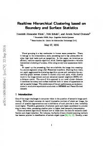
ECG procesing time
Real-Time Classification of ECGs on a PDA. Rodríguez, Goñi and Illarramendi
800 700 600 500 400 300 200 100 0
III. IMPLEMENTATION OF THE CLASSIFIER ON A PDA
9 For this experiment, we simulate the ECG sensors with software in a PC that sends the ECG data of the MIT-DB to the PDA, via Bluetooth.
61
129
199
281
368
454
542
634
ECG adquisition time PDA_1
PC_1
700
ECG procesing time
After having developed an ECG beat and rhythm classifier it was necessary to prove if the classifier could be run, in real time, into a PDA, because it is known that the most powerful current PDAs, even with the latest technological advances, are environments with limited computing resources if compared to PCs. Moreover, the processing tasks that a monitoring system implies require a high computation cost: the signal acquisition, processing and visualization (see figure 3). The signal acquisition9 implies: the picking up of the sample (in our case with frequency of 360 samples per second that is equivalent to 21,600 samples per minute), the conversion of the digital samples into a format understandable by the rest of the system (signal preprocessor and classifier) and their grouping together into signal packages with a defined size. The signal processing implies running several threads: the thread that performs the preprocessing and classification, the thread that stores the signal and classification results in a local database; the thread that manages the alarms and finally another thread in charge of the communications between the PDA and a control center. The hard restriction here is that the thread that preprocesses and classifies the signal has to finish before the signal acquisition obtains the next signal package. In this section, we will answer the next question: which is the appropriate size of a signal package? or, in other words, how often does the signal process have to be executed? We will call “processing cycle duration” to that time. It is obvious that the greater the processing cycle duration is, the greater the rhythm detection delay is. The rhythm detection delay grows with the signal package size because at least four beats are needed in order to classify the rhythm, and some beats may delay until the package is completed according to the processing cycle duration. However, the processing cycle duration cannot be very small because the system would get overloaded: the threads of the signal processing have to synchronize with the thread that performs the signal acquisition. And, moreover, it does not have much sense to start a new processing cycle if a new signal package with at least one beat has not yet arrived: no new rhythm can be detected.
10
600 500 400 300 200 100 0 66
132
196
260
324
388
452
518
582
646
ECG adquision time PDA-2
PC-2
Fig. 4. Time in which an acquired signal starts its processing, for signal packets of one and two seconds
Therefore, in order to establish the optimal processing cycle duration we tested the system performance for processing cycles of one and two seconds respectively. Both types of test were performed in the PDA10 and in a PC (with the goal of pointing up their different performance). The experiment consisted on: 1) running several threads into each device: a signal acquisition thread and all threads involved in the signal processing and classification, storing and visualization; and 2) measuring when the signal package, provided by the signal acquisition, started its processing in the signal processor and classifier. Notice that only processing times of those threads into PC and PDA had an influence on that time and not communication times from the sensors to PC and PDA. Figure 4 shows four functions: PC-1, PDA-1, PC-2 and PDA-2. Every point (x,y) of all those functions indicates that the signal package provided by the signal acquisition thread at the second x is processed by the processor thread at the second y. For PC-1 and PDA-1 functions, the processing cycle duration is of 1 second, and for PC-2 and PDA-2 functions it is of 2 seconds. In PC-1 and PC-2, the processing cycle is performed in the PC, and, obviously PDA-1 and PDA-2 in the PDA. As it can be observed in the figures, in both cases the system running in the PC achieves a stable state since the corresponding functions are very close to the diagonal function 10 The platform used for the implementation of classifier has been the next PDA: an iPaq 3970 with a 400Mhz XScale processor, 64MB SDRAM and 48 MB Flash memory. The PC configuration was Pentium IV (512MB RAM, 2.4GHHz).
Real-Time Classification of ECGs on a PDA. Rodríguez, Goñi and Illarramendi (that would mean that the signal packet received at second x is processed by the system at second x). The stability comes from the fact that the difference between the diagonal and the PC-x functions does not grow with time. In other words, the system performs all the tasks before the next signal package has been arrived. In the PDA case, for processing cycles of one second, this property is not achieved but it was achieved with processing cycles of two seconds. Therefore, the optimal processing cycle duration would be of 2 seconds for the case of the PDA. In that case the average rhythm detection delay would be of 6.66 seconds. In the PC case, the optimal processing cycle duration would be of 1 second being the average rhythm detection delay of 4.43 seconds.
[3]
[4]
[5] [6] [7] [8]
[9]
[10]
IV. CONCLUSION AND FUTURE WORK Monitoring systems that perform a complete ECG analysis in a local device near the patients are of great interest because they allow to improve the quality of life of persons that suffer from arrhythmias and reduce communication costs. For an anywhere and at anytime monitoring system, used devices have to be actually mobile. That is why we advocate for using PDAs as the core of these kinds of monitoring systems. In this paper, we have presented the steps followed in order to build a complete ECG beat and rhythm classifier for a PDA. The obtained results for the classifier have shown that it is comparable to other ECG classifiers found in the literature. In particular, it provides a very good accuracy for classifying rhythms (100% for arrhythmias that require medical assistance in less than 3 minutes, 97.95% for arrhythmias that require medical assistance in less than an hour and 95% for arrhythmias that usually happen before a worse arrhythmia). Finally, we have incorporated that classifier into a PDA, and performed a set of experiments that show its feasibility. Moreover, those experiments have shown that the ECG signal processing and the classification can be performed in real-time on the PDA by using a processing cycle duration of 2 seconds, that is, if it is performed every 2 seconds. In that case, the rhythm detection delay would be of 6.66 seconds. As future work, we plan to incorporate this PDA into a realtime monitoring system that acquires and analyzes ECG of people that may be moving, and sends alarms to a hospital center when high-risk arrhythmias are found.
[11]
[12]
[13] [14] [15]
[16] [17]
[18]
[19] [20]
[21]
[22]
[23]
ACKNOWLEDGMENT
[24]
We thank I. Beaumont, L. Dranca and I. Inza for their help in the implementation and the use of classification tools, and Dr. Montes for his help with some cardiologic aspects.
[25]
REFERENCES [1] [2]
Farreras and Rozman, Medicina Interna. 13rd ed. in CD-ROM, ch. 3, pp. 395-523. Oct, 2001. Medtronic Reveal® Insertable Loop Recorder. "The World's First Implantable Diagnostic Device". Available: www.medtronic.com.
[26]
[27]
11
N. Daja, I. Relgin, and B. Reljin, “Telemonitoring in cardiology–ECG transmission by mobile phone”. Journal Annals of the Academy of Studenica 4, 2001. QRS Diagnostic (2004, Sep 8). PDAs-Mobile Diagnostic Workstations. QRS Diagnostic [Online]. QRS White Paper. Available: http://www.qrsdiagnostic.com. Cardio Control (2004, Sep 8). Cardio Perfect CE. Cardio Control [Online]. Product. Available: http://www.cardiocontrol.com/cardio.htm. ActiveECG (2004, Sep 8). Available: http://www.activecenter.com. Pulse Medical Limited (2004, Sep 8). ECG System, MeditSense 100 H. Pulse Medical Limited [Online]. Available: www.pulsemedical.co.uk I. Sachpazidis, “@Home: a modular telemedicine system.” in Proc. 2nd workshop on Mobile Computing in Medicine, Heidelberg, Germany, April 2002. D. Konstansas, V. Jones and R. Hersog, “MobiHealth- innovative 2.5/3G mobile services and applications for healthcare”, in Workshop on Standardization in E-Health. Geneva, Italy. May 2003. C. Kunze, U. Grossmann, W. Stork and K.D. Müller-Glaser, “Application of ubiquitous computing in personal health monitoring systems.” in Proc. 36th Ann. meeting of the German Society for Biomed. Eng., 2002. J. Bai, Y. Zhang, D. Shen, L. Wen, C. Ding, Z. Cui, F. Tian, B. Yu, B. Dai and J. Zhang. A portable ECG and blood pressure telemonitoring system. IEEE Engineering in Medicine and Biology Magazine, 18(4): pp 63-70, Jul-Aug 1999 J. Bai, Y. Zhang, B. Dai, Z. Zhu, J. Zhang, Z. Cui, J. Lin and D. Ye, 1996, The design and preliminary evaluation of home ECG/BP monitoring network. J.Telemedicine and Telecare, 2(1):100-106 R. Le Blanc, “Quantitative analysis of cardiac arrhythmias.” CRC: Critical Review in Biomedical Engineering, 14(1):1-43, 1986. T. M. Mitchell, Machine Learning. ISBN 0-07-042807-7. J.L. Talmon, R.M. Dassen and V. Karthaus. “Neural nets and classification trees: A comparison in the domain of ECG analysis.” In Gelsema and Kanal (edss) [118], pages 415-423. Physionet (2004, Sep 8). The research resource for complex physiologic signals. PhysioNet [Online]. Available: http://www.physionet.org/. P. Jané, A. Blasi, J. García and P. Laguna. “Evaluation of an Automatic Threshold Based Detector of Waveform Limits in Holter ECG with the QT database”. in Computers in Cardiology 1997, vol. 24, pp. 295-298. I. H. Witten and E. Frank. Data Mining: Practical Machine Learning Tools and Techniques with Java Implementations. Morgan Kaufmann Publishers, 1999. AnswerTree (2004, Sep 8). AnswerTree. SPSS [Online]. Available: http://www.spss.com/spssbi/answertree/. J. R. Quinlan, “An empirical comparison of genetic and decision tree classifiers.” in 15th Int. Conf on Machine Learning, pp 135-141, Ann Arbor, Michigan, 1988. J. R. Quinlan. “Comparing connectionist and symbolic learning methods.” In Proc. of Workshop on Computational Learning Theory and Natural Learning Systems. vol 1. Constraints and Prospects. MIT Press, 1993. G. Krishna Prasad and J. S. Sahambi. “Classification of ECG Arrhythmias using multi-resolution analysis and neural network”. IEEE TENCON [OnLine] Available: http://www.ewh.ieee.org/ecc/r10/ Tencon2003/Articles/739.pdf. 2003 P. Aguirre, J. Cardelina and N. Loeff, “CARDIDENT: sistema de detección, clasificación e identificación en línea de complejos QRS.” InfoSuis, 13(2). ISSN 1510-2173. M. Lagerholm and C. Peterson. Clustering ECG Complex Using Hermite Funtion and self-organizing maps. IEEE Trans Biomed Eng. 2000 Jul;47(7):838-48 S. Osowski, L. T. Hoai and T. Markiewicz. Support Vector Machine based expert system for reliable heart beat recognition. TBME-0036612002. U. Ayesta, L. Serrano and I. Romero. Complexity Measure revisited: A new algorithm for classifying cardiac arrhythmias. in 23rd Ann. Int. Conf. of the IEEE Engineering in Medicine and Biology Society, 2001. D. Ge, N. Srinivasan and S. M. Krishnan. (2004, Sep 8). “Cardiac arrhythmia classification using autoregressive modeling. BioMed Eng” [Online].Available:http://www.pubmedcentral.nih.gov/articlerender.fcgi ?artid=149374.
Real-Time Classification of ECGs on a PDA. Rodríguez, Goñi and Illarramendi [28] R. Kohavi. “A Study of Cross-Validation and Bootstrap for Accuracy Estimation and Model Selection”. in international joint Conf on Artificial Intelligence (IJCAI), 1995. Jimena Rodríguez was born in San Luis, Argentina, in 1975. She received the BSc degree in Computer Science from the Blas Pascal University of Cordoba, Argentina in 1999. Since 2000, she is researcher member in the Interoperable Database Group at the Computer Science Faculty of the Basque Country University where she is a PhD candidate. Her research interests are in classification and management of biomedical signals into mobile computing environment. Alfredo Goñi was born in Pamplona, Spain, in 1967. He received the BSc degree and PhD in Computer Science from the University of the Basque Country in 1990 and 1996 respectively. He has been working at the Public University of Navarra, University of Zaragoza and University of the Basque Country where he is now an Associate Professor. His main interests are data management in mobile computing, telemedicine and information systems. He has published several articles in international journals and conferences. Arantza Illarramendi was born in San Sebastián, Spain. She received the BSc degree from the Computer Science Faculty of the Technical University of Madrid, Spain, and her PhD in computer science from the Basque Country University, Spain. Since January 1995, she is a Full Professor at the Basque Country University. Her main research interests are data management in mobile environments and semantic interoperability among information systems. Her publications include several journal and conference articles. She is member of the IFIP WG 2.6.
12