letters to nature ..............................................................
Recognition memory may be supported by two independent types of retrieval, conscious recollection of a specific experience and a sense of familiarity gained from previous exposure to particular stimuli1,2. In humans, signal detection techniques have been used to distinguish recollection and familiarity, respectively, in asymmetrical and curvilinear components of their receiver operating characteristic (ROC) curves, standard curves that represent item recognition across different levels of confidence or bias. To determine whether animals also employ multiple processes in recognition memory and to explore the anatomical basis of this distinction, we adapted these techniques to examine odour recognition memory in rats. Their ROC curve had asymmetrical and curvilinear components, indicating the existence of both recollection and familiarity in rats. Furthermore, following selective damage to the hippocampus the ROC curve became entirely symmetrical and remained curvilinear, supporting the view that the hippocampus specifically mediates the capacity for recollection. When meeting people on a second occasion, we sometimes recall
the previous encounter fully but at other times experience only a sense of familiarity without recollective experience. Studies on human amnesic patients and functional neuroimaging in normal human subjects have suggested that the hippocampus may be critical to recollection whereas familiarity may be mediated by the surrounding cortical areas3–5. However, this proposal is controversial6,7, and definitive evidence on the specific role of the hippocampus is beyond the anatomical resolution of current neuropsychological and functional imaging studies on humans. Animal models are well suited for investigations of the specific contribution of the hippocampus in recognition memory because they allow circumscribed brain damage to be generated experimentally, and previous studies on animals have indicated functional distinctions between the hippocampus and adjacent cortical areas8,9. However, studies on animals have also failed to provide consensus in that the severity and rate of forgetting in recognition deficits following hippocampal damage is highly variable across different tests10–17. A possible explanation is that the hippocampus mediates only the recollective component of recognition and any residual accuracy in performance is due to extra-hippocampal familiarity processes. Our strategy in testing this hypothesis takes advantage of differences in the retrieval dynamics of recollection and familiarity. In several theoretical models, familiarity is indexed by graded memory strength supporting a precise match between a current stimulus and stored memory, whereas recollection involves a threshold for retrieval of associative and contextual information about an event (for review see ref. 1). An approach that has been particularly useful in distinguishing recollection and familiarity on the basis of these differences is the application of signal detection theory in the analysis of ROCs. In these studies, subjects are initially presented
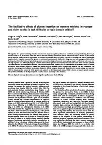
Figure 1 ROCs for recognition performance in humans and rats. a–c, Performance of humans in verbal recognition (adapted from ref. 20). d–f, Performance of rats in odour recognition. d, Normal rats tested with a 30-min delay. Insets: recollection (R) and familiarity (F) estimates. e, Postoperative performance with a 30-min delay, including an
estimated curve for controls based on familiarity alone (con F). f, Control rats tested with a 75-min memory delay. Diagonal dotted lines represent chance performance across criterion levels. C, control group; H, hippocampal group. Error bars, ^s.e.m.; asterisk, P , 0.05.
Recollection-like memory retrieval in rats is dependent on the hippocampus Norbert J. Fortin, Sean P. Wright & Howard Eichenbaum Center for Memory and Brain, Boston University, 2 Cummington Street, Boston, Massachusetts 02215, USA .............................................................................................................................................................................
188
©2004 Nature Publishing Group
NATURE | VOL 431 | 9 SEPTEMBER 2004 | www.nature.com/nature
letters to nature with a list of items to remember, then following a delay, are presented with the same (old) items as well as new items. Recognition performance is scored in terms of hits (correct identification of old items) and false alarms (misidentification of new items, as though they were old). ROC curves relate the proportion of hits to false alarms across a range of confidence levels or response criteria18. In the latter case, the curve varies from a ‘liberal’ response criterion, where subjects make a high proportion of hits but also generate many false alarms (Fig. 1a, upper right corner), to a ‘conservative’ criterion, where subjects generate few false alarms but also make a smaller proportion of hits (lower left corner). Chance performance at intermediate criteria falls along the diagonal whereas successful recognition results in an ROC function above the chance line, reflecting more hits than false alarms. Supporting the theoretical distinctions in retrieval dynamics, ROC analyses have revealed an overall asymmetric recognition performance pattern (Fig. 1a), composed of two independent processes: a symmetrical, curvilinear component associated with familiarity (Fig. 1b) and an asymmetrical, linear component marked by above-zero threshold recollection even under the most stringent bias conditions (Fig. 1c). Here we adapted signal detection theory for testing recognition memory in rats to address three questions. First, can recognition memory be assessed in rats using ROC analyses? Second, do rats have distinct processes for recollection and familiarity in recognition memory? Third, is the hippocampus selectively involved in recollection? We designed a recognition memory task that exploits rats’ superb memory capacity for odours. In each daily session, rats initially sampled a list of common household scents. Then, following a 30-min delay, old and new odours were individually presented in a random order. Across sessions the rats’ response criterion was biased by altering the difficulty of responding to the test odour and the pay-off ratio for correct ‘new’ and ‘old’ responses (see Methods). Initially, we tested 12 intact rats across the full range of
Figure 2 Lesions of the hippocampus reconstructed on coronal sections of the rat brain. Numbers indicate anterior–posterior (AP) distances (in mm) from bregma. Dark grey, smallest lesion; light grey, largest lesion. NATURE | VOL 431 | 9 SEPTEMBER 2004 | www.nature.com/nature
bias levels and then plotted mean scores for each bias level (1–5) (Fig. 1d). The ROC curves were then derived by fitting the data points by using a least-squares model with recollection and familiarity as parameters (see Supplementary Information). The ROC was significantly curvilinear (F Quad 2,2 ¼ 30.60, P , 0.05), suggesting the contribution of a familiarity process3,19. Furthermore, the ROC was asymmetric, with a positive Y-intercept (R ¼ 0.40) and the slope of the z-transformed linear approximation was less than 1 (t 11 ¼ 23.17; P , 0.05; see Supplementary Information), indicating the presence of a recollection component as well (see ref. 19). This pattern closely matches the overall ROC of humans in verbal recognition performance (compare to Fig. 1a; ref. 20). On the basis of estimates of recollection and familiarity components of performance calculated for each subject, we then separated the rats into two matched groups (Fig. 1d, inset). One group received selective lesions to the hippocampus and the other received sham control operations (Fig. 2). After recovery, overall control performance was 73% correct and animals with hippocampal lesions were modestly impaired (66%; t 10 ¼ 22.39, P , 0.05). The deficit was not attributable to a general shift in response bias (overall response to test cup ¼ 46% for controls, 50% for hippocampal rats; t 10 ¼ 0.766). The ROC of control rats continued to reflect both the recollective and familiarity components (Fig. 1e). By contrast, the ROC of animals with selective hippocampal lesions became fully symmetrical (R ¼ 0; z-transformed slope not different from 1, t 5 ¼ 0.10) and remained curvilinear (F Quad 2,2 ¼ 155.47, P , 0.05), characteristic of recognition based solely on familiarity (compare to Fig. 1b). Furthermore, when the contribution of the recollective component was algebraically removed from the ROC of control animals, the resulting curve superimposed onto the ROC of rats with hippocampal lesions (see ‘con F’ in Fig. 1e; see Supplementary Information). Additional analyses of the raw scores also provide compelling evidence that recollection is severely impaired in rats with hippocampal lesions, whereas familiarity is intact. The hit rates of the lesioned group were significantly lower than those of the control group (F 1,10 ¼ 6.61, P , 0.05; group £ bias level interaction: F 4,40 ¼ 2.87, P , 0.05) but not from those of the con F group (F 1,10 ¼ 0.26; group £ bias level interaction: F 4,40 ¼ 0.54). False alarm rates did not differ between the groups (F 1,10 ¼ 0.13; group £ bias level interaction: F 4,40 ¼ 0.14; see Supplementary Information). Subsequent planned comparisons also showed that familiarity estimates did not differ between the two groups (t 10 ¼ 0.65; Fig. 1e inset; see Supplementary Information) and remained significantly greater
Figure 3 Odour recognition task. a, Sequence of odour presentations. b, Test cup heights and reward pay-offs for each bias level.
©2004 Nature Publishing Group
189
letters to nature than 0 (controls: t 5 ¼ 4.12, P , 0.05; hippocampus: t 5 ¼ 5.04, P , 0.05), whereas recollection estimates for the hippocampal group were significantly lower than those of the control group (t 10 ¼ 2.13, P , 0.05) and did not differ significantly from 0 (t 5 ¼ 1.70; controls .0: t 5 ¼ 3.37, P , 0.05). Given that any performance deficit must result in an ROC closer to the diagonal (chance performance), it is possible that the alteration in the ROC pattern of hippocampus-damaged animals is not specific to recollection, but reflects a generalized decline in memory. To test this hypothesis, we challenged control rats by increasing the memory delay to 75 min. This manipulation succeeded in reducing their overall performance to 64% correct, numerically below that of the rats with hippocampal lesions at the 30-min delay. Under those conditions, the ROC of controls became more linear (R 2linear 30 min ¼ 0.86, R 2linear 75 min ¼ 0.99; t 5 ¼ 23.36, P , 0.05), suggesting that familiarity contributed substantially less to performance. However, unlike the ROC of animals with hippocampal lesions, the ROC of controls continued to have an asymmetrical recollective component (Y-intercept .0; t 5 ¼ 2.13, P , 0.05; z-transformed slope ,1, t 5 ¼ 24.49, P , 0.05; Fig. 1f). The degree of curvature (R 2Quad 2 R 2Linear) was also higher in the hippocampal group at 30 min than the control group at 75 min (t 10 ¼ 2.71, P , 0.05), confirming that the curves are qualitatively different. This pattern of performance indicates that the deficit observed in rats with hippocampal damage is specific to recollection and not the consequence of a general decrease in performance. Furthermore, these findings are consistent with previous reports that familiarity decays more than recollection shortly after learning in humans21,22. An alternative model of recognition memory argues that an asymmetrical ROC curve can be explained by the combination of two parameters: the difference in memory strength between old and new item distributions (d 0 ), and a larger variance of the old item distribution than that for the new items (Vold . V new ¼ 1; ref. 18). The unequal-variance model can account for the ROC of normal rats before surgery (V old ¼ 1.27 and d 0 ¼ 1.76) and the postoperative ROC of control rats (Vold ¼ 1.33 and d 0 ¼ 1.63). This model can also account for the performance of rats with hippocampal lesions, but the variances become equal (Vold ¼ 0.97; not significantly different from V new ¼ 1.0, t 5 ¼ 0.63) leaving a signaldetection process governed by only a single variable (d 0 ¼ 1.01), as in the case of the dual-process model. Thus, the unequal-variance model confirms that hippocampal lesions selectively eliminate the specific factor that mediates the identification of old items under the most conservative response criterion, analogous to the effect of removing recollection in the dual-process model. Our results could explain the pattern of findings from previous studies on monkeys and rats performing the delayed non-match to sample (DNMS) recognition task10–15,23. The behavioural protocol used here is quite similar to the DNMS task, and indeed revealed the typical modest impairment in recognition following hippocampal lesions. The present analyses allow us to understand the modest overall deficit as reflecting the combination of a severe and selective impairment in recollection contrasted with normal familiarity. Also, the present results suggest that mixed findings across different types of recognition task10–17 may be a consequence of differential loading of recollective and familiarity memory demands. Our findings do not shed light on the subjective experience of recollection and familiarity in animals24, but do provide the first objective evidence of a distinction between the two processes in a non-human species. Furthermore, our results also reveal that the threshold retrieval process characteristic of human episodic recollection is dependent on the hippocampus. Other studies of recollection in humans and hippocampal-dependent memory in animals have emphasized the importance of memory for associations between items4,25,26, for spatial16,17 and temporal context27,28, for bridging temporal gaps29 and for the flow of events in unique 190
experiences30. The present observations add to that list of properties a fundamental role for the hippocampus in retrieval dynamics associated with recollective experience. A
Methods Subjects were 225–250-g male Long Evans rats maintained at a minimum of 85% of normal body weight. They were initially trained to dig for quartered Cheerio cereal rewards in 125-ml plastic cups filled with playground sand scented with distinct odours (see ref. 30 for details), which were presented individually in the front of the home cage. Then the rats were trained on a non-matching task in which a single odour was presented as a sample, followed by two test odours presented consecutively. On each test, the animal obtained an additional reward by digging in the test cup if the odour was ‘new’ (that is, non-match) or by refraining from digging in the test cup and approaching an alternate empty cup at the back of the cage if the odour was ‘old’ (that is, match). Each animal was trained to a criterion of 80% correct over three consecutive sessions (six trials per session; average sessions to criterion ¼ 11.25 ^ 2.83 s.d.). In subsequent training, each session consisted of a presentation of a list of five sample odours followed by five new and five old test odours presented in a random order. Subjects reached a criterion of 80% correct over three consecutive sessions (average sessions to criterion ¼ 37.83 ^ 6.32). For the final training, five different response criteria (conservative to liberal) were generated by biasing the animal’s choices through varying the height of the test cup (4 cm, 6.5 cm and 8.5 cm) and the amount of Cheerios provided at the correct choice (between a quarter and three pieces; see Fig. 3). Initially, each session consisted of a 10-odour list at bias level two (same as previous training) for 21.75 ^ 2.52 sessions, then animals were acclimated to the five bias levels for 15 sessions (three per bias level). ROC analyses were performed on the subsequent 20 preoperative and 20 postoperative sessions (four sessions per bias level for each) using a 30-min delay between the end of the sample list and the beginning of the tests. In each session, the first four test odours allowed the animal to set its response criterion, then performance on the remaining 16 test odours was used to generate ROC curves for each subject. In addition, estimates of recollection and familiarity were calculated on the basis of the Y-intercepts and the d 0 s (transformed into a probability to facilitate comparison with recollection, see Supplementary Information), respectively, for individual subjects. Also, an idealized ROC curve for familiarity alone was calculated by algebraically removing the estimated recollection component from the control curve (compare with Fig. 1c; see Supplementary Information). Finally, controls were again tested for 20 sessions with a 75-min delay. Following preoperative training, animals were operated on and received either selective lesions to the hippocampus or sham lesions. Anaesthesia was administered using nitrous oxide and was supplemented by 1% halothane. Atropine sulphate (0.081 mg) was injected to prevent respiratory difficulties, and body temperature was maintained at 37 8C. At each of 12 sites bilaterally, the dura was pierced, a 100-mm nichrome electrode (0.7-mm uninsulated tip) was lowered into the hippocampus, and lesions were made by passing a 7–11-mA radiofrequency current (Radionics RFG-4A) for 1 min. Sham controls underwent the same surgery, except that the electrode was not lowered into the brain after puncturing the dura. Animals recovered for two weeks and regained their preoperative weights. Lesioned animals lost 32–61% (mean ¼ 42%) of the total volume of the hippocampus, sparing the adjacent subiculum and perirhinal, postrhinal and entorhinal cortices. The cortex overlying the hippocampus was slightly damaged in some animals. Two animals had slight damage to the optic tract and one to the medial geniculate nucleus. Received 6 May; accepted 12 July 2004; doi:10.1038/nature02853. 1. Yonelinas, A. P. The nature of recollection and familiarity: a review of 30 years of research. J. Mem. Lang. 46, 441–517 (2002). 2. Sherman, S. J., Atri, A., Hasselmo, M. E., Stern, C. E. & Howard, M. W. Scopolamine impairs human recognition memory: data and modeling. Behav. Neurosci. 117, 526–539 (2003). 3. Yonelinas, A. P. et al. Effects of extensive temporal lobe damage or mild hypoxia on recollection and familiarity. Nature Neurosci. 5, 1236–1241 (2002). 4. Davachi, L. & Wagner, A. D. Hippocampal contributions to episodic encoding: insights from relational and item-based learning. J. Neurophysiol. 88, 982–990 (2002). 5. Ranganath, C., Johnson, M. K. & D’Esposito, M. Prefrontal activity associated with working memory and episodic long-term memory. Neuropsychologia 41, 378–389 (2003). 6. Manns, J. R., Hopkins, R. O., Reed, J. M., Kitchener, E. G. & Squire, L. R. Recognition memory and the human hippocampus. Neuron 37, 171–180 (2003). 7. Stark, C. E. & Squire, L. R. Functional magnetic resonance imaging (fMRI) activity in the hippocampal region during recognition memory. J. Neurosci. 20, 7776–7781 (2000). 8. Eichenbaum, H., Otto, T. & Cohen, N. J. Two functional components of the hippocampal memory system. Behav. Brain Sci. 17, 449–472, 472–518 (1994). 9. Brown, M. W. & Aggleton, J. P. Recognition memory: what are the roles of the perirhinal cortex and hippocampus? Nature Rev. Neurosci. 2, 51–61 (2001). 10. Beason-Held, L. L., Rosene, D. L., Killiany, R. J. & Moss, M. B. Hippocampal formation lesions produce memory impairment in the rhesus monkey. Hippocampus 9, 562–574 (1999). 11. Zola, S. M. et al. Impaired recognition memory in monkeys after damage limited to the hippocampal region. J. Neurosci. 20, 451–463 (2000). 12. Nemanic, S., Alvarado, M. C. & Bachevalier, J. The hippocampal/parahippocampal regions and recognition memory: insights from visual paired comparison versus object-delayed nonmatching in monkeys. J. Neurosci. 24, 2013–2026 (2004). 13. Mumby, D. G. Perspectives on object recognition memory following hippocampal damage: lessons from studies on rats. Behav. Brain Res. 127, 159–181 (2001). 14. Dudchenko, P. A., Wood, E. R. & Eichenbaum, H. Neurotoxic hippocampal lesions have no effect on odor span and little effect on odor recognition memory but produce significant impairments on spatial span, recognition, and alternation. J. Neurosci. 20, 2964–2977 (2000).
©2004 Nature Publishing Group
NATURE | VOL 431 | 9 SEPTEMBER 2004 | www.nature.com/nature
letters to nature 15. Steckler, T., Drinkenburg, W. H., Sahgal, A. & Aggleton, J. P. Recognition memory in rats II. Neuroanatomical substrates. Prog. Neurobiol. 54, 313–332 (1998). 16. Mumby, D. G., Gaskin, S., Glenn, M. J., Schramek, T. E. & Lehmann, H. Hippocampal damage and exploratory preferences in rats: memory for objects, places, and contexts. Learn. Mem. 9, 49–57 (2002). 17. Eacott, M. J. & Norman, G. Integrated memory for object, place, and context in rats: a possible model of episodic-like memory? J. Neurosci. 24, 1948–1953 (2004). 18. Macmillan, N. A. & Creelman, C. D. Detection Theory: A User’s Guide (Cambridge Univ. Press, New York, 1991). 19. Yonelinas, A. P., Kroll, N. E., Dobbins, I., Lazzara, M. & Knight, R. T. Recollection and familiarity deficits in amnesia: convergence of remember-know, process dissociation, and receiver operating characteristic data. Neuropsychology 12, 323–339 (1998). 20. Yonelinas, A. P. Components of episodic memory: the contribution of recollection and familiarity. Phil. Trans. R. Soc. Lond. B 356, 1363–1374 (2001). 21. Hockley, W. E. Item versus associative information: further comparison of forgetting rates. J. Exp. Psychol. Learn. Mem. Cogn. 18, 1321–1330 (1992). 22. Yonelinas, A. P. & Levy, B. J. Dissociating familiarity from recollection in human recognition memory: different rates of forgetting over short retention intervals. Psychon. Bull. Rev. 9, 575–582 (2002). 23. Murray, E. A. & Mishkin, M. Object recognition and location memory in monkeys with excitotoxic lesions of the amygdala and hippocampus. J. Neurosci. 18, 6568–6582 (1998). 24. Tulving, E. Episodic memory: from mind to brain. Annu. Rev. Psychol. 53, 1–25 (2002). 25. Bunsey, M. & Eichenbaum, H. Conservation of hippocampal memory function in rats and humans. Nature 379, 255–257 (1996). 26. Day, M., Langston, R. & Morris, R. G. Glutamate-receptor-mediated encoding and retrieval of pairedassociate learning. Nature 424, 205–209 (2003). 27. Wood, E. R., Dudchenko, P. A., Robitsek, R. J. & Eichenbaum, H. Hippocampal neurons encode information about different types of memory episodes occurring in the same location. Neuron 27, 623–633 (2000). 28. Kesner, R. P. in Neurobiology of Comparative Cognition (eds Kesner, R. P. & Olton, D. S.) 179–203 (Lawrence Erlbaum, New Jersey, 1990). 29. Clark, R. E. & Squire, L. R. Classical conditioning and brain systems: the role of awareness. Science 280, 77–81 (1998). 30. Fortin, N. J., Agster, K. L. & Eichenbaum, H. Critical role of the hippocampus in memory for sequences of events. Nature Neurosci. 5, 458–462 (2002).
during nerve growth. By contrast, myelination proceeds normally. The capacity of wild-type and mutant Schwann cells to elongate is cell-autonomous, indicating that passive stretching can account for the lengthening of the internode during limb growth. As predicted on theoretical grounds, decreased internodal distances strikingly decrease conduction velocities and so affect motor function. We propose that microtubule-based transport in the longitudinal bands of Cajal permits internodal Schwann cells to lengthen in response to axonal growth, thus ensuring rapid nerve impulse transmission. Nodes of Ranvier in peripheral nerves are flanked by Schwann
Supplementary Information accompanies the paper on www.nature.com/nature. Acknowledgements We thank J. O’Connell, A. Milewski, L. Giocomo, J. Estes, D. Tosa, R. Kline, J. Davidson and B. Goldberg for help with behavioural testing. We also thank R. Komorowski for histological work, A. Yonelinas for providing the least-squares curve-fitting algorithm and J. Manns for comments on an earlier version of this manuscript. Supported by NIA and NIMH. Competing interests statement The authors declare that they have no competing financial interests. Correspondence and requests for materials should be addressed to H.B.E. (
[email protected]).
..............................................................
Restricted growth of Schwann cells lacking Cajal bands slows conduction in myelinated nerves Felipe A. Court, Diane L. Sherman, Thomas Pratt, Emer M. Garry, Richard R. Ribchester, David F. Cottrell, Susan M. Fleetwood-Walker & Peter J. Brophy Centre for Neuroscience Research, University of Edinburgh, Edinburgh EH9 1QH, UK .............................................................................................................................................................................
Nerve impulses are propagated at nodes of Ranvier in the myelinated nerves of vertebrates. Internodal distances have been proposed to affect the velocity of nerve impulse conduction1; however, direct evidence is lacking, and the cellular mechanisms that might regulate the length of the myelinated segments are unknown. Ramo´n y Cajal described longitudinal and transverse bands of cytoplasm or trabeculae in internodal Schwann cells and suggested that they had a nutritive function2. Here we show that internodal growth in wild-type nerves is precisely matched to nerve extension, but disruption of the cytoplasmic bands in Periaxin-null mice impairs Schwann cell elongation NATURE | VOL 431 | 9 SEPTEMBER 2004 | www.nature.com/nature
Figure 1 Longitudinal and transverse bands of Cajal in Schwann cells and their disruption in quadriceps nerve of Periaxin-null (KO) mice at 3 weeks. a, Longitudinal and transverse protoplasmic bands stained with silver by Ramo´n y Cajal (reproduced with permission)2. b, Teased fibres double-labelled with TRITC–phalloidin (green) and an antibody against DRP2 (red). Schwann cell cytoplasm is excluded from spheroidal clusters immunopositive for DRP2. c, Immunostaining of fibres from WT and Prx KO mice for the Schwann cell cytoplasmic protein S100. Scale bar, 20 mm in b and c. d–f, Electron micrographs of transverse sections of WT and KO quadriceps nerves. d, WT Schwann cell cytoplasm (asterisks) is restricted to regions delimited by appositions between the Schwann cell plasma membrane and the abaxonal layer of the myelin sheath. e, The sharp transition between the apposition and cytoplasmic zones is shown by the arrow. Scale bar, 0.2 mm. The inset shows a high-power view of the transition zone. f, In the absence of appositions in the KO, the Schwann cell cytoplasm forms a concentric ring around the myelin sheath. Scale bar, 1 mm (d and f ). g, The proportion of abaxonal appositions per Schwann cell in WT and KO mice (n ¼ 3 for WT and KO).
©2004 Nature Publishing Group
191