Fisher-Hoch, S. P., J. A. Khan, S. Rehman, S. Mirza, M. Khurshid, and J. B.. McCormick. 1995. ... An outbreak at the Rashid Hospital. Lancet ii:939â941. 21.
JOURNAL OF CLINICAL MICROBIOLOGY, May 2002, p. 1587–1591 0095-1137/02/$04.00⫹0 DOI: 10.1128/JCM.40.5.1587–1591.2002 Copyright © 2002, American Society for Microbiology. All Rights Reserved.
Vol. 40, No. 5
Recombinant Nucleoprotein-Based Enzyme-Linked Immunosorbent Assay for Detection of Immunoglobulin G Antibodies to Crimean-Congo Hemorrhagic Fever Virus Masayuki Saijo,1 Tang Qing,2 Masahiro Niikura,1 Akihiko Maeda,1 Tetsuro Ikegami,1 Christophe Prehaud,3 Ichiro Kurane,1 and Shigeru Morikawa1* Special Pathogens Laboratory, Department of Virology 1, National Institute of Infectious Diseases, Gakuen 4-7-1, Musashimurayama, Tokyo 208-0011, Japan1; Second Division of Viral Hemorrhagic Fever, Institute of Epidemiology and Microbiology, Chinese Academy of Preventive Medicine, Changping, Beijing 102206, People’s Republic of China2; and Unité de Neuroimmunologie Virale, Institut Pasteur, 75724 Paris Cedex 15, France3 Received 23 August 2001/Returned for modification 27 September 2001/Accepted 22 January 2002
The full-length nucleoprotein of Crimean-Congo hemorrhagic fever virus (CCHFV; 482 amino acid residues) was expressed as a His-tagged recombinant protein (His-CCHFV rNP) in the baculovirus system. The HisCCHFV rNP was efficiently expressed in insect cells and purified by Ni2ⴙ column chromatography. Using this substrate, an immunoglobulin G (IgG) enzyme-linked immunosorbent assay (ELISA) was developed. We evaluated the sensitivity and specificity of the IgG ELISA, using serum samples previously determined to be antibody positive or negative by immunofluorescence tests on CCHFV-infected Vero E6 cells. We found very good correlation between the two tests: 87% for the positive sera (13 of 15) and 99% for the negative sera (107 of 108). These results indicate that the new IgG ELISA using His-CCHFV rNP has high sensitivity and specificity for detecting CCHFV antibodies. The CCHF patients’ sera with high titers reacted only with the NP fragment containing amino acid residues between 201 and 306 in Western blotting. It is known that amino acid homologies are high in this region among various isolates. Thus, it is expected that this ELISA can detect antibodies not only for Chinese strains of CCHFV but also for other strains circulating in the world. These results suggest that the IgG ELISA system developed with the recombinant CCHFV NP is a valuable tool for diagnosis and epidemiological investigations of CCHFV infections. break-free areas through CCHFV-infected ticks, humans, and animals. In the present study, we developed an enzyme-linked immunosorbent assay (ELISA) to detect CCHFV-directed immunoglobulin G (IgG) by using the recombinant nucleoprotein (rNP). We demonstrated that this new ELISA system has high sensitivity and specificity in detecting CCHFV antibody in human sera in comparison to the indirect immunofluorescence (IIF) method using authentic viral antigen. The results suggest the usefulness of this IgG ELISA for serological diagnosis and epidemiological studies of CCHFV infections.
Crimean-Congo hemorrhagic fever virus (CCHFV) belongs to the family Bunyaviridae (genus Nairovirus) and causes severe hemorrhagic symptoms in humans (7). CCHF is distributed worldwide with the exception of the American continents. There have been many outbreaks of CCHF in countries from South Africa to the western part of China, including Eastern European and Middle Eastern countries (8). CCHFV is a tick-borne virus which is transmitted by ticks from the genus Hyalomma (7). Humans are usually infected with CCHFV either through the bites of infected ticks or by direct contact with virus-contaminated tissues or blood. CCHF outbreaks have been reported among agricultural workers, abattoir workers, and shepherds who handle livestock animals such as sheep, goats, and ostriches (10, 21). Furthermore, nosocomial, or in-house, CCHF infections have also been reported among caregivers (2, 6, 20, 23). It was reported that the epidemic of CCHF in the United Arab Emirates was caused by imported livestock and ticks from Somalia and Nigeria (17). Although there has been no definite evidence that CCHFV is imported from an outbreak area to CCHFV-free countries through CCHFV-infected humans, it is possible that the virus could be introduced to out-
MATERIALS AND METHODS Cells and viruses. The Vero E6 cell line was purchased from the American Type Culture Collection and cultured in Eagle’s minimum essential medium containing 10% fetal bovine serum and antibiotics (penicillin and streptomycin). Tn5 insect cells were also used for the expression of CCHFV rNP in a baculovirus system. Tn5 insect cells were cultured in TC-100 (Life Technologies, Rockville, Md.) supplemented with 10% fetal bovine serum, 2% tryptose phosphate broth (Becton Dickinson Microbiology Systems, Sparks, Md.), and kanamycin. CCHFV (Chinese strain 66019) isolated from a patient with CCHF in the western part of the Xinjiang Autonomous Region, People’s Republic of China, in 1966 was used in the study (24). Sera. Twenty-five serum samples were collected from human subjects in the area where CCHF is endemic, the western part of the Xinjiang Autonomous Region. Two serum samples collected from patients with CCHF in the convalescent phase were provided to us by T. G. Ksiazek, Special Pathogens Branch, National Center for Infectious Diseases, Centers for Disease Control and Prevention (CDC), Atlanta, Ga. Ninety-six serum samples collected from Japanese volunteers who had no past history of travel to the area where CCHF is endemic were used as controls. An anti-CCHFV rNP polyclonal rabbit serum was raised in a rabbit previously immunized with purified CCHFV rNP in the form of a
* Corresponding author. Mailing address: Special Pathogens Laboratory, Department of Virology 1, National Institute of Infectious Diseases, Gakuen 4-7-1, Musashimurayama, Tokyo 208-0011, Japan. Phone: 81-42-561-0771, ext. 791. Fax: 81-42-561-2039. E-mail: morikawa @nih.go.jp. 1587
1588
SAIJO ET AL.
J. CLIN. MICROBIOL.
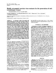
FIG. 1. The entire NP and truncated NP fragments of CCHFV. The truncated NP fragments were expressed as fusion proteins with GST on the N-terminal side. The designations and amino acid positions of the truncated NP fragments are indicated.
mixture with adjuvant (Inject Alum; Pierce, Rockford, Ill.). Further, a monkey (Macaca fascicularis) was immunized with purified CCHFV rNP using the adjuvant Inject Alum, and serum was collected and used as a positive control. The monkey serum collected before immunization was used as a negative control. The method of expression and purification of CCHFV rNP is described below. A monoclonal antibody directed against glutathione S-transferase (GST) protein was generated by fusing the spleen cells collected from BALB/c mice immunized with purified GST-tagged protein with mouse myeloma cells (the P3/Ag568 cell line) and was used in these experiments. Recombinant transfer vector. In order to construct our transfer vector, an entire cDNA clone of NP from CCHFV Chinese strain 8402 was used. The DNA of CCHFV NP was amplified by PCR from the source using primers CCHF-NP/ F(HI) (5⬘-GTGCTGGATCCATGGAGAATAAAATCG-3⬘) and CCHF-NP/ R(HI) (5⬘-CGGATCCTCAGATGATGTTGGCACTG-3⬘) (the BamHI restriction sites are underlined in both sequences). The PCR conditions were 5 cycles of denaturation at 94°C for 40 s, annealing at 40°C for 30 s, annealing at 50°C for 30 s, and extension at 72°C for 2 min; 15 cycles of denaturation at 94°C for 40 s, annealing at 55°C for 30 s, and extension at 72°C for 2 min; and additional extension at 72°C for 5 min. The amplified DNA (1.6 kbp) was digested with BamHI and subcloned into the BamHI site of pQE30 vector DNA (QIAGEN GmbH, Hilden, Germany) to construct pQE30-CCHFV NP. The inserted CCHFV NP DNA was sequenced using appropriate primers with ABI PRISMTM 310 genetic analyzer (PE Applied Biosystems, Foster City, Calif.) and was confirmed to be in proper orientation to the promoter and identical to the original sequence. The DNA fragment of CCHFV NP with a histidine (His) tag was isolated from the plasmid pQE30-CCHFV NP by digestion with EcoRI and SalI. Then, it was repaired for blunting with Klenow enzyme and ligated into the blunt-ended cloning site of pAcYM1 (15). The resultant recombinant transfer vector with the correct orientation to the promoter was designated pAcYM1His-CCHFV NP. Generation of recombinant baculovirus. Tn5 insect cells were transfected with mixtures of purified Autographa californica nuclear polyhedrosis virus (AcMNPV) DNA and recombinant pAcYM1-His-CCHFV NP following the procedures described by Kitts et al. (11) with the modification of Matsuura et al. (15). Recombinant baculovirus was isolated. The baculovirus, which expresses a His-tagged recombinant NP of CCHFV (His-CCHFV rNP), was designated Ac-His-CCHFV NP. A baculovirus (Ac-⌬P) which lacks polyhedrin expression was used as a control virus. Expression of His-CCHFV NP. Ac-His-CCHFV NP-infected Tn5 cells were incubated at 26°C for 72 h. Then, the cells were washed twice with cold phosphate-buffered saline (PBS) solution and lysed in cold PBS solution containing 1% Nonidet P-40 (NP-40). The cell lysate was centrifuged at 13,000 ⫻ g at 4°C for 10 min. The supernatant fraction was collected as a source of His-CCHFV NP for purification. The His-CCHFV rNP was purified using an Ni2⫹ resin purification system (QIAGEN GmbH) according to the manufacturer’s instructions. Expression of His-CCHFV rNP was analyzed on sodium dodecyl sulfatepolyacrylamide gel electrophoresis (SDS-PAGE) gels (12% polyacrylamide) stained with Coomassie blue. Expression of truncated NPs. In order to determine the antigenic regions within the NP, we expressed overlapping fragments of NP (Fig. 1). The DNA corresponding to each of the truncated NP fragments was amplified with the designed primer sets. The amplified DNA was subcloned into the BamHI and EcoRI cloning sites of plasmid pGEX-2T (Amersham Pharmacia Biotech, Little Chalfont, Buckinghamshire, England). Each insert was sequenced and confirmed
to be in a correct frame and identical to the original sequence. The GST-tagged NP fragments were expressed in an Escherichia coli (BL21) system. Western blotting. His-CCHFV rNP and the recombinant NP fragments were tested for reactivity to the serum samples by Western blotting. E. coli (BL21) cell lines expressing each of the NP fragments were resuspended in an appropriate amount of PBS solution and sonicated at full power for 1 min. The sonicated cell suspensions and His-CCHFV rNP were then adjusted to 1⫻ SDS sample buffer (0.0625 M Tris [pH 6.8], 2% SDS, 10% glycerol, 5% -mercaptoethanol, 0.001% bromophenol blue) and boiled for 10 min. Equal quantities of samples were loaded onto an SDS–12% PAGE gel (4% stacking gel). The protein bands were then electroblotted onto nitrocellulose membrane. The blots were incubated in PBS containing 0.05% Tween-20 (TPBS) and 5% skim milk (M-TPBS) at room temperature for 1 h to block nonspecific sites. The blots were incubated with CCHF patients’ sera (1:100 dilution), with the rabbit serum to His-CCHFV rNP (1:500 dilution), or with the monoclonal antibody to GST protein for 1 h at room temperature. After being washed in TPBS, the membrane was incubated with secondary antibodies (horseradish peroxidase [HRPO]-conjugated goat antihuman IgG antibody, HRPO-conjugated goat anti-rabbit IgG, and HRPO-conjugated goat anti-mouse IgG [1:1,000 dilution]; ZYMED Laboratories, San Francisco, Calif.). Antigen levels were visualized by reaction with POD immunostain set (Wako BioProducts, Inc., Tokyo, Japan). ELISA. ELISA was performed as previously described except for the antigen preparation (12, 18). Briefly, ELISA plates were coated with the predetermined optimal quantity of purified His-CCHFV rNP (approximately 100 ng/well) at 4°C overnight. Then, each well of the plates was inoculated with 200 l of M-TPBS and incubated for 1 h for blocking. The plates were washed three times with TPBS and then inoculated with the test samples (100 l/well), which were diluted fourfold from 1:100 to 1:6,400 with M-TPBS. After a 1-h incubation period, the plates were washed three times with TPBS, and then the plates were inoculated with goat anti-human IgG antibody labeled with HRPO (1:1,000 dilution; ZYMED Laboratories). After a 1-h incubation period, the plates were washed and 100 l of ABTS [2,2⬘-azinobis(3-ethylbenzthiazolinesulfonic acid)] solution (Roche Diagnostics, Mannheim, Germany) was added to each well. The plates were incubated for 30 min at 37°C, and the optical density (OD) was measured at 405 nm with reference at 490 nm. The adjusted OD was calculated by subtracting the OD of the noncoated wells from that of the corresponding wells. The mean and standard deviation (SD) were calculated from those of 96 control sera. The cutoff value for the assay was defined as the mean plus 3 SD. Positive- and negative-control monkey sera were tested in each ELISA for verification. IIF. The antibody titer to CCHFV was determined by the IIF method, using CCHFV (strain 66019)-infected Vero E6 cells. Briefly, Vero E6 cells infected with CCHFV were washed with PBS and air dried on 14-well HT-coated slide glasses (AR Brown Co., Ltd., Tokyo, Japan). The cells were then fixed in cold acetone for 10 min and used as antigens. Serum samples were serially twofold diluted with PBS from 1:20 to 1:640. The diluted samples were put on the antigens and incubated for 1 h at 37°C under humidified conditions. The slides were washed with PBS and inoculated with fluorescein isothionate-labeled goat anti-human IgG antibody (1:70 dilution; ZYMED Laboratories). After the slides were washed with PBS, fluorescein isothionate signal was observed under an immunofluorescent microscope (Olympus, Tokyo, Japan). Positive- and negative-control monkey sera were also tested by the IIF method in each IIF test for verification.
VOL. 40, 2002
IgG ELISA USING NUCLEOPROTEIN OF CCHFV
1589
TABLE 1. Relationship between results of IgG ELISA with His-CCHFV rNP and the IIF method using CCHFV-infected Vero E6 cells ELISA result
Positive Negative
No. of IIF results that were: Positive
Negative
13 2b
1a 107
a This serum sample, which showed a negative reaction by the IIF method but a positive reaction by IgG ELISA, was collected from a subject in Xinjiang Autonomous Region, People’s Republic of China. b Antibody titers of these two sera to CCHFV were 40 and 80.
FIG. 2. SDS-PAGE (a) and Western blot (b) analyses of the expression of His-CCHFV NP in insect cells upon infection with Ac-HisCCHFV rNP and purified His-CCHFV rNP. (a) Crude material from the supernatant fraction of 1% NP-40–PBS lysate (lane 1) and purified His-CCHFV rNP (lane 2) are shown. M, molecular mass markers. (b) His-CCHFV rNP in 1% NP-40–PBS lysate was stained by Western blotting with CCHFV antibody-positive human serum (lane 1), while nothing in 1% NP-40–PBS lysate of Ac-⌬P-infected control insect cells was stained with the same serum (lane 2).
determined to be CCHFV antibody positive or negative by IIF testing with CCHFV-infected Vero E6 cells. Fifteen serum samples consisting of 13 sera collected in the Xinjiang Autonomous Region and the 2 sera from CDC were determined to be positive by IIF testing, and 108 serum samples were determined to be negative by IIF testing. The mean and SD obtained using 96 Japanese sera at 1:400 dilution were 0.078 and 0.045, respectively. Therefore, the cutoff value of ODs at 1:400 dilution was determined to be 0.213. Thirteen of the 15 IIF-positive sera and 1 of the 108 IIFnegative sera were determined to be positive by IgG ELISA (Table 1). The 1 serum sample which showed a negative reaction by the IIF test but a positive reaction by IgG ELISA was among the 12 IIF-negative Chinese sera. All the Japanese control sera were determined to be antibody negative by this criterion. The sensitivity and specificity were thus calculated to be 87 and 99%, respectively. Relationships between OD values determined by IgG ELISA and CCHFV antibody titers determined by the IIF method.
Statistical analysis. The antibody titers determined by the IIF test and the OD values determined by IgG ELISA with His-CCHFV rNP were compared by Spearman’s correlation coefficient by rank. Nucleotide sequence accession number. The complete nucleotide sequence of the Crimean-Congo hemorrhagic fever virus (8402 strain) NP gene used in this paper is registered in GenBank under accession no. AJ010649.
RESULTS Expression and purification of His-CCHFV rNP. The expression of His-CCHFV rNP in Ac-His-CCHFV NP-infected insect cells was demonstrated by SDS-PAGE analysis. The His-CCHFV rNP was purified almost to homogeneity, as shown in Fig. 2a. The His-CCHFV rNP was specifically detected by Western blotting with CCHF patient’s serum, indicating that the protein band visualized in Fig. 2a (lane 2) is the expressed recombinant His-CCHFV rNP with the antigenicity of the rNP of CCHFV (Fig. 2b). Sensitivity and specificity of IgG ELISA with His-CCHFV rNP. The positive-control monkey sera showed positive reactions in an IIF test with a titer of 160, while the negativecontrol monkey serum showed a negative reaction. The OD value of the positive-control monkey serum by ELISA with His-CCHFV rNP was 0.885 at 1:400 dilution. Using 123 sera, this IgG ELISA with His-CCHFV rNP was tested for sensitivity and specificity compared with those of an IIF test with authentic CCHFV antigen. These serum samples had been
FIG. 3. Relationship between OD values at 1:400 by IgG ELISA with His-CCHFV rNP and antibody titers determined by the IIF method. Fifteen CCHFV antibody-positive sera and one positive-control monkey serum raised against His-CCHFV rNP serum were plotted.
1590
SAIJO ET AL.
J. CLIN. MICROBIOL.
TABLE 2. Reactions of anti-CCHFV serum samples to full-length rNP of CCHFV (His-CCHFV rNP) and truncated NP fragments
Sample
Reaction of NP fragmentb Antibody titer to GSTHisSpecies CCHFV CCHFV CCHFV NPc by IIF rNP 1 2 3 4 5
MAba to GST Mouse Anti-His-CCHFV rNP Rabbit Serum 1 Human Serum 2 Human Serum 3 Human Serum 4 Human
⬎640 ⬎640 320 ⬎640 ⬎640
⫺ ⫹ ⫹ ⫹ ⫹ ⫹
⫹ ⫺ ⫺ ⫺ ⫺ ⫺
⫹ ⫺ ⫺ ⫺ ⫺ ⫺
⫹ ⫹ ⫹ ⫹ ⫹ ⫹
⫹ ⫺ ⫺ ⫺ ⫺ ⫺
⫹ ⫺ ⫺ ⫺ ⫺ ⫺
a
MAb, monoclonal antibody. Negative (⫺) and positive (⫹) reactions of tested sera to full-length or truncated NP fragments in Western blotting. c The numbers 1 to 5 indicate the name of the truncated fragment (Fig. 1). b
OD values determined by IgG ELISA were compared with antibody titers determined by the IIF method, using the 15 IIF-positive human sera and 1 positive-control monkey serum (Fig. 3). There was a significant positive correlation between the ODs at 1:400 dilution and the CCHFV antibody titers (P ⬍ 0.01). Antigenicities of truncated CCHFV NPs. We attempted to localize the antigenic regions recognized by CCHFV antibodies. We selected four human serum samples, which had high antibody titers to CCHFV, and the rabbit polyclonal serum to His-CCHFV rNP. The full-length CCHFV rNP and the five overlapping truncated NP fragments were examined for antigenicity using these serum samples in Western blotting (Fig. 1). As shown in Table 2, all of the tested sera reacted only to GST-CCHFV NP3. No sera reacted to the other truncated NP fragments. These results indicate that GST-CCHFV NP3, the central region of CCHFV NP, has high antigenicity and suggest that the epitopes recognized by CCHFV antibodies within CCHFV NP are present only in this fragment. DISCUSSION Various methods, including ELISA, IIF testing, complement fixation, and reversed passive hemagglutination and inhibition methods, have been used for detecting CCHFV antibodies (1, 3, 5, 9, 16, 22). Nevertheless, all these methods require at one stage that live virus be manipulated, which necessitates the use of biosafety level 4 containment. The CCHFV rNP from CCHFV strain AP92 (GenBank accession no. U04958) expressed in the baculovirus system had been reported to be useful for the detection of specific antibody to the CCHFV NP, but the sensitivity and specificity of the IgG ELISA were not evaluated (14). In the present study, we expressed CCHFV rNP from CCHFV Chinese strain 8402. The amino acid homologies in CCHFV NP and CCHFV NP3 from CCHFV strain 8402 were 91.9 and 92.5%, respectively, with those from CCHFV strain AP92. We confirmed that the CCHFV rNP from a Chinese strain was also useful as an ELISA antigen for detecting specific antibodies in CCHF patients’ sera. Furthermore, the sensitivity and specificity of the IgG ELISA using His-CCHFV rNP were determined in comparison to IIF testing with authentic viral antigen in the present study. Marriott and colleagues revealed that the antibodies to Dugbe and Hazara viruses, related nairoviruses, did not cross-react with
CCHFV rNP in ELISA (14). Therefore, His-CCHFV rNP is considered not to be cross-reactive with the antibodies to these viruses in ELISA. The advantage of our IgG ELISA system is that the antigen can be prepared in a facility without a biosafety level 4 laboratory. Furthermore, as shown in Fig. 2, His-CCHFV rNP was soluble in 1% NP-40–PBS solution, and the His tag on the N terminus allowed purification of this protein. Based on those characteristics, this ELISA is a very attractive system compared to other methods commonly used worldwide. The main antigenic region of the NP was located on GSTCCHFV NP3, the central fragment of the NP between amino acids 201 and 306 (Table 2). The amino acid residues in this region were compared among CCHFV isolates, which included Chinese strains (GenBank accession no. AY029157, M86625, AF354296, AF358784, AF362080, AJ010648, and AJ010649) and non-Chinese strains (GenBank accession no. U88410, U88411, U88412, U88413, U88414, U88415, U88416, and U04958). The homologies were between 92 and 100%, indicating that this region is extremely highly conserved. The CCHFV NP3 had 97 to 100% homology to the other Chinese strains and 92 to 96% homology to the non-Chinese strains IbAr 10200, DAK8194, and UGANDA3010. Thus, it is likely that our IgG ELISA prepared with the rNP of CCHFV Chinese strains can detect antibodies not only to CCHFV Chinese strains but also to the other stains circulating in the world. We believe that IgG ELISA alone is not enough for the accurate diagnosis of CCHF. Detection of IgM specific to CCHFV and CCHFV antigens is also important for early diagnosis of CCHF. Detection of IgM by the IIF method or by IgM capture ELISA using CCHFV rNP is in the process of validation in our laboratories. Virus isolation, CCHFV antigen detection ELISA, and reverse transcription-PCR are also useful methods for the diagnosis of CCHF (4, 13, 17, 19). In summary, we developed an IgG ELISA system using CCHFV rNP and demonstrated that it has high sensitivity and specificity. Therefore, it is expected that this IgG ELISA will be useful for diagnosis and for seroepidemiological studies of CCHFV infections. ACKNOWLEDGMENTS We thank T. G. Ksiazek and C. J. Peters, Special Pathogens Branch, CDC, Atlanta, Ga., for providing us with some CCHF patients’ serum samples. We are grateful to M. Ogata, Department of Virology 1, National Institute of Infectious Diseases, Tokyo, Japan, for her technical assistance in this work. We also thank X. Zhao and X. Tao in the Second Division of Viral Hemorrhagic Fever, Institute of Epidemiology and Microbiology, Chinese Academy of Preventive Medicine, for their technical assistance. This work is supported by a grant-in-aid from the Ministry of Health, Labor and Welfare of Japan; Japan Food Hygiene Association, Tokyo, Japan; and Japan Health Science Foundation, Tokyo, Japan. REFERENCES 1. Al Tikiriti, S. K., F. K. Hassan, I. M. Moslih, F. Jurji, M. I. A. Mahmud, and H. H. Tantawi. 1981. Congo/Crimean haemorrhagic fever in Iraq: a seroepidemiological survey. J. Trop. Med. Hyg. 84:117–120. 2. Burney, M. I., A. Ghafoor, M. Saleen, P. A. Webb, and J. Casals. 1980. Nosocomial outbreak of viral hemorrhagic fever caused by Crimean hemorrhagic fever-Congo virus in Pakistan, January 1976. Am. J. Trop. Med. Hyg. 29:941–947. 3. Burt, F. J., P. A. Leman, J. C. Abbott, and R. Swanepoel. 1994. Serodiagnosis of Crimean-Congo haemorrhagic fever. Epidemiol. Infect. 113:551–562. 4. Burt, F. J., P. A. Leman, J. F. Smith, and R. Swanepoel. 1998. The use of a
VOL. 40, 2002
5.
6. 7. 8. 9.
10.
11. 12. 13.
14. 15.
reverse transcription-polymerase chain reaction for the detection of viral nucleic acid in the diagnosis of Crimean-Congo haemorrhagic fever. J. Virol. Methods 70:129–137. Burt, F. J., R. Swanepoel, and L. E. O. Braack. 1993. Enzyme-linked immunosorbent assays for the detection of antibody to Crimean-Congo haemorrhagic fever virus in the sera of livestock and wild vertebrates. Epidemiol. Infect. 111:547–557. Fisher-Hoch, S. P., J. A. Khan, S. Rehman, S. Mirza, M. Khurshid, and J. B. McCormick. 1995. Crimean Congo-haemorrhagic fever treated with oral ribavirin. Lancet 346:472–475. Gonzalez-Scarano, F., and N. Nathanson. 1996. Bunyaviridae, p. 1473–1504. In B. Fields, D. M. Knipe, and P. Howley (ed.), Fields virology, 3rd ed. Lippincott-Raven, Philadelphia, Pa. Hoogstraal, H. 1979. The epidemiology of tick-borne Crimean-Congo hemorrhagic fever in Asia, Europe, and Africa. J. Med. Entomol. 15:307–417. Johnson, B. K., D. Ocheng, A. Gichogo, M. Okiro, D. Libondo, P. M. Tukei, M. Ho, M. Mugambi, G. L. Timms, and M. French. 1983. Antibodies against haemorrhagic fever viruses in Kenya populations. Trans. R. Soc. Trop. Med. Hyg. 77:731–733. Khan, A. S., G. O. Maupin, P. E. Rollin, A. M. Noor, H. H. Shurie, A. G. Shalabi, S. Wasef, Y. M. Haddad, R. Sadek, K. Ijaz, C. J. Peters, and T. G. Ksiazek. 1997. An outbreak of Crimean-Congo hemorrhagic fever in the United Arab Emirates, 1994–1995. Am. J. Trop. Med. Hyg. 57:519–525. Kitts, P. A., M. D. Ayres, and R. D. Possee. 1990. Linearization of baculovirus DNA enhances the recovery of recombinant virus expression vectors. Nucleic Acids Res. 18:5667–5672. Ksiazek, T. G., C. P. West, P. E. Rollin, P. B. Jahling, and C. J. Peters. 1999. ELISA for the detection of antibodies to Ebola viruses. J. Infect. Dis. 179(Suppl. 1):S192-S198. Logan, T. M., K. J. Linthicum, J. R. Moulton, and T. G. Ksiazek. 1993. Antigen-capture enzyme-linked immunosorbent assay for detection and quantification of Crimean-Congo hemorrhagic fever virus in the tick Hyalomma truncatum. J. Virol. Methods 42:33–44. Marriott, A. C., T. Polyzoni, A. Antoniadis, and P. A. Nuttall. 1994. Detection of human antibodies to Crimean-Congo haemorrhagic fever virus using expressed viral nucleocapsid protein. J. Gen. Virol. 75:2157–2161. Matsuura, Y., R. D. Possee, H. A. Overton, and D. Bishop. 1987. Baculovirus
IgG ELISA USING NUCLEOPROTEIN OF CCHFV
16.
17.
18.
19.
20. 21. 22. 23.
24.
1591
expression vectors: the requirements for high level expression of proteins, including glycoproteins. J. Gen. Virol. 68:1233–1250. Morrill, J. C., A. K. Soliman, I. Z. Iman, B. A. M. Botros, M. I. Moussa, and D. M. Watts. 1990. Serological evidence of Crimean-Congo haemorrhagic fever viral infection among camels imported into Egypt. J. Trop. Med. Hyg. 93:201–204. Rodriguez, L. L., G. O. Maupin, T. G. Ksiazek, P. E. Rollin, A. S. Khan, T. F. Schwarz, R. S. Lofts, J. F. Smith, A. M. Noor, C. J. Peters, and S. T. Nichol. 1997. Molecular investigation of a multisource outbreak of Crimean-Congo hemorrhagic fever in the United Arab Emirates. Am. J. Trop. Med. Hyg. 57:512–518. Saijo, M., M. Niikura, S. Morikawa, T. G. Ksiazek, R. F. Meyer, C. J. Peters, and I. Kurane. 2001. Enzyme-linked immunosorbent assays for detection of antibodies to Ebola and Marburg viruses using recombinant nucleoproteins. J. Clin. Microbiol. 39:1–7. Schwarz, T. F., H. Nsanze, M. Longson, H. Nitschko, S. Gilch., H. Shurie, A. Ameen, A. R. M. Zahir, U. G. Acharya, and G. Jager. 1996. Polymerase chain reaction for diagnosis and identification of distant variants of CrimeanCongo hemorrhagic fever virus in the United Arab Emirates. Am. J. Trop. Med. Hyg. 55:190–196. Suleiman, M. N., J. M. Muscat-Baron, J. R. Harries, A. G. Satti, G. S. Platt, E. T. Bowen, and D. I. Simpson. 1980. Congo/Crimean haemorrhagic fever in Dubai. An outbreak at the Rashid Hospital. Lancet ii:939–941. Swanepoel, R., A. J. Shepherd, P. A. Leman, and S. P. Shepherd. 1985. A common-source outbreak of Crimean-Congo haemorrhagic fever on a dairy farm. S. Afr. Med. J. 68:635–637. Swanepoel, R., J. K. Struthers, and G. M. McGillivary. 1983. Reversed passive hemagglutination and inhibition with Rift Valley fever and CrimeanCongo hemorrhagic fever viruses. Am. J. Trop. Med. Hyg. 32:610–617. van Eeden, P. J., J. R. Joubert, B. W. van de Wal, J. L. King, A. de Kock, and J. H. Groenewald. 1985. A nosocomial outbreak of viral hemorrhagic fever caused by Crimean-Congo haemorrhagic fever at Tygerberg Hospital. Part I. Clinical features. S. Afr. Med. J. 68:711–717. Yen, Y. C., L. X. Kong, L. Lee, Y. Q. Zhang, F. Li, B. J. Cai, and S. Y. Gao. 1985. Characteristics of Crimean-Congo hemorrhagic fever virus (Xinjiang strain) in China. Am. J. Trop. Med. Hyg. 34:1179–1182.