IJA E
Vo l . 118 , n . 1 (Su p p l .): 4 - 6 , 2013 I TA L I A N J O U R N A L O F A N ATO M Y A N D E M B RYO LO G Y
Recombinant RXFP1-LDL-A module does not form dimers Emma J. Petrie1, Matthew A. Periguini1, Ross A.D. Bathgate2, Paul R. Gooley1,* 1 2
The Department of Biochemistry & Molecular Biology, The Bio21 Institute, The University of Melbourne, Parkville, Victoria, Australia 3010 Florey Neuroscience Institute, The University of Melbourne, Parkville, Victoria, Australia 3010
Abstract The Relaxin receptor, RXFP1, is a complex G-protein coupled receptor (GPCR). It has a rhodopsin-like 7 transmembrane helix region and a large ecto-domain containing Leucine-rich repeats and a Low Desnsity Lipoprotein Class-A module at the N-terminus. RXFP1 and the closely related receptor for INSL3, RXFP2 are the only mammalian GPCRs to contain an LDL-A module. The LDL-A module has been shown to be essential for receptor signal activation. RXFP1, like other GPCRs, has been shown to form dimers however the interface upon association is currently unknown. As LDL-A modules are commonly found as repeats we hypothesized that the LDL-A module may associate at the dimer interface and play a role in receptor activation. To this end we analyzed the ability for the LDL-A module to oligomerise via Analytical Ultracentrifugation (AUC).
Many G-protein coupled receptors (GPCRs) have been observed to form functional homo- and hetero-oligomeric associations. The relaxin receptor, RXFP1 along with the closely related receptor for INSL3, RXFP2, have also been shown to form dimers(Kern et al. 2008; Svendsen et al. 2008; Svendsen et al. 2008; Svendsen et al. 2009). RXFP1 and RXFP2 have large ectodomains containing Leucine-rich repeats (LRRs) and a single Low Density Lipoprotein Class – A (LDL-A) module at the N-terminus. RXFP1 and RXFP2 are unique as they are the only mammalian GPCRs to contain a LDL-A module. LDL-A modules are usually involved in protein-protein interactions, and typically occur as repeating units in proteins relating to lipoprotein metabolism such as the LDL receptor and LDL related protein (LRP). The function of the LDL-A module in RXFP1/2 appears to be essential to signal activation. Removal or replacement of the RXFP1 module does not effect cell surface expression of the receptors or the affinity of ligand. However receptors that lack a LDL-A module or have it replaced with another LDL-A module are unable to signal even with ligand fully bound (Hopkins et al. 2007). The molecular detail of this signaling event including the interface where LDL-A module binds remains unclear. LDL-A modules are often found as functional repeating units, however as RXFP1 and RXFP2 contain a single module, we hypothesized that these LDL-A modules may play a role in the oligomerisation of the RXFP1 receptor. * Corresponding Author: Dept of Biochemistry & Molecular Biology, The Bo21 Molecular Science and Biotechnology Institute, The University of Melbourne, 30 Flemington Road, Parkville, Victoria, 3010 Australia. Tel: 61-3-8344-2273; Fax:61-3-9348-1421; E-mail:
[email protected].
© 2013 Firenze University Press ht tp://w w w.fupress.com/ijae
Recombinant RXFP1-LDL-A module does not form dimers
5
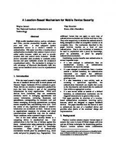
Protein Expression and Purification – The RXFP1 LDL-A module was expressed and purified as reported previously(Hopkins et al. 2005; Hopkins et al. 2007). Analytical Ultracentrifugation – Sedimentation Equilibrium experiments were conducted using a Beckman XLA Analytical Ultracentrifuge. Samples were analyzed at 0.1 mg/ml, 0.3 mg/ml and 1.0 mg/ml. Runs were conducted at 20 °C at 30,000 g and radial scans (minimum 6.8 cm, maximum 7.2 cm) were measured at 230 and 235 nm. Data was analysed using software SEDPHAT (Vistica et al. 2004). The molecular mass calculated for the three samples at 0.1 mg/ml, 0.3 mg/ml and 1.0 mg/ml post centrifugation was measured to be 4.6 kDa, which correlated well to the monomeric molecular weight of the recombinant protein protein (Figure 1). These results clearly show that even at high concentrations of up to 1 mg/ml the LDL-A module in its recombinant form does not form dimers. This observation contributes to the model of RXFP1 dimerization and receptor activation, in that the LDL-A module is unlikely to associate at the dimer interface. This leads to speculation; that firstly the TM region and/or the LRRs are the primary dimer interface, and that the LDL-A module exerts its function to elicit signal activation through an interaction with the transmembrane domain. The identification of the RXFP1 dimer interface requires further investigation as does the molecular detail of how the LDL-A drives signal activation.
Figure 1. Sedimentation equilibrium analytical ultracentrifugation data of RXFP1 LDL-A at 0.3 mg/mL and 1.0 mg/mL shows that the module does not oligomerise. The data fits to a molecular weight of 4.6 kDa correlating well with size of monomeric recombinant LDL-A.
6
Emma J Petrie, Matthew A Periguini, Ross AD Bathgate, Paul R Gooley
References Hopkins, E. J., R. A. Bathgate, et al. (2005). “The human LGR7 low-density lipoprotein class A module requires calcium for structure.” Ann N Y Acad Sci 1041: 27-34. Hopkins, E. J., S. Layfield, et al. (2007). “The NMR solution structure of the relaxin (RXFP1) receptor lipoprotein receptor class A module and identification of key residues in the N-terminal region of the module that mediate receptor activation.” J Biol Chem 282(6): 4172-4184. Kern, A., D. Hubbard, et al. (2008). “Cloning, expression, and functional characterization of relaxin receptor (leucine-rich repeat-containing g protein-coupled receptor 7) splice variants from human fetal membranes.” Endocrinology 149(3): 1277-1294. Svendsen, A. M., M. Vrecl, et al. (2008). “Cooperative binding of insulin-like Peptide 3 to a dimeric relaxin family peptide receptor 2.” Endocrinology 149(3): 1113-1120. Svendsen, A. M., M. Vrecl, et al. (2009). “Dimerization and negative cooperativity in the relaxin family peptide receptors.” Ann N Y Acad Sci 1160: 54-59. Svendsen, A. M., A. Zalesko, et al. (2008). “Negative cooperativity in H2 relaxin binding to a dimeric relaxin family peptide receptor 1.” Mol Cell Endocrinol 296(1-2): 10-17. Vistica, J., J. Dam, et al. (2004). “Sedimentation equilibrium analysis of protein interactions with global implicit mass conservation constraints and systematic noise decomposition.” Analytical biochemistry 326(2): 234-256.