between families, species, age, sex, left and right eye, as well as the 2 body positions were ... differed significantly by body position in tawny owls (P 5 .01) and common buzzards (P 5 .04). .... appearance as nestling, juvenile, or adult (includ-.
Reference Intervals for Intraocular Pressure Measured by Rebound Tonometry in Ten Raptor Species and Factors Affecting the Intraocular Pressure Author(s) :Anne Reuter, Dr Med Vet, Kerstin Müller, Dr Med Vet, Dipl ECZM, Gisela Arndt, Dr Rer Pol, and Johanna Corinna Eule, Dr Med Vet, Dipl ECVO Source: Journal of Avian Medicine and Surgery, 25(3):165-172. 2011. Published By: Association of Avian Veterinarians DOI: 10.1647/2009-056.1 URL: http://www.bioone.org/doi/full/10.1647/2009-056.1
BioOne (www.bioone.org) is a a nonprofit, online aggregation of core research in the biological, ecological, and environmental sciences. BioOne provides a sustainable online platform for over 170 journals and books published by nonprofit societies, associations, museums, institutions, and presses. Your use of this PDF, the BioOne Web site, and all posted and associated content indicates your acceptance of BioOne’s Terms of Use, available at www.bioone.org/page/terms_of_use. Usage of BioOne content is strictly limited to personal, educational, and non-commercial use. Commercial inquiries or rights and permissions requests should be directed to the individual publisher as copyright holder.
BioOne sees sustainable scholarly publishing as an inherently collaborative enterprise connecting authors, nonprofit publishers, academic institutions, research libraries, and research funders in the common goal of maximizing access to critical research.
Journal of Avian Medicine and Surgery 25(3):165–172, 2011 ’ 2011 by the Association of Avian Veterinarians
Original Studies
Reference Intervals for Intraocular Pressure Measured by Rebound Tonometry in Ten Raptor Species and Factors Affecting the Intraocular Pressure Anne Reuter, Dr Med Vet, Kerstin Mu ¨ ller, Dr Med Vet, Dipl ECZM, Gisela Arndt, Dr Rer Pol, and Johanna Corinna Eule, Dr Med Vet, Dipl ECVO Abstract: Intraocular pressure (IOP) was measured with the TonoVet rebound tonometer in 10 raptor species, and possible factors affecting IOP were investigated. A complete ophthalmic examination was performed, and IOP was assessed in 2 positions, upright and dorsal recumbency, in 237 birds belonging to the families Accipitridae, Falconidae, Strigidae, and Tytonidae. Mean IOP values of healthy eyes were calculated for each species, and differences between families, species, age, sex, left and right eye, as well as the 2 body positions were evaluated. Physiologic fluctuations of IOP were assessed by measuring IOP serially for 5 days at the same time of day in 15 birds of 3 species. Results showed IOP values varied by family and species, with the following mean IOP values (mm Hg 6 SD) determined: white-tailed sea eagle (Haliaeetus albicilla), 26.9 6 5.8; red kite (Milvus milvus), 13.0 6 5.5; northern goshawk (Accipiter gentilis), 18.3 6 3.8; Eurasian sparrowhawk (Accipiter nisus), 15.5 6 2.5; common buzzard (Buteo buteo), 26.9 6 7.0; common kestrel (Falco tinnunculus), 9.8 6 2.5; peregrine falcon, (Falco peregrinus), 12.7 6 5.8; tawny owl (Strix aluco), 9.4 6 4.1; long-eared owl (Asio otus), 7.8 6 3.2; and barn owl (Tyto alba), 10.8 6 3.8. No significant differences were found between sexes or between left and right eyes. In goshawks, common buzzards, and common kestrels, mean IOP was significantly lower in juvenile birds than it was in adult birds. Mean IOP differed significantly by body position in tawny owls (P 5 .01) and common buzzards (P 5 .04). By measuring IOP over several days, mean physiologic variations of 62 mm Hg were detected. Differences in IOP between species and age groups should be considered when interpreting tonometric results. Physiologic fluctuations of IOP may occur and should not be misinterpreted. These results show that rebound tonometry is a useful diagnostic tool in measuring IOP in birds of prey because it provides rapid results and is well tolerated by birds. Key words: trauma, intraocular pressure, rebound tonometry, TonoVet, body position, ophthalmology, avian, birds of prey
ophthalmic examination should be performed on every free-ranging bird of prey presented for clinical evaluation. One part of the ophthalmic examination is the assessment of intraocular pressure (IOP) by tonometry. Thus far, only one device for reliable measurement of IOP has been validated in birds. The Tono-Pen (Reichert Technologies, Depew, NY, USA) relies on the principle of applanation tonometry, measuring the pressure required to flatten a predefined area of the cornea.5 This tonometer is restricted to use in eyes with a corneal diameter exceeding 9.0 mm6 and requires the application of a local anesthetic.7 Reference
Introduction Free-ranging birds of prey are frequently presented to veterinarians because of traumatic injuries, including car accidents and collisions with buildings.1–3 In addition to wounds and fractures, these birds often suffer from ocular injuries.4 The aim of rehabilitating wild birds is to ultimately release them back into the wild. Vision is crucial for these birds; therefore, a thorough From the Small Animal Clinic (Reuter, Mu¨ller, Eule) and the Institute of Biometrics and Information Processing (Arndt), Faculty of Veterinary Medicine, Freie Universita¨t Berlin, Oertzenweg 19b, 14163 Berlin, Germany.
165
166
JOURNAL OF AVIAN MEDICINE AND SURGERY
values have been established for a wide range of avian species.8–12 Comparison of results from the Tono-Pen to a manometric method revealed good correlation of manometry and tonometry between 10 and 25 mm Hg; below 10 mm Hg, the TonoPen provided falsely high results, whereas in the pressure range above 25 mm Hg, results were falsely lowered.9 A new instrument to determine IOP has been developed and is now commercially available for veterinary use. The concept of the TonoVet (Icare Finland Oy, Espoo, Finland) tonometer is based on rebound tonometry.13 A magnetic probe is inserted into the tonometer’s collar, which is equipped with electric coils. During measurement, a magnetic field is induced that propels the probe against the cornea, where it is rejected and returned, provoking a voltage change in the coils according to the probe’s velocity. This voltage change is converted into an electric signal and used to calculate IOP. The magnetic probes are manufactured for single use to prevent transmission of pathogens. A plastic-covered tip prevents corneal damage. Diameter of the contact surface measures only 1.4 mm. Measurements can be taken without topical anesthesia and results are provided within a few seconds. For reliable IOP evaluation, 6 consecutive measurements are necessary. Cancelling the lowest and highest value, the tonometer displays the mean of the remaining 4 measurements. The TonoVet possesses 3 internal calibration settings: d, for dogs and cats; h, for horses; and p, for other species. The accuracy and reproducibility of the TonoVet has been determined for birds of prey on the d setting.14 Compared with manometric methods, the TonoVet was less accurate than the Tono-Pen in rock pigeons (Columba livia).10 In chickens, the opposite was found.15 In Eastern screech owls (Megascops asio), the TonoVet gave higher values than the Tono-Pen on the d setting (for dogs and cats), whereas on the p setting (for other species), TonoVet values were lower than those obtained with the Tono-Pen.8 In another study, measurement results of TonoVet and Tono-Pen differed significantly in various species of raptors. Additionally, a strong correlation between TonoVet results and corneal thickness was observed.16 In all studies, the TonoVet was well tolerated by birds. The IOP in mammals may be influenced by a variety of factors, such as age, sex, time of the day, and anesthesia.17–25 Chickens and pigeons have been proven to have a circadian IOP rhythm,9,26 whereas that rhythm was not present
in common buzzards (Buteo buteo) and tawny owls (Strix aluco).9 Isoflurane anesthesia administered via the air sacs is known to lower IOP.27 In mammals, IOP varies depending on body position.28–34 These factors need to be taken into consideration when interpreting tonometry results. The purpose of this study was to establish IOP reference intervals for different species of birds of prey with the TonoVet, as well as to evaluate physiologic fluctuations and the effect of age, sex, and body position on tonometry results. Materials and Methods All birds studied were diseased or orphaned free-ranging birds of prey that were admitted for treatment and rehabilitation. Our study population comprised 10 different species of the 4 families of Accipitridae, Falconidae, Strigidae, and Tytonidae. Age was classified by physical appearance as nestling, juvenile, or adult (including immature and subadult white-tailed sea eagles [Haliaeetus albicilla]).35–37 A total of 237 birds were examined between April 2008 and April 2009. A complete ophthalmologic examination by slit-lamp biomicroscopy (SL-15, Kowa Company, Tokyo, Japan) and indirect ophthalmoscopy (BETA 200, Heine Optotechnik, Herrsching, Germany) was conducted on each bird as part of the routine clinical examination. The IOP was measured bilaterally with the TonoVet (Figs 1 and 2) in an upright position. The birds were manually restrained, and neither topical anesthesia nor sedation was used. Age and sex were assessed according to morphologic criteria whenever possible. All measurements were determined between 7:30 AM and 6:00 PM; to minimize the birds’ stress, we did not adhere to a predefined, consistent schedule. For assessment of IOP reference intervals, data were used from ophthalmologically unremarkable eyes. To identify physiologic fluctuations in IOP, tonometry was repeated in 15 birds of 3 species on 4 additional days at the same time each day. To avoid additional stress, those days were chosen in accordance with treatment regimens and were, therefore, not consecutive. In 197 birds, IOP was measured consecutively in 2 different body positions under the same conditions. After measuring IOP in upright position, animals were positioned in dorsal recumbency with the head held at heart level. After 5 seconds of adaptation, the dorsal recum-
REUTER ET AL—REFERENCE VALUES FOR IOP IN RAPTORS
167
repeated measurements was obtained. Significance was set at P , .05. For all analyses, SPSS 15.0 for Windows (formerly SPSS Inc, now IBM Corporation, Armonk, NY, USA) was used. Results
Figure 1. The rebound tonometer TonoVet (Icare Finland Oy) with a magnetic probe.
bent IOP was measured. For all tonometric measurements, the TonoVet setting d for dogs and cats was used. Because results were not normally distributed (Kolmogorov-Smirnov test), a descriptive analysis was performed calculating the medians and ranges. Distribution was evaluated graphically by histograms. To allow comparison with findings in the current literature, means and standard deviations were calculated as well. Differences in IOP by bird family, species, age groups, and sex were evaluated by the Mann-Whitney-Wilcoxon test for independent variables. Potential IOP differences between right and left eyes and between upright position and dorsal recumbency were assessed by the Wilcoxon test for dependent variables. Statistical analyses were only performed on species that comprised more than 10 members. To evaluate physiologic fluctuations of IOP, the mean variance of the 5 values measured on different days was calculated. By extracting the root of those values, the standard variation for
A total of 237 free-ranging birds of prey were included in the study. Table 1 shows the distribution by age and sex within the study population. Of those birds, 44 (18.6%) were diagnosed with unilateral ophthalmologic disease. Values obtained from those eyes were not included in this study. For assessment of IOP reference intervals, data from 430 healthy eyes were evaluated. Table 2 shows the results of tonometric measurements. We detected significant differences between Accipitridae birds and birds of the other families (P , .001) and between Falconidae and Strigidae birds (P 5 .025). Except for the white-tailed sea eagle and the common buzzard (P 5 .94), significant differences in IOP could be shown between each of the species of the Accipitridae family (P , .05). In northern goshawks (Accipiter gentilis), common buzzards, and common kestrels (Falco tinnunculus), significant differences in IOP between juvenile and adult birds were found (P # .001), but not in Eurasian sparrowhawks (Accipiter nisus) (P 5 .10) and tawny owls (P 5 .26). The IOP was not significantly influenced by sex in white-tailed sea eagles (P 5 .92), northern goshawks (P 5 .05), Eurasian sparrowhawks (P 5 .29), common buzzards (P 5 .21), or common kestrels (P 5 .20). In barn owls (Tyto alba), longeared owls (Asio otus), pereguine falcons (Falco perequinus), red kites (Milvus milvus), and tawny owls, differences between sexes could not be assessed because of low sample size or unknown sex. There were no significant differences in IOP between left and right eyes in any species (P . .05). To assess possible physiologic fluctuations of IOP, we calculated the standard deviations of the repeated measurements performed in 15 birds. These standard deviations indicate the variation of measurement results. For the 15 birds examined, the mean standard deviation of 5 replicated measurements was 1.71 mm Hg (SD range, 0.89–3.35). The birds’ IOP values in upright and dorsal recumbent positions were compared (Table 3). Significant differences of minor clinical value were only detected in common buzzards and tawny owls.
168
JOURNAL OF AVIAN MEDICINE AND SURGERY
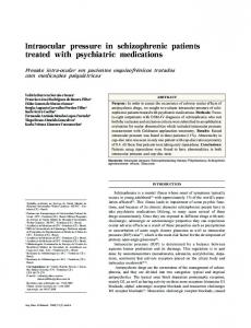
Figure 2. Measuring intraocular pressure in a long-eared owl with the TonoVet (Icare Finland Oy) rebound tonometer.
Because of the high prevalence of ocular diseases in free-ranging birds of prey, a complete ophthalmologic examination should be performed on each bird presented to a clinician, including an assessment of IOP. With the TonoVet rebound tonometer, reliable results can be obtained quickly (less than 1 second per corneal touch) and, because of the small contact
Discussion The aim of this study was to evaluate the use of the TonoVet rebound tonometer in birds of prey. Reference intervals for intraocular pressure were determined in 10 species after a complete ophthalmologic examination was performed to verify ocular health.
Table 1. Distribution by age and sex of 237 birds of prey in a study of intraocular pressure measured by TonoVet (Icare Finland Oy) rebound tonometry. Age, n
Sex, n
Species
Nestling
Juvenile
Adult
Female
Male
Unknown
Total
Barn owl Common buzzard Common kestrel Northern goshawk Long-eared owl Peregrine falcon Red kite Eurasian sparrowhawk Tawny owl White-tailed sea eagle Total
0 1 7 0 0 1 0 0 2 0 11
1 15 55 23 2 1 1 13 6 2 119
3 32 10 11 11 2 3 14 7 14 107
0 7 5 16 3 3 1 24 1 8 68
0 16 8 18 1 1 0 3 1 8 56
4 25 59 0 9 0 3 0 13 0 113
4 48 72 34 13 4 4 27 15 16 237
169
REUTER ET AL—REFERENCE VALUES FOR IOP IN RAPTORS
Table 2. Intraocular pressure (IOP) from the healthy eyes of 10 species of birds of prey as determined by TonoVet (Icare Finland Oy) rebound tonometer (setting d). IOP, mm Hg Species
Range
Median
Mean
SD
6 2 4
5.0–16.0 9.0–10.0 5.0–16.0
11.0 9.5 12.5
10.8 9.5 11.5
3.8 0.7 4.7
86 2 28 56
14.0–44.0 16.0 14.0–32.0 17.0–44.0
27.0 — 21.5 30.0
26.9 — 21.6 29.9
7.0 — 4.8 6.1
141 13 109 19
4.0–15.0 4.0–10.0 4.0–15.0 5.0–15.0
10.0 7.0 10.0 12.0
9.8 7.1 9.8 11.6
2.5 1.9 2.3 2.7
Northern goshawk Juvenile Adult
58 40 18
12.0–29.0 12.0–29.0 16.0–25.0
18.0 16.0 21.0
18.3 17.0 21.2
3.8 3.6 2.4
Long-eared owl Juvenile Adult
21 4 17
4.0–13.0 4.0–5.0 4.0–13.0
7.0 4.5 8.0
7.8 4.5 8.5
3.2 0.6 3.0
Peregrine falcon Nestling Juvenile Adult
7 1 2 4
5.0–21.0 5.0 10.0–13.0 10.0–21.0
10.0 — 11.5 15.0
12.7 — 11.5 15.3
5.8 — 2.1 6.1
Red kite Juvenile Adult
8 2 6
4.0–19.0 14.0 4.0–19.0
14.5 — 15.5
13.0 — 12.7
5.5 — 6.5
Eurasian sparrowhawk Juvenile Adult
47 21 26
10.0–23.0 10.0–19.0 11.0–23.0
16.0 15.0 16.0
15.5 14.8 16.0
2.5 2.0 2.8
Tawny owl Nestling Juvenile Adult
27 4 11 12
3.0–17.0 5.0–8.0 3.0–17.0 4.0–15.0
10.0 5.5 6.0 11.5
9.4 6.0 8.7 11.1
4.1 1.4 4.9 3.1
White-tailed sea eagle Juvenile Adult
29 4 25
17.0–41.0 17.0–24.0 18.0–41.0
28.0 22.0 28.0
26.9 21.3 27.8
5.8 3.1 5.7
Barn owl Juvenile Adult Common buzzard Nestling Juvenile Adult Common kestrel Nestling Juvenile Adult
No. of eyes tested
area, application of a topical anesthetic is not required, whereas applanation tonometry necessitates local anesthesia.7 When used in small eyes, repeated measurements are needed with the applanation tonometer to gain valid results,9 leading to increased stress and possible reduction of IOP because of the effect of tonography.5 In our study, differences in IOP intervals by families and species were observed. In general, IOP values were higher in the Accipitridae species compared with that of other families. These findings might be related to differences in corneal
thickness, which was shown to correlate positively ´ n et al16 found mean IOP values with IOP.16 Bayo to be 10.0 mm Hg in common kestrels, 9.31 mm Hg in little owls (Athene noctua), and 11.96 mm Hg in Eurasian eagle owls (Bubo bubo), which are concordant with our results in common kestrels and the owl species. Tonometry in different species of eagles (booted eagle [Aquila pennata], Bonelli’s eagle [Aquila fasciata], and short-toed snake-eagle [Circaetus gallicus]) revealed higher IOP values (up to 40.26 mm Hg) associated with thicker corneas.11 That finding
170
JOURNAL OF AVIAN MEDICINE AND SURGERY
Table 3. Median intraocular pressure (IOP) and P values of the differences between IOP upright and dorsal recumbent positions as determined with the TonoVet (Icare Finland Oy) rebound tonometer (setting d).
Species
No. of eyes tested
Upright median IOP, mm Hg
Dorsal recumbent median IOP, mm Hg
P
Common buzzard Common kestrel Northern goshawk Long-eared owl Eurasian sparrowhawk Tawny owl White-tailed sea eagle
69 131 36 14 30 20 20
27.0 10.0 16.0 6.0 15.0 9.5 28.0
26.0 10.0 17.0 8.5 15.0 11.5 28.5
.04a .85 .92 .42 .60 .01a .25
a
Significant at P , .05 by Wilcoxon test for dependant variables.
agrees with the high IOP values we found in the Accipitridae species (white-tailed sea eagles, common buzzards, and northern goshawks). These species-specific differences highlight the need for species-specific reference intervals, especially because reference intervals assessed for applanation tonometry cannot be transferred to rebound tonometry.8,11 Sex did not have a significant effect on IOP, which confirms the findings of other authors.8,9 In contrast, age differences have to be considered because we found significant differences between juvenile and adult northern goshawks, common buzzards, and common kestrels. In these species, younger birds had lower IOP values than did older birds. The same trend could be observed in the other species, with juvenile birds showing lower median values than adult birds. Results of an earlier study revealed a comparable rise in IOP with age in common kestrels, Muscovy ducks (Cairina moschata), and greylag geese (Anser anser) measured by applanation tonometry,9 whereas the same could not be proven for eastern screech owls.8 The increase of IOP with age may be caused by age-dependent changes of ocular rigidity. In humans, an increase of ocular rigidity was observed with increasing age and was positively correlated to IOP.38 Moreover, increased corneal thickness was age dependent and progressed until the 70th day of life and then remained constant.39 Because IOP measurements by rebound tonometry are influenced by corneal thickness,16 that might be another factor for the observed increase in IOP. In humans, however, IOP was no longer correlated with age after adjusting the IOP for confounders, like systolic blood pressure,40 and in dogs, no differences in IOP could be detected between 6-week-old puppies and 1-year-old dogs.41 These results emphasize a multifactorial development of IOP with age.
In 3 species, IOP was measured at the same time of day for 5 days to evaluate physiologic fluctuations of IOP. In the clinical setting, mean physiologic variations of 62 mm Hg should be considered. These findings are supported by IOP measurements in chickens at intervals of several hours.15 The variations evaluated in that study were linked to stress, diurnal variations, or even measurement errors. In humans, such unpredictable variations were seen when monitoring patients with glaucoma, and no causality could be found.42 Possible factors may include external pressure on the eye, alterations in blood pressure by changing body positions, or pressure on the neck.43,44 In rabbits, telemetric surveys revealed changes in IOP during normal handling, during tonometry, and even when drinking water.45 In the birds in this study, we did not apply pressure on the eyes or neck during handling, but stress may have played an important role. In tawny owls and common buzzards, body position had a significant effect on IOP. The results may be caused by the study design, eg, the degree of dorsal recumbency. In mice, cats, and horses, an increase of IOP was achieved when the animal’s head was positioned under its heart level.29,30,34 Anatomic differences between birds and mammals need to be considered. The Schlemm canal is wider in birds, and therefore, the facility of aqueous outflow is higher.46 Other important factors of IOP regulation in birds might exist, and further studies are needed. Rebound tonometry is a new method to measure IOP and offers a stress-minimizing, well-tolerated tool for ophthalmic diagnostic testing in birds. Species-specific differences should always be considered, as well as age-dependant variations. The different IOP values in upright and dorsal recumbent positions found in common buzzards and tawny owls are of minor clinical
REUTER ET AL—REFERENCE VALUES FOR IOP IN RAPTORS
impact; therefore, IOP measurement in both positions appears to be appropriate. 15.
References 1. Deem SL, Terrell SP, Forrester DJ. A retrospective study of morbidity and mortality of raptors in Florida: 1988–1994. J Zoo Wildl Med. 1998;29(2): 160–164. 2. Fix AS, Barrows SZ. Raptors rehabilitated in Iowa during 1986 and 1987: a retrospective study. J Wildl Dis. 1990;26(1):18–21. 3. Wendell MD, Sleeman JM, Kratz G. Retrospective study of morbidity and mortality of raptors admitted to Colorado State University Veterinary Teaching Hospital during 1995 to 1998. J Wildl Dis. 2002;38(1):101–106. 4. Korbel RT. Disorders of the posterior eye segment in raptors—examination procedures and findings. In: Lumeij JT, Remple JD, Redig PT, et al, eds. Raptor Biomedicine III. Lake Worth, FL: Zoological Education Network; 2000:179–193. 5. Rumberger E. Physiologische und messtechnische Prinzipien der Augendruckmessung. In: Rumberger DJ, ed. Tonometrie. Neubrandenburg, Germany: Rethra Verlag Neubrandenburg; 2008. 6. Korbel R, Braun J. Tonometrie beim Vogel mit dem Tonopen XL. Tiera¨rztl Prax. 1999;27(K): 208–217. 7. Tono-Pen XL instruction manual. Medtronic Solan Web site. http://www.pitt.edu/˜ mercyres/ Tonopen.pdf. Updated 2000. Accessed July 22, 2011. 8. Harris MC, Schorling JJ, Herring IP, et al. Ophthalmic examination findings in a colony of screech owls (Megascops asio). Vet Ophthalmol. 2008;11(3):186–192. 9. Braun J. Weiterfu¨hrende Untersuchungen zur Bestimmung des Intraokulardrucks bei Vo¨geln mit einem Elektronischen Tonometer [Further investigation to determine the intraocular pressure in birds with an electronic tonometer (in German)]. Munich, Germany: Ludwig-Maximilians-Universita¨t; 1995. 10. Go¨rig C, Schoemaker NJ, Stades FC, Boeve MH. Evaluation of different tonometers in exotic animals [abstract]. Vet Ophthalmol. 2005;8(6):430. 11. Jeong MB, Kim YJ, Yi NY, et al. Comparison of the rebound tonometer (TonoVet) with the applanation tonometer (TonoPen XL) in normal Eurasian eagle owls (Bubo bubo). Vet Ophthalmol. 2007;10(6):376–379. 12. Stiles J, Buyukmihci NC, Farver TB. Tonometry of normal eyes in raptors. Am J Vet Res. 1994;55(4): 477–479. 13. Kontiola AI. A new induction-based impact method for measuring intraocular pressure. Acta Ophthalmol Scand. 2000;78(2):142–145. 14. Reuter A, Muller K, Arndt G, Eule JC. Accuracy and reproducibility of the TonoVet rebound
16.
17.
18.
19.
20.
21.
22.
23.
24.
25.
26.
27. 28.
29.
30.
171
tonometer in birds of prey. Vet Ophthalmol. 2010; 13(suppl):80–85. Prashar A, Guggenheim JA, Erichsen JT, et al. Measurement of intraocular pressure (IOP) in chickens using a rebound tonometer: quantitative evaluation of variance due to position inaccuracies. Exp Eye Res. 2007;85(4):563–571. Bayo´n A, Vecino E, Albert A, et al. Evaluation of intraocular pressure obtained by two tonometers, and their correlations with corneal thickness obtained by pachymetry in raptors [abstract]. Vet Ophthalmol. 2006;9(6):432. Chen CL, Gelatt KN, Gum GG. Serum hydrocortisone (cortisol) values in glaucomatous and normotensive beagles. Am J Vet Res. 1980;41(9): 1516–1518. Del Sole MJ, Sande PH, Bernades JM, et al. Circadian rhythm of intraocular pressure in cats. Vet Ophthalmol. 2007;10(3):155–161. Gelatt KN, MacKay EO. Distribution of intraocular pressure in dogs. Vet Ophthalmol. 1998;1(2–3): 109–114. Ofri R, Steinmetz A, Thielebein J, et al. Factors affecting intraocular pressure in lions. Vet J. 2008; 177(1):124–129. Sugimoto E, Aihara M, Ota T, Araie M. Effect of light cycle on 24-hour pattern of mouse intraocular pressure. J Glaucoma. 2006;15(6):505–511. Ofri R, Horowitz I, Jacobson S, Kass PH. The effects of anesthesia and gender on intraocular pressure in lions (Panthera leo). J Zoo Wildl Med. 1998;29(3):307–310. Ofri R, Shub N, Galin Z, et al. Effect of reproductive status on intraocular pressure in cats. Am J Vet Res. 2002;63(2):159–162. Hofmeister EH, Mosunic CB, Torres BT, et al. Effects of ketamine, diazepam, and their combination on intraocular pressures in clinically normal dogs. Am J Vet Res. 2006;67(7):1136–1139. Jia L, Cepurna WO, Johnson EC, Morrison JC. Effect of general anesthetics on IOP in rats with experimental aqueous outflow obstruction. Invest Ophthalmol Vis Sci. 2000;41(11):3415–3419. Nickla DL, Wildsoet C, Wallman J. The circadian rhythm in intraocular pressure and its relation to diurnal ocular growth changes in chicks. Exp Eye Res. 1998;66(2):183–193. Korbel RT. Tonometrie beim Vogel mit dem Tonopen XL. Tiera¨rztl Prax. 1999;27(K):208–217. Broadwater JJ, Schorling JJ, Herring IP, Elvinger F. Effect of body position on intraocular pressure in dogs without glaucoma. Am J Vet Res. 2008; 69(4):527–530. Aihara M, Lindsey JD, Weinreb RN. Episcleral venous pressure of mouse eye and effect of body position. Curr Eye Res. 2003;27(6):355–362. Kindler S, Schieszler A. The influence of head and body position on the intraocular pressure of the feline eye. Vet Ophthalmol. 2009;12(1):66.
172
JOURNAL OF AVIAN MEDICINE AND SURGERY
31. Setogawa A, Kawai Y. Measurement of intraocular pressure by both invasive and noninvasive techniques in rabbits exposed to head-down tilt. Jpn J Physiol. 1998;48(1):25–31. 32. Chiquet C, Custaud MA, Le Traon AP, et al. Changes in intraocular pressure during prolonged (7-day) head-down tilt bedrest. J Glaucoma. 2003; 12(3):204–208. 33. Hirooka K, Shiraga F. Relationship between postural change of the intraocular pressure and visual field loss in primary open-angle glaucoma. J Glaucoma. 2003;12(4):379–382. 34. Komaromy AM, Garg CD, Ying GS, Liu C. Effect of head position on intraocular pressure in horses. Am J Vet Res. 2006;67(7):1232–1235. 35. Forsman D. The Raptors of Europe and the Middle East: A Handbook to Field Identification. London, England: Poyser; 2007. 36. Glutz von Blotzheim UN, Bauer KM. Handbuch der Vo¨gel Mitteleuropas. Vol 9. Wiesbaden, Germany: AULA-Verlag; 1980. 37. Glutz von Blotzheim UN, Bauer KM, Bezzel E. Handbuch der Vo¨gel Mitteleuropas. Vol 4. Wiesbaden, Germany: AULA-Verlag; 1989. 38. Pallikaris IG, Kymionis GD, Ginis HS, et al. Ocular rigidity in living human eyes. Invest Ophthalmol Vis Sci. 2005;46(2):409–414. 39. Montiani-Ferreira F, Cardoso F, Petersen-Jones S. Postnatal development of central corneal thickness
40.
41.
42.
43.
44.
45.
46.
in chicks of Gallus gallus domesticus. Vet Ophthalmol. 2004;7(1):37–39. Rochtchina E, Mitchell P, Wang JJ. Relationship between age and intraocular pressure: the Blue Mountains Eye Study. Clin Experiment Ophthalmol. 2002;30(3):173–175. Mughannam AJ, Cook CS, Fritz CL. Change in intraocular pressure during maturation in Labrador retriever dogs. Vet Ophthalmol. 2004;7(2): 87–89. Sit AJ. Continuous monitoring of intraocular pressure: rationale and progress toward a clinical device. J Glaucoma. 2009;18(4):272–279. Adler FH. Intraocular pressure. In: Alm A, Kaufman PL, eds. Adler’s Physiology of the Eye: Clinical Application. 10th ed. Maryland Heights, MO: Mosby; 2002. Pauli AM, Bentley E, Diehl KA, Miller PE. Effects of the application of neck pressure by a collar or harness on intraocular pressure in dogs. J Am Anim Hosp Assoc. 2006;42(3):207–211. Dinslage S, McLaren J, Brubaker R. Intraocular pressure in rabbits by telemetry, II: effects of animal handling and drugs. Invest Ophthalmol Vis Sci. 1998;39(12):2485–2489. Tripathi RC, Tripathi BJ. The mechanism of aqueous outflow in birds, I: an ultrastructural study of normal eyes. Exp Eye Res. 1973;15(3): 409–423.