Drug Metabolism Reviews, 37:379^04, 2005 Copyright © Taylor & Francis Inc. ISSN: 0360-2532 print / 1097-9883 online DOI: 10.1081/DMR-200046136
I Taylor & Francis Taylor 6. Francis Croup
REGULATION OF CYTOCHROME P450 BY POSTTRANSLATIONAL MODIFICATION Mike Aguiar Applied R&D, MDS Pharma Services, St. Laurent (Montreal), Quebec, Canada Department of Medicine, McGill University, Montreal, Quebec, Canada
Robert Masse Applied R&D, MDS Pharma Services, St. Laurent (Montreal), Quebec, Canada
Bernard F. Gibbs Applied R&D, MDS Pharma Services, St. Laurent (Montreal), Quebec, Canada Department of Medicine, McGill University, Montreal, Quebec, Canada Cytochrome P450s are a family of enzymes represented in all kingdoms with expression in many species. Over 3,000 enzymes have been identified in nature. Humans express 57 putatively functional enzymes with a variety of critical physiological roles. They are involved in the metabolic oxidation, peroxidation, and reduction of many endogenous and exogenous compounds including xenobiotics, steroids, bile acids, fatty acids, eicosanoids, environmental pollutants, and carcinogens [Nelson, D. R., Kamataki, T., Waxman, D. J., Guengerich, F. P., Estabrook, R. W., Feyereisen, R., Gonzalez, F. J., Coon, Af. J., Gunsalus, I. C, Gotoh, O. (1993) The P450 superfamily: update on new sequences, gene mapping, accession numbers, early trivial names of enzymes, and nomenclature. DNA Cell Biol. 12(1):I—51.] The development of numerous diseases and disorders including cancer and cardiovascular and endocrine dysfunction has been linked to P450s. Several levels of regulation, including transcription, translation, and posttranslational modification, participate in maintaining the proper function of P450s. Modifications including phosphorylation, glycosylation, nitration, and ubiquitination have been described for P450s. Their physiological significance includes modulation of enzyme activity, targeting to specific cellular compartments, and tagging for proteasomal degradation. Knowledge of P450 posttranslational regulation is derived from studies with relatively few enzymes. In many cases, there is only enough evidence to suggest the occurrence and a possible role for the modification. Thus, many P450 enzymes have not been fully characterized. With the introduction of current proteomics tools, we are primed to answer many important questions regarding regulation of P450 in response to a posttranslational modification. This review considers regulation ofP450 in a context that describes the potential role and physiological significance of each modification. Key Words: Cytochrome P450; Posttranslational modification; Phosphorylation; Ubiquitination; Nitration; Glycosylation.
Address correspondence to Bernard F. Gibbs, Applied R&D, MDS Pharma Services, 2350 Cohen Street, St. Laurent (Montreal), Quebec, H4R 2N6, Canada and Department of Medicine, McGill University, Montreal, Quebec, Canada; E-mail:
[email protected] 379
380
M. AGUIAR ET AL.
INTRODUCTION Background Information on P450 Since the introduction of modern molecular biology techniques allowing for the sequencing of entire genomes, researchers have identified and continue to discover many P450 enzymes in a wide variety of organisms. In order to systematically identify and categorize this growing family of enzymes, a leading group of researchers in the field established the current system of nomenclature (Nelson et al., 1996). P450s are named with the prefix CYP or P450 followed by an Arabic number defining the family, an uppercase letter defining the subfamily, and another Arabic number defining an individual enzyme (e.g., P450 3A4). In cases where only a single member exists in a given family, it may be identified simply as P450 followed only by the family number (e.g., P450 51). The reader may refer to the cytochrome P450 home page (http:// dmelson.utmem.edu/CytochromeP450.html) for the latest nomenclature updates and links to related sites. All mammalian tissues examined express some P450 enzyme system (Porter and Coon, 1991). In addition, mammals express multiple enzymes simultaneously in a variety of tissues, including liver, kidney, lung, and adrenal (Bhagwat et al., 1999a; Guengerich, 2001; Lohr et al., 1998; Parker and Schimmer, 1997). It is also appreciated that various enzymes are found not only in different cell and tissue types, but also in different subcellular compartments, such as the outer nuclear membrane, endoplasmic reticulum (ER), mitochondria, golgi, peroxisome, and plasma membrane. Certain enzymes are found in several different subcellular compartments simultaneously (Guengerich, 2001). All cytochrome P450s, with the exception of bacterial enzymes, are membrane bound. Microsomal enzymes are tethered to the membrane through a hydrophobic transmembrane helix at the N-terminus of the protein, which also serves as a targeting sequence for the signal recognition particle dependent cotranslational incorporation of a nascent P450 into the ER membrane (Bar-Nun et al., 1980; Sakaguchi et al., 1984). Insertion continues until a halt-transfer signal is reached that effectively anchors the P450 to the membrane with the bulk of the protein exposed on the cytosolic side (Monier et al., 1988; Sakaguchi et al., 1987; Szczesna-Skorupa and Kemper, 1989; Szczesna-Skorupa et al., 1988). Because detergents are required for solubilizing membrane-bound P450s, all native mammalian P450s have evaded crystallization and x-ray crystallographic study. To avoid this problem, researchers have modified several P450s resulting in soluble mutants that can be crystallized and that have structures that were solved. The first mammalian P450 structure reported was that of rabbit P450 2C5 (Williams et al., 2000), which was also crystallized in complexes with two different substrates (Wester et al., 2003a,b). Interestingly, one of the two substrates binds in two different orientations (Wester et al., 2003a). These studies of P450 2C5 offer insightful information on the fiexibility of the enzyme and shed light on the ability of P450s to accommodate a variety of substrates with different shapes and sizes. The structure of human P450 2C8 was recently reported, revealing an active site volume twice that of P450 2C5, consistent with the size of its preferred substrates (Schoch et al., 2004). Human P450 2C9 has also been crystallized, with and without bound warfarin, a known anticoagulant substrate (Williams et al., 2003). The structure reveals a new binding pocket that may accommodate several substrate molecules simultaneously and possibly
REGULATION OF CYTOCHROME P450
381
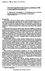
account for some complex drug-drug interactions. Another group has also reported a structure for P450 2C9 complexed with flurbiprofen (Wester et al,, 2004), The structure was obtained without modifications to the catalytic domain, revealing some significant conformational differences. The more recent structure helps explain some experimental observations in terms of the substrate selectivity of the enzyme, which were not easily explained with the earlier structure, A crystal structure for rabbit P450 2B4, one of the first P450s to be purified and studied in detail, has been reported in a wide open conformation (Scott et al,, 2003) and in a closed form complexed with the specific inhibitor 4-(4-chlorophenyl)-imidazole (Scott et al,, 2004), These structures may help to understand how mammalian P450s "open/close," allowing substrate to access the buried active site (Poulos, 2003), Two groups have recently reported crystal structures for human P450 3A4: one unliganded structure from each group and two structures hound to different substrates (Williams et al, 2004; Yano et al,, 2004a), The collection of structures should provide a better understanding of the substrate selectivity and unusual kinetics of this important enzyme. Finally, a structure for P450 2A6 has been reported in a meeting abstract (Yano et al,, 2004b), Humans express 57 putatively functional genes and 58 pseudogenes (Nelson et al,, 2004), These may be divided roughly into two groups based on their substrate specificity, P450s involved in the metabolism of most drugs and carcinogens are derived from families 1-3, This group demonstrates wide substrate specificity, with certain enzymes acting on a large number of structurally varied substrates such as in the case of human P450 3A4, The second group demonstrates high substrate specificity, catalyzing the biosynthesis and metabolism of endogenous substrates including cholesterol, steroids, vitamins, and eicosanoids. Several steroidogenic P450 enzymes are outlined in Fig, 1,
Lanosterol
I P450 51 Cholesterol ^ ^ Pregnenolone ^ ^ * 17-OH Pregnenolone ^ ^ * Dehydroepiandrosterone P450 11AI P45017A1 P450 17A1
J P450 7A1
I 3P HSD
7-OH Cholesterol
Progesterone
^ ^ ^ P450 17A1
Bile Acids
11-Deoxycorticosterone |P45O11BI Corticosterone
J 3P HSD 17-OH Progesterone
i 3P HSD ^ ^ * Androstenedione ^ ^ * Estrone P450 17A1 P45019A1
11-Deoxycortisol
Testosterone
^^^^ Estradiol P450 19AI
JP45O11B1 Cortisol
Aldosterone |P45O11B2 18-OH Corticosterone
Figure 1. Cholesterol, bile acid, and steroid hormone biosynthesis: phosphorylated P450 enzymes are indicated in boldface.
382
M. AGUIAR ET AL.
Classes of P450 P450s fall into three classes hased on the reduction system transferring electrons. Class I P450s are associated with the inner mitochondrial memhrane and some hacterial systems. Reducing equivalents from NADPH or NADH are transferred two electrons at a time to redoxin reductase, which carries a flavin adenine dinucleotide (FAD) prosthetic group. The isoalloxazine ring of FAD may exist in several oxidation states, which allows for the subsequent transfer of electrons individually to a mobile Fe2S2-containing protein called redoxin. Reduced redoxin is thought to shuttle electrons to the P450, returning to the reductase in the oxidized form for additional cycles. Class II P450s are those that reside in the ER and receive reducing equivalents from NADPH via P450 reductase or cytochrome b^ (a heme protein associated with the ER), P450 reductase is a membrane-bound protein containing a FAD and a flavin mononucleotide (FMN) prosthetic group that also has an isoalloxazine ring with properties similar to FAD, An electron pair from NADPH is received by FAD, which relays the electrons to FMN, finally transferring electrons in single file to the P450, In certain reactions, the first electron is transferred from P450 reductase, while the second electron is transferred from cytochrome b^. Cytochrome b^ may be reduced either by P450 reductase, or cytochrome b^ reductase. Interestingly, apo-^s (cytochrome ^5 devoid of heme), which cannot transfer electrons, is required for optimal activity in a number of P450 enzymes and only with certain substrates (Hlavica and Lewis, 2001; Yamazaki et al,, 2001), Class III P450s are isomerases instead of monooxygenases. They do not require the participation of any redox partners or donors of reducing equivalents. Water is not produced, and therefore, molecular oxygen is not required for catalysis. Substrates of class III P450s are simply rearranged into products. Examples of P450s from this class include P450 8A1 [prostacyclin [PGI2] synthase] and P450 5 [thromboxane [TXA2] synthase].
POSTTRANSLATIOIMAL MODIFICATION A posttranslational modification may be defined as "any difference between a functional protein and the linear polypeptide sequence encoded between the initiation and the termination codons of its structural gene" (Han and Martinage, 1992), Examples of noncovalent modifications include incorporation of cofactors such as heme, protein folding, and the association of subunits to form an oligomeric protein, Allosteric phenomena manifested as deviations from Michaelis-Menten kinetics have been demonstrated for numerous P450 enzymes. Various components known to interact with P450, including substrates, inhibitors, membrane lipids, and redox partners (such as the previously mentioned cytochrome ^5), have been shown to act as homotropic and heterotropic effectors (Hlavica and Lewis, 2001), P450 2E1 is stabilized by ethanol, leading to increased cellular levels of the P450 (Roberts et al,, 1995), Covalent modifications, including cleavage of a signal peptide, formation of disulfide bonds, and an array of modifications to amino acid residues, including phosphorylation, nitration, glycosylation, methylation, sulfation, acetylation, and prenylation, provide another means of posttranslational modification. The remainder of the present article is dedicated to the identification and characterization of covalent P450 posttranslational modifications with an emphasis placed on describing the physiological role of each modification.
REGULATION OF CYTOCHROME P450
383
PHOSPHORYLATIOIM An article by Cohen (2002), opens with the following statement, "Protein phosphorylation regulates most aspects of cell life, whereas abnormal phosphorylation is a cause or consequence of disease." Cascades that activate the production of cyclic adenosine monophosphate (cAMP), leading to activation of protein kinase A (PKA) in eukaryotic cells illustrate a remarkable example. Once activated, PKA can phosphorylate many target proteins, resulting in a wide variety of cellular responses, including regulation of gene transcription, modulation of enzyme activity, targeting of protein for degradation, and targeting to various intracellular locations.
MODULATION OF P450 ACTIVITY BY PHOSPHORYLATION A number of reports over the last two decades describe the phosphorylation of over twenty P450 enzymes in microsomes, intact hepatocytes, cell culture, and in vivo (Table 1). Whereas some studies demonstrate stimulation of phosphorylation by addition of hormones and intracellular second messengers, other reports correlate phosphorylation with modulation of enzyme activity (Koch and Waxman, 1989; Oesch-Bartlomowicz et al., 1998, 2001; Pyerin and Taniguchi, 1989). Much of the work has focused on members of family 1-3 P450s. Unfortunately, studies regarding regulation by posttranslational modification of the major drug metabolizing P450s, with the exception of P450 3A4 and P450 2E1, which will be discussed below (i.e., P450 1A2, P450 2B6, P450 2C8, P450 2C19, P450 2C9, P450 2D6), have not appeared in the literature. However, evidence for the modification and regulation of P450 enzymes involved specifically in cholesterol and steroid homeostasis has been reported.
PHOSPHORYLATION OF STEROID HORMONE SYNTHESIZING P450 ENZYMES Steroid hormones play an essential role in stress response, water and electrolyte balance, sexual differentiation and reproduction, bone and tissue homeostasis, cognitive function, and numerous other key physiological processes (Evans, 1988; Parker and Schimmer, 1993). Androgens and estrogens are known to participate in the development of breast and prostate cancer. Given their potent effects, steroid hormone levels must be very carefully regulated. The adrenal cortex and the gonads are the key sites of glucocorticoid, mineralocorticoid, and sex steroid biosynthesis. Synthesis of steroids requires the concerted action of a number of cytochrome P450 steroid hydroxylases (Fig. 1). Transcdptional regulation of the genes encoding these steroid hydroxylases occurs in response to trophic hormones and transcription factors such as adrenocorticotropin (ACTH) and steroidogenic factor 1 (SF-1), with selective expression in steroidogenic cells (Parker and Schimmer, 1993, 1996, 1997; Stocco, 2000). In addition to transcdptional regulation, they are regulated by posttranslational modification including phosphorylation. The underlying theme involves signal transduction pathways resulting in the activation of cyclic nucleotide and other second messenger molecules ultimately activating kinase/phosphatases that act on P450s as well as ferredoxin/adrenodoxin.
384
M. AGUIAR ET AL.
Table 1 Phosphorylated P450 enzymes P450
Species
P450 1A2
Rat Rabbit Rat
P450 2B1
P450 2B2
Rat
P450 2B4 P450 2C6
Rabbit Rat
P450 P450 P450 P450
Rat Rat Rat Mouse
2C7 2C11 2C12 2E1
Liver microsomes Liver microsomes Liver microsomes Hepatocytes Liver (in vivo) Liver microsomes Hepatocytes
Quail
Liver {in vivo) Liver microsomes Liver microsomes Liver {in vivo) Liver microsomes Liver microsomes Liver microsomes COS 7 V79 Liver microsomes Hepatocytes Liver {in vivo) Hepatocytes, Liver microsomes E-coli expressed Liver microsomes Liver microsomes E-coli expressed E-coli expressed Corpus luteum mitochondria Adrenal cortex mitochondria! inner membrane Adrenal cortex mitochondrial inner membrane NCI-H295, COS-1, Kin 8 expressed, Adrenal mierosomes Testis microsomes NCI-H295R, NCI-H295R expressed Placenta MCF-7 cells Brain
Rat Chicken
Liver microsomes Kidney mitochondria
Rat P450 3A1
Rat
P450 3A4 P450 3A6 P450 7A1
Human Rabbit Rat
P450 llAl
Human Bovine
P450 UBl
Bovine
P450 17A1
Human
P450 19A1
Rat Human Human*
P450 51 P450 27A1**
Source
Reference Pyerin et al., 1987 Pyerin et al., 1987 Jansson et al., 1990 Oesch-Bartlomowicz et al., Oesch-Bartlomowicz et al., Pyerin et al., 1987 Oesch-Bartlomowicz and Oesch, 1990 Koch and Waxman, 1989 Epstein et al., 1989 Epstein et al., 1989 Koch and Waxman, 1989 Epstein et al., 1989 Pyerin et al., 1987 Epstein et al., 1989 Freeman and Wolf, 1994 Oesch-Bartlomowicz et al., Menez et al., 1993 Oesch-Bartlomowicz et al., Koch and Waxman, 1989 Eliasson et al., 1994 Wang et al., 2001 Pyerin et al, 1987 Tang and Chiang, 1986 Nguyen et al., 1996 Nguyen et al., 1996 Caron et al., 1975 Vilgrain et al., 1984
2001 2001
1998 1998
Dcfaye et al., 1982 Zhang et al., 1995 Lohr and Kuhn-Velten, 1997 Biason-Lauber, 2000 Bellino and Holben, 1989 Yue et al., 2003 Balthazart et al., 2001a; Balthazart et al., 2001b Sonoda et al., 1995 Ghazarian et al., 1985
*Putatively phosphorylated. **Chicken 25-OH Vitamin D 1-hydroxylase sequence not yet reported.
Cholesterol Biosynthesis The uptake and synthesis of cholesterol is carefully regulated by cholesterol feedback mechanisms on a number of different enzymes, including acetoacetyl-CoA reductase, HMG-CoA synthase, HMG-CoA reductase, prenyl transferase, squalene
REGULATION OF CYTOCHROME P450
385
synthetase, squalene epoxidase, and low-density lipoprotein (LDL) receptor (Sonoda et al,, 1995), When dietary cholesterol is low, various organisms including humans synthesize cholesterol entirely from Acetyl-CoA, One of the biosynthetic steps leading up to cholesterol involves the removal of the ]4a-methyl group from lanosterol and 24, 25-dihydroxylanosterol (DHL), Sterol 14a-demethylase (P450 51) is the microsomal cytochrome P450 that catalyzes this reaction. The human analog of this enzyme is expressed in testis, ovary, adrenal, prostate, liver, kidney, and lung. Experiments performed with preparations of purified rat liver P450 51 have demonstrated that enzyme activity increases when dephosphorylated. When purified enzyme is pretreated with type III bacterial alkaline phosphatase, followed by reconstitution with the remaining system components (NADPH-P450 reductase, NADPH, and 24,25-dihydroxylanosterol), the result is an increase in enzyme activity relative to a control (preparation that was not treated with phosphatase). Thus, it has been proposed that phosphorylation-dephosphorylation of P450 51 may be involved in regulation of the enzyme activity (Sonoda et al,, 1995), and consequently, may represent an important regulatory mechanism of cholesterol biosynthesis and potentially a molecular target for cholesterol-lowering drugs.
Cholesterol Metabolism and the Synthesis of Bile Acids Cholesterol is the precursor of all steroids, hence its requirement for maintaining proper endocrine function. However, excessive blood cholesterol levels may lead to medical disorders such as gallstones. The conversion of cholesterol to hydrophilic bile acids in the liver provides an important pathway for its elimination. The rate-limiting step in the synthesis of bile acids from cholesterol is catalyzed by cholesterol 7a-hydroxylase (P450 7A]), a "rheostat" in cholesterol homeostasis. Researchers have established the regulation of P450 7A1 activity by hormones, cytosolic factors, bile acids, and diurnal rhythm (Myant and Mitropoulos, 1977), In addition, literature has appeared mostly arguing for, but with a few reports against, the modulation of P450 7A1 activity by posttranslational phosphorylation (Berglund et al,, 1986; Diven et al, 1988; Einarsson et al,, 1986; Holsztynska and Waxman, 1987; Nguyen et al,, 1996), The main shortcoming of all but the most recent report (Nguyen et al,, 1996) was that the findings were all based on indirect evidence, relating perturbations to the enzyme preparation with kinases, phosphatases, phosphatase inhibitors, etc, to changes in enzyme activity without directly observing corresponding changes to the phosphorylation state of P450 7AI, The study employing Escherichia coll {E. coll) expressed rat and human P450 7A1 has demonstrated modulation of enzyme activity in vitro by phosphorylation and dephosphorylation with a direct corresponding change in phosphorylation state of P450 7A1 as demonstrated hy incorporation of •'^P phosphate into purified enzyme applied to sodium dodecyl-sulfate polyacrylamide gel electrophoresis (SDS-PAGE) (Nguyen et al,, 1996), Taken together, these results demonstrate that phosphorylation and dephosphorylation are reversible modes of regulation of P450 7A1 activity in vitro. The physiological significance of these phenomena remains to be established, as no reports have appeared demonstrating the importance of this mode of regulation in whole cells or in vivo.
386
M. AGUIAR ET AL.
Biosynthesis of Pregnenolone, a Checkpoint in Steroid Hormone Biosynthesis The first step in the conversion of cholesterol to steroids is the synthesis of pregnenolone. The reaction involves three separate hydroxylation steps, with three equivalents of O2 and NADPH. The enzyme that catalyzes this reaction (P450 l l A l ) is expressed in all tissues that synthesize steroids from cholesterol. P450 11 Al resides in the inner mitochondrial membrane. Transcriptional regulation of P450 l l A l and other steroidogenic P450s is well documented (Parker and Schimmer, 1993, 1996, 1997; Stocco, 2000). Activity of P450 11 Al is also regulated by phosphorylation. Initial evidence for stimulation of P450 l l A l activity by phosphorylation was reported several years ago (Caron et al., 1975). A crude preparation of P450 l l A l was isolated from bovine corpus luteum mitochondria. Limiting P450 was reconstituted with excess redoxin and redoxin reductase, and the reconstituted system was subjected to treatment with PKA, cAMP, and ATP. The resulting system demonstrated increased P450 l l A l activity. Decisive evidence for the posttranslational phosphorylation of P450 l l A l came nearly a decade later (Vilgrain et al., 1984). A demonstration with purified bovine adrenocortical P450 l l A l showed that the P450 is efficiently phosphorylated by Ca^"^-activated phospholipid-sensitive protein kinase C (PKC). Four moles of phosphate were incorporated per mole of P450 11 Al, with serine and threonine as the target amino acids phosphorylated in a ratio of 1:1 as revealed by amino acid analysis. Interestingly, PKC activity is also found to be associated with bovine adrenocortical inner mitochondrial membrane (Vilgrain et al., 1984). Thus, it appears that phosphorylation of P450 l l A l in a reconstituted system results in increased enzyme activity. Additional experiments would be required to demonstrate the physiological relevance of the phenomenon.
Adrenarche, Androgen Biosynthesis, and Cancer P450 17A1 is a Class II P450 expressed in all primary steroidogenic tissues. Activity of this enzyme is required for the biosynthesis of precursors of glucocorticoids and androgens (Fig. 1). Much interest has been focused on developing inhibitors of P450 17A1, because activity of this one enzyme can direct the course of an androgen-dependent malignancy, such as prostate cancer. Understanding all factors, including posttranslational modification, that regulate P450 17A1 activity is therefore of great importance. Two consecutive reactions in steroid biosynthesis are catalyzed by this enzyme; the 17a-hydroxylation of pregnenolone and progesterone and the subsequent cleavage of the 17-20 steroid bond (lyase activity) of 17-OH-pregnenolone and 17-OH-progesterone (Nakajin and Hall, 1981). Both of these reactions are catalyzed at a single bifunctional active site (Nakajin et al., 1981). The two catalytic steps may be coupled (17hydroxylation followed immediately by 17-20 lyase activity), or catalysis may stop after the initial hydroxylation step, resulting in the formation of androgens and precursors of glucocorticoids, respectively. The adrenals of children between one and eight years of age secrete cortisol (a C21 steroid) but very little sex steroid precursors (C19 steroids). This is due to the adrenal 17ahydroxylation activity of P450 17A1 with a lack of lyase activity. Between seven and nine years of age, the adrenals begin to produce and secrete increasing levels of C19
387
REGULATION OF CYTOCHROME P450
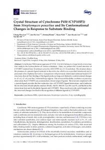
Steroids (Fig, 2) without a corresponding increase in levels of cortisol or adrenocorticotropin secretion (Cutler et al,, 1978; Korth-Schutz et al,, 1976; Parker and Odell, 1980), The secretion of increasing levels of sex steroids continues until the age of 25-35, after which time they begin to drop gradually until they return to childhood levels at 70-80 years of age (Orentreich et al,, 1984), This programmed development of adrenal P450 17A1 17,20-lyase activity is known as adrenarche. In an effort to explain this phenomenon, experiments were performed to determine if the change in lyase activity could be associated with an increase in posttranslational modification of P450 17A1, Human P450 17A1 is phosphorylated on serine and threonine residues by PKA, Phosphorylation increases 17,20-lyase activity, while dephosphorylation has the opposite effect (Zhang et al,, 1995), Phosphorylation of rat testicular P450 17A1 increases the ligand-binding efficiency of the enzyme for its natural ligand (progesterone) as well as the rate of P450 17A1 proteolytic degradation (Lohr and Kuhn-Velten, 1997), These findings have led researchers to propose that pituitary hormones (corticotropin, lutropin, or human chorionic gonadotropin) known to activate the G protein-coupled receptor (GPCR)/adenylate cyclase/PKA signal transduction pathway, may account for adrenarche and the proteolytic regulation of P450 17A1 levels (Lohr and Kuhn-Velten, 1997), Recently, leptin has been identified as a hormone capable of stimulating the lyase activity of P450 17A1 in intact human adrenocortical carcinoma cells in a manner consistent with the development of adrenarche. As may he observed in Fig, 3, physiological levels of leptin, acting through its receptor and the downstream signal
400-
GIRLS: Closed symbols BOYS: Open symbols
^
° a
LU' • a
300-
O DC LLI IV> O CC
o
a.
a
0
t/) ai
•
e
250*
•
200' 160 140
fr
/
CC Q
I
UJ Q
100 80 60 40 20
O
'
A
0
n
*
•
/ J a 0
, o
0
LU
O
•
/ 0 o
^
'
D
•
t,
0
^
12
i //
3
CA MONTHS
•
.
o"
• • •8* O • "
3
4
5
0