ANTICANCER RESEARCH 32: 2347-2352 (2012)
Review
Regulatory Mechanisms of the HB-EGF Autocrine Loop in Inflammation, Homeostasis, Development and Cancer KOHEI MIYATA1,2, FUSANORI YOTSUMOTO2,3, SUNG OUK NAM1,2, MASAHIDE KUROKI2,3 and SHINGO MIYAMOTO1,2
Departments of 1Obstetrics and Gynecology, 2Central Research Institute for Advanced Molecular Medicine and 3Biochemistry, Faculty of Medicine, Fukuoka University, Fukuoka, Japan
Abstract. Heparin binding epidermal growth factor-like growth factor (HB-EGF) is involved in development and homeostasis as well as in pathological processing of chronic diseases, especially cancer. Enhancement of HB-EGF expression is directly or indirectly regulated by transcriptional factors, including activator protein-1 (AP-1), specificity protein (SP)1, SP3, nuclear factor kappa B (NF-κB), hypoxia inducible factor 1, alpha subunit (HIF-1α, myogenic differentiation 1 (MyoD), Wilms tumor 1 (WT-1) and snail homolog 1 (Snail), and also by microRNAs. These transcription or posttranscription factors may communicate to form an autocrine HB-EGF amplification loop. Emerging evidence has indicated that HB-EGF is a rational target for the therapy of cancer and atherosclerosis. In this review, we discuss the relationship between the HB-EGF autocrine loop and HB-EGF transcriptional factors, and we highlight HB-EGF as a therapeutic target in diverse diseases. The epidermal growth factor receptor family of receptor tyrosine kinases is composed of four members in mammals; ERBB1/HER1 (also EGFR), ERBB2/HER-2, ERBB3/HER3 and ERBB4/HER-4 (1, 2). There are several ERBB-specific ligands that can be categorized into three groups depending on receptor binding specificity. The first group includes epidermal growth factor (EGF), amphiregulin (AREG), and transforming growth factor alpha (TGF-α), which all bind specifically to ERBB1. The second group includes betacellulin (BTC), heparin-binding EGF-like growth factor
Correspondence to: Shingo Miyamoto, MD, Ph.D., Department of Obstetrics and Gynecology, Faculty of Medicine, Fukuoka University, 7-45-1 Nanakuma, Jonan-ku, Fukuoka 814-0180, Japan. Tel: +81 928011011, Fax: +81 928654114, e-mail:
[email protected] Key Words: HB-EGF, transcription factor, autocrine, cancer, inflammation, homeostasis, review.
0250-7005/2012 $2.00+.40
(HB-EGF) and epiregulin (EREG), which all exhibit dual specificity for ERBB1 and ERBB4. The third group includes neuregulin (NRG), which binds to ERBB3 or ERBB4 (3). ERBB1-mediated intracellular signaling controls many of the functions required for growth, migration and proliferation (4). The activation of ERBB1 through binding to its ligands transmits signals for cell growth [dependent on the Kirsten rat sarcoma viral oncogene (Ras)- v-raf-1 murine leukemia viral oncogene homolog 1 (Raf)-extracellular signal-regulated kinases (ERK) pathway], cell survival [dependent on the phosphoinositide-3-kinase (PI3K) v-akt murine thymoma viral oncogene homolog 1 (AKT) pathway], and transcriptional control [dependent on signal transducer and activator of transcription 3 (Stat3)]. Activated ERBB1 forms a heterodimer complex with other ERBBs or other receptors, resulting in a variety of different signals. ERBB1 ligands, which are located on the cell membrane, are proteolytically cleaved and released upon action of various stimuli, including growth factors, cytokines, ultraviolet light, hypoxia and anticancer agents. HB-EGF, an ERBB1 ligand, is synthesized as a transmembrane precursor (pro-HB-EGF) that can serve as a juxtacrine growth factor. ERBB1 transactivation is induced by a soluble form of HB-EGF through ectodomain shedding in a paracrine manner. In lipid metabolism and cancer progression, HB-EGF expression is up-regulated in an autocrine manner. This means that HB-EGF is a promising target for the therapy of cancer and atherosclerosis (5, 6). Thus, it is plausible that HB-EGF contributes to diverse biological events through its own amplification loop via unique transcriptional control. In this review, we focus on the relationship between HB-EGF expression and transcription factors through the autocrine loop.
Nuclear Factor Kappa B (NF-κB) in Inflammation HB-EGF is a 22 kDa protein that can be purified from conditioned medium, in which macrophage-like U-937 cells have been cultured (7). Initial reports suggested that pro-
2347
ANTICANCER RESEARCH 32: 2347-2352 (2012) inflammatory cytokines, such as tumor necrosis factor (TNF)-α and interleukin-1β (IL-1β), induced HB-EGF expression in human umbilical vein endothelial cells (HUVEC) and A549 cells (8, 9). In addition, TNF-α and lipopolysaccharide (LPS) augment HB-EGF by up-regulating its transcription, thereby contributing to cell growth and tumor progression. Pro-inflammatory cytokines, including TNF-α and IL-6, activate NF-κB, a key transcriptional regulator of inflammation. NF-κB also induces pro-inflammatory cytokines, including TNF-α and IL-6 (10). This autocrine cytokine loop, which is important in chronic inflammatory disease, seems to be regulated by the transcriptional activity of NF-κB. In principle, activated NF-κB regulates the transcription of over 150 genes, including many related to inflammation, such as that for HB-EGF (11, 12). However, the complexities of the molecular relationship between HBEGF and NF-κB remain to be resolved. Studies from our laboratories have shown that an increase of activated AKT enhanced the expression of HB-EGF in fibroblasts, and the suppression of HB-EGF attenuated the activation of AKT in cancer cells (13). In addition, the suppression of HB-EGF expression resulted in inhibition of NF-κB activation, whereas an inhibitor of NF-κB promoted HB-EGF expression (manuscript in preparation). ERBB1 transactivation mediated by HB-EGF may induce NF-κB activation, leading to enhanced expression of IL-6 and IL-8 (14). In A549 cells, ERBB1 transactivation mediated by HBEGF may result in NF-κB activation, leading to enhanced expression of matrix metallopeptidase 9 (MMP-9) (9). On the other hand, HB-EGF attenuated NF-κB activation in colon cancer cells, an action mediated by IL-1β and interferon gamma (IFN-γ). According to these lines of evidence, it is plausible that HB-EGF activates AKT via ERBB1 transactivation and that the activated AKT induces NF-κB activation, resulting in a variety of genes being up-regulated. Thus, the activation of NF-κB seems to be indirectly regulated by HB-EGF, while NF-κB activation may not always be associated with HBEGF expression.
AP-1, SP1 and SP3 in Homeostasis HB-EGF plays a pivotal role in wound repair, inducing migration and proliferation of keratinocytes, fibroblasts, and smooth muscle cells to fill the injured area and to promote re-epithelialization and granulation tissue formation in the wound (15, 16). Estrogen also contributes to wound healing. Estrogen promotes granulation tissue formation and collagen deposition (17) and inhibits excessive inflammation in the wound by inhibiting neutrophil influx into the wound and by inhibiting production of a pro-inflammatory cytokines in monocytes/macrophages (18, 19). 17β-Estradiol
2348
(E2) participates in wound healing by up-regulating the expression of HB-EGF, which potentiates wound repair (20). This augmentation of HB-EGF expression is due to transcriptional activation of the HB-EGF promoter by the binding of activator protein-1 [AP-1, composed of FBJ murine osteosarcoma viral oncogene homolog (FOS) and jun proto-oncogene (JUN)] and SP1 (20). HB-EGF and ERBB1 have been recognized as direct AP-1 target genes (21-23). The establishment of pregnancy requires an intimate physical interaction, as well as a molecular dialogue, between the conceptus and the maternal reproductive tract. In any species, HB-EGF is an essential molecule for implantation. In mice and rats, maternal ovarian estrogen and progesterone (P4) are indispensable for implantation. In hamsters, P4 alone is enough to achieve blastocyst implantation and to induce HB-EGF expression in the uterine luminal epithelium surrounding the blastocyst. On the other hand, SP1 or SP3 is involved in the expression of HB-EGF and of 17β-hydroxysteroid dehydrogenase type 2, which is a key target of progesterone action (24, 25). In addition to this evidence, SP1 may mediate the regulation of endometrial epithelial gene expression during early pregnancy (26). Accordingly, progesterone and HB-EGF may act during implantation through a mutual amplification loop via SP1 or SP3. The molecular mechanism of insulin-induced HB-EGF expression is similar to that of E2-induced HB-EGF expression. The insulin response element of HB-EGF is able to bind AP-1, SP1 and NFκB, and this element is also detected in other promoters of EGF ligand genes, such as EPI and AREG (27). These three transcriptional factors are consequently activated by PI3K. Gastrin is a peptide hormone that is important as both an acid secretagogue (28) and as a trophic factor for the gastrointestinal mucosa (29). Studies have shown that gastrin stimulates the cleavage of pro-HB-EGF into sHBEGF and stimulates the expression of HB-EGF at the mRNA and protein levels, in both whole rat stomach and rat gastric epithelial cell lines (30, 31). Gastrin regulates HBEGF expression via PKC/ERBB1 signaling (32). Helicobacter pylori infection up-regulates levels of HB-EGF mRNA and protein and increases HB-EGF shedding (33). In addition, Helicobacter pylori infection of gastric epithelial cells activates AP-1 and NF-κB, which may lead to MMP-7 overexpression (34) and to the induction of the endogenous gastrin gene through AP-1 signaling, and not through NF-κB, SP1 or SP3 (35, 36). Although there is no evidence that gastrin promotes HB-EGF expression via certain transcriptional factors, H. pylori enhances the expression of gastrin and HB-EGF through AP-1. Collectively, AP-1, SP1 and SP3 may directly regulate HBEGF expression through different signaling pathways.
Miyata et al: Transcriptional and Post-transcriptional Control of HB-EGF Expression (Review)
MyoD, PDX1 and WT1 in Development Several transcriptional factors are involved in spatiotemporal development through HB-EGF expression. Direct interaction between myogenic differentiation 1 (MyoD) and the HBEGF promoter is transiently found during skeletal muscle cell differentiation and the membrane form of HB-EGF (proHB-EGF) is expressed preferentially in myotubes (37). The pancreas is an organ composed of two distinct cell populations: exocrine cells that secrete digestive enzymes and endocrine cells that secrete hormones. HB-EGF is responsible for developing exocrine and endocrine cells. Colocalization of pancreatic and duodenal homeobox-1 (PDX1) and HB-EGF is detected in pancreatic ductal cells, and PDX-1 is identified as a direct regulator of HB-EGF (38). The Wilms’ tumor gene (WT1) encodes a zinc finger transcriptional factor that is vital during the development of several organs. The embryos of WT1-null mice die in utero with agenesis of kidneys, gonads, adrenal glands and spleen (39). WT1 contributes to the regulation of the EGF family of growth factors during nephrogenesis and its binding site is located upstream (-1580/-1170 bp) of the HB-EGF transcriptional start site (40). On the other hand, analysis of the HB-EGF knock-out mouse has also shown that HB-EGF plays an important role in various developmental and homeostasis processes. HB-EGF-null mice exhibit abnormal lung and eyelid development, poor skin wound healing, retinoid-induced skin hyperplasia, and heart chamber and valve malformation (41). These findings indicate that HB-EGF is an essential ligand of ERBB1 during development. However, there is no evidence that HB-EGF directly activates MyoD or WT1 genes.
Transcription Factors in Cancer HB-EGF has been reported to be a promising therapeutic target for ovarian, breast, gastric and endometrial cancer (42, 43). Although overexpression of HB-EGF is found in several types of cancer, the underlying molecular mechanisms remains unclear. RAS and RAF oncogenes participate in diverse responses to extracellular stimuli. Deferential display analysis shows that forced expression of RAS and RAF results in upregulated HB-EGF mRNA levels (44). In fibroblasts, the expression of ERBB1, ERBB2 and AKT enhance HB-EGF expression among EGFRs ligands (13). In principle, HB-EGF can activate ERBB1, HER-2, RAS, RAF, ERK and AKT. Accordingly, HB-EGF forms an autocrine amplification loop for itself via molecules dependent on cell growth or cell survival signals. However, there is no information concerning the transcription factors involved in the HB-EGF autocrine amplification loop. On the other hand, there are some reports that refer to a relationship between HB-EGF and the wellknown tumor suppressor, p53. Wild-type p53 functions as a transcriptional factor and induces cell cycle arrest, apoptosis and
senescence (45, 46). HB-EGF is a transcriptional target of p53 (47, 48). On the other hand, mutations in p53 have been detected in a variety of human cancer types (45). Most p53 alterations are point missense mutations that lead to the synthesis of stable proteins that accumulate in the nuclei of tumor cells (49). Mutant, but not wild-type, p53 initiates a feedback loop that involves transactivation of early growth response 1 (EGR-1), which in turn increases the secretion of EGFRs ligands and stimulates the EGFRs signaling pathway (50). In ovarian, breast, gastric and endometrial cancer, overexpression of HB-EGF might be partially linked to mutation of p53. Highly malignant tumors are characterized by their response to hypoxia. Hypoxia in cancer cells predominantly induces the aberrant expression of pro-angiogenic factors, such as vascular endothelial growth factor (VEGF), via an increase in the non-hydroxyl form of hypoxia inducible factor 1, alpha subunit (HIF-1α) from the hydroxyl form (51). Hypoxia also enhances the expression of HB-EGF related molecules, including SP1, HB-EGF and a disintegrin and metalloproteinase domain-containing protein 17 (ADAM17) (52, 53). Under hypoxic conditions, HB-EGF also provokes the production of VEGF and activation of endothelial nitric oxide synthase (eNOS) via HIF-1α (54). Accordingly, it is plausible that hypoxia evokes HB-EGF and ADAM 17 expression via Sp1 and that VEGF production and eNOS activation are stimulated by HB-EGF through HIF-1α. Overexpression of NF-κB is found in some types of human cancers (55). To identify NF-κB-related chemotherapy genes, microarray analysis was performed by Wang et al. The authors found that HB-EGF is up-regulated in colorectal cancer cell lines by treatment with SN38, an activated form of irinotecan (56). In this study, AP-1, as well as NF-κB, was implicated in the enhancement of HB-EGF expression. In addition, they also reported that the transcription factor, SP1, is involved in NPI-0052 (secondgeneration proteasome inhibitor)-induced HB-EGF transcription in pancreatic cancer cells (57). HB-EGF regulates the expression of MMP-9 and VEGF via the transcription factor, Snail, through epithelial-mesenchymal transition (58).
Post-transcriptional Control The post-transcriptional silencing of target mRNAs by small RNAs was a revolutionary discovery in the understanding of genetic information control. Accumulating evidence has revealed that microRNAs, which regulate most genes in the human genome, are members of a large class of non-coding RNAs of approximately 22 nucleotides in length and that microRNAs are strongly conserved between vertebrates, invertebrates and plants (59, 60). It is thought that microRNA mutants can appear apparently normal but can
2349
ANTICANCER RESEARCH 32: 2347-2352 (2012)
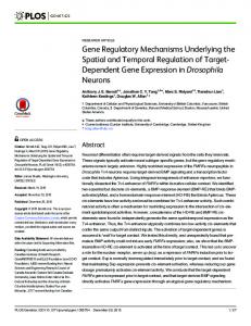
Figure 1. Schematic representation showing that Heparin binding epidermal growth factor-like growth factor (HB-EGF) expression is regulated by a variety of transcriptional factors in inflammation (NF-κB and AP-1), homeostasis (AP-1 and SP1) and development (PDX-1, WT1 and MyoD). In cancer biology, although many transcriptional regulations have been described, such as mutant p53 and EGR1, it is possible that other unknown factors may affect the HB-EGF autocrine loop and that the established factors may also contribute to the regulation of HB-EGF expression.
exhibit phenotype crisis under stress conditions (61). Genome-wide microRNA expression profiling studies using high-throughput technologies have demonstrated that almost all cancer types present a specific profile of up-regulated and down-regulated microRNAs (62, 63). De-regulated microRNA expression is recognized as an early event in tumorigenesis, and the levels of circulating microRNAs are regarded as reliable cancer biomarkers (64). In addition, clinical studies have demonstrated the potential use of microRNAs as predictors of sensitivity to radiotherapy and to anticancer agents (65). Inhibition of HB-EGF by the addition of an miR-212 mimic can induce cetuximab sensitivity in cetuximab-resistant cell lines, suggesting that increased expression of HB-EGF due to down-regulation of miR-212 is a possible mechanism of cetuximab resistance (66). Although microRNAs play indispensable roles in stress responses, these molecular mechanisms should be further investigated in order to elucidate the fundamental roles of microRNAs in controlling mRNA regulation during stress, including chronic disease and cancer.
Conclusion Accumulating evidence suggests that AP-1, SP1 and SP3 are directly involved in the HB-EGF autocrine amplification loop and that NF-κB, HIF-1α, and other transcriptional factors indirectly participate in this loop (Figure 1). Unfortunately, it remains unknown how post-transcriptional regulation is associated with the HB-EGF autocrine loop.
2350
Recently, the development of anticancer agents against HBEGF has advanced. CRM197 is a diphtheria toxin mutant that binds directly to the EGF-like domain and represses the mitogenic activity of HB-EGF (67). A phase II study of CRM197 will start shortly including patients with recurrent ovarian cancer in several University Hospitals in Japan under the approval of the Institutional Ethical Committees. In addition to CRM197, a neutralizing antibody targeting HBEGF is also being developed (68). It will be interesting to discover whether these agents completely block the HB-EGF autocrine loop. In the near future, the development of RNAi or small molecular weight compounds against HB-EGF-related transcriptional factors will allow us to improve the clinical outcome for HB-EGF-related chronic diseases.
References 1 Carpenter G: The EGF receptor: a nexus for trafficking and signaling. Bioassays 22: 697-707, 2000. 2 Riese DJ 2nd and Stem DF: Specificity within the EGF family/ErbB receptor family signaling network. Bioassays 20: 41-48, 1998. 3 Nautiyal J, Rishi AK and Majumdar AP: Emerging therapies in gastrointestinal cancers. World J Gastroenterol 12: 7440-7450, 2006. 4 Krause DS and Van Etten RA: Tyrosine kinases as targets for cancer therapy. N Engl J Med 353: 172-187, 2005. 5 Miyamoto S, Yagi H, Yotsumoto F, Kawarabayashi T and Mekada E: Heparin-binding epidermal growth factor-like growth factor as a novel targeting molecule for cancer therapy. Cancer Sci 97: 341347, 2006. 6 Bakken AM, Protack CD, Roztocil E, Nicholl SM and Davies MG: Cell migration in response to the amino-terminal fragment
Miyata et al: Transcriptional and Post-transcriptional Control of HB-EGF Expression (Review)
7
8
9
10
11 12
13
14
15
16
17
18 19
20
21
of urokinase requires epidermal growth factor receptor activation through an ADAM-mediated mechanism. J Vasc Surg 49: 12961303, 2009. Moscatello DK, Holgado-Madruga M, Emlet DR, Montgomery RB and Wong AJ: Constitutive activation of phosphatidylinositol 3-kinase by a naturally occurring mutant epidermal growth factor receptor. J Biol Chem 273: 200-206, 1998. Yoshizumi M, Kourembanas S, Temizer DH, Cambria RP, Quertermous T and Lee ME: Tumor necrosis factor increases transcription of the heparin-binding epidermal growth factor-like growth factor gene in vascular endothelial cells. J Biol Chem 267: 9467-9469, 1992. Cheng CY, Kuo CT, Lin CC, Hsieh HL and Yang CM: IL-1beta induces expression of matrix metalloproteinase-9 and cell migration via a c-SRC-dependent, growth factor receptor transactivation in A549 cells. Br J Pharmacol 160: 1595-1610, 2010. Kao CY, Chen Y, Thai P, Wachi S, Huang F, Kim C, Harper RW and Wu R: IL-17 markedly up-regulates beta-defensin-2 expression in human airway epithelium via JAK and NF-kappaB signaling pathways. J Immunol 173: 3482-3491, 2004. Pahl HL: Activators and target genes of REL/NF-κB transcription factors. Oncogene 18: 6853-6866, 1999. Murthy A, Defamie V, Smookler DS, Di Grappa MA, Horiuchi K, Federici M, Sibilia M, Blobel CP and Khokha R: Ectodomain shedding of EGFR ligands and TNFR1 dictates hepatocyte apoptosis during fulminant hepatitis in mice. J Clin Invest 120: 2731-2744, 2010. Yotsumoto F, Oki E, Tokunaga E, Maehara Y, Kuroki M and Miyamoto S: HB-EGF orchestrates the complex signals involved in triple-negative and trastuzumab-resistant breast cancer. Int J Cancer 127: 2707-2717, 2010. Pan Z, Wang Z, Yang H, Zhang F and Reinach PS: TRPV1 activation is required for hypertonicity-stimulated inflammatory cytokine release in human corneal epithelial cells. Invest Ophthalmol Vis Sci 52: 485-493, 2011. Ellis PD, Hadfield KM, Pascall JC and Brown KD: Heparinbinding epidermal growth factor like growth factor gene expression is induced by scrape-wounding epithelial cell monolayers: involvement of mitogen-activated protein kinase cascades. Biochem J 354: 99-106, 2001. Iwamoto R and Mekada E: Heparin-binding EGF-like growth factor: a juxtacrine growth factor. Cytokine Growth Factor Rev 11: 335-344, 2000. Ashcroft GS, Greenwell-Wild T, Horan MA, Wahl SM and Ferguson MW: Topical estrogen accelerates cutaneous wound healing in aged humans associated with an altered inflammatory response. Am J Pathol 155: 1137-1146, 1999. Ashcroft GS and Ashworth JJ: Potential role of estrogens in wound healing. Am J Clin Dermatol 4: 737-743, 2003. Ashcroft GS, Mills SJ, Lei K, Gibbons L, Jeong MJ, Taniguchi M, Burow M, Horan MA, Wahl SM and Nakayama T: Estrogen modulates cutaneous wound healing by down-regulating macrophage migration inhibitory factor. J Clin Invest 111: 1309-1318, 2003. Kanda N and Watanabe S: 17β-Estradiol enhances heparin-binding epidermal growth factor-like growth factor production in human keratinocytes. Am J Physiol Cell Physiol 288: C813-C823, 2005. Fu S, Bottoli I, Goller M and Vogt PK: Heparin-binding epidermal growth factor-like growth factor, a v-Jun target gene, induces oncogenic transformation. Proc Natl Acad Sci USA 96: 5716-5721, 1999.
22 Johnson AC, Murphy BA, Matelis CM, Rubinstein Y, Piebenga EC, Akers LM, Neta G, Vinson C and Birrer M: Activator protein-1 mediates induced but not basal epidermal growth factor receptor gene expression. Mol Med 6: 17-27, 2000. 23 Park JM, Adam RM, Peters CA, Guthrie PD, Sun Z, Klagsbrun M and Freeman MR: AP-1 mediates stretch-induced expression of HB-EGF in bladder smooth muscle cells. Am J Physiol 277: C294301, 1999. 24 Edwards DP: Regulation of signal transduction pathways by estrogen and progesterone. Annu Rev Physiol 67: 335-376, 2005. 25 Cheng YH, Imir A, Suzuki T, Fenkci V, Yilmaz B, Sasano H and Bulun SE: SP1 and SP3 mediate progesterone-dependent induction of the 17β hydroxysteroid dehydrogenase type 2 gene in human endometrium. Biol Reprod 75: 605-614, 2006. 26 Simmen RC, Zhang XL, Zhang D, Wang Y, Michel FJ and Simmen FA: Expression and regulatory function of the transcription factor SP1 in the uterine endometrium at early pregnancy: implications for epithelial phenotype. Mol Cell Endocrinol 159: 159-170, 2000. 27 Ornskov D, Nexo E and Sorensen BS: Insulin induces a transcriptional activation of epiregulin, HB-EGF and amphiregulin, by a PI3K-dependent mechanism: identification of a specific insulin-responsive promoter element. Biochem Biophys Res Commun 354: 885-891, 2007. 28 Chen D, Zhao CM, Dockray GJ, Varro A, Van Hoek A, Sinclair NF, Wang TC and Koh TJ: Glycine-extended gastrin synergizes with gastrin 17 to stimulate acid secretion in gastrin-deficient mice. Gastroenterology 119: 756-765, 2000. 29 Koh TJ and Chen D: Gastrin as a growth factor in the gastrointestinal tract. Regul Pept 93: 37-44, 2000. 30 Miyazaki Y, Shinomura Y, Tsutsui S, Zushi S, Higashimoto Y, Kanayama S, Higashiyama S, Taniguchi N and Matsuzawa Y: Gastrin induces heparin-binding epidermal growth factor-like growth factor in rat gastric epithelial cells transfected with gastrin receptor. Gastroenterology 116: 78-89, 1999. 31 Tsutsui S, Shinomura Y, Higashiyama S, Higashimoto Y, Miyazaki Y, Kanayama S, Hiraoka S, Minami T, Kitamura S, Murayama Y, Miyagawa J, Taniguchi N and Matsuzawa Y: Induction of heparin-binding epidermal growth factor-like growth factor and amphiregulin mRNAs by gastrin in the rat stomach. Biochem Biophys Res Commun 235: 520-523, 1997. 32 Sinclair NF, Ai W, Raychowdhury R, Bi M, Wang TC, Koh TJ and McLaughlin JT: Gastrin regulates the heparin-binding epidermal-like growth factor promoter via a PKC/EGFRdependent mechanism. Am J Physiol Gastrointest Liver Physiol 286: G992-999, 2004. 33 Dickson JH, Grabowska A, El-Zaatari M, Atherton J and Watson SA: Helicobacter pylori can induce heparin-binding epidermal growth factor expression via gastrin and its receptor. Cancer Res 66: 7524-7531, 2006. 34 Wroblewski LE, Noble PJ, Pagliocca A, Pritchard DM, Hart CA, Campbell F, Dodson AR, Dockray GJ and Varro A: Stimulation of MMP-7 (matrilysin) by Helicobacter pylori in human gastric epithelial cells: role in epithelial cell migration. J Cell Sci 116: 3017-3026, 2003. 35 Tamara P, Tucker TP, Gray BM, Eaton KA and Merchant JL: Helicobacter pylori induction of the gastrin promoter through GC-rich DNA elements. Helicobacter 15: 438-448, 2010. 36 Kovac S, Xiao L, Shulkes A, Patel O and Baldwin GS: Gastrin increases its own synthesis in gastrointestinal cancer cells via the CCK2 receptor. FEBS Lett 584: 4413-4418, 2010.
2351
ANTICANCER RESEARCH 32: 2347-2352 (2012) 37 Chen X, Raab G, Deutsch U, Zhang J, Ezzell RM and Klagsbrun M: Induction of heparin-binding EGF-like growth factor expression during myogenesis. Activation of the gene by MyoD and localization of the transmembrane form of the protein on the myotube surface. J Biol Chem 270: 18285-18294, 1995. 38 Kaneto H, Miyagawa J, Kajimoto Y, Yamamoto K, Watada H, Umayahara Y, Hanafusa T, Matsuzawa Y, Yamasaki Y, Higashiyama S and Taniguchi N: Expression of heparin-binding epidermal growth factor-like growth factor during pancreas development. A potential role of PDX-1 in transcriptional activation. J Biol Chem 272: 29137-29143, 1997. 39 Hammes A, Guo JK, Lutsch G, Leheste JR, Landrock D, Ziegler U, Gubler MC and Schedl A: Two splice variants of the Wilms' tumor 1 gene have distinct functions during sex determination and nephron formation. Cell 106: 319-329, 2001. 40 Kim HS, Kim MS, Hancock AL, Harper JC, Park JY, Poy G, Perantoni AO, Cam M, Malik K and Lee SB: Identification of novel Wilms' tumor suppressor gene target genes implicated in kidney development. J Biol Chem 282: 16278-16287, 2007. 41 Iwamoto R and Mekada E: ERBB and HB-EGF signaling in heart development and function. Cell Struct Funct 31: 1-14, 2006. 42 Miyamoto S, Hirata M, Yamazaki A, Kageyama T, Hasuwa H, Mizushima H, Tanaka Y, Yagi H, Sonoda K, Kai M, Kanoh H, Nakano H and Mekada E: Heparin-binding EGF-like growth factor is a promising target for ovarian cancer therapy. Cancer Res 64: 5720-5727, 2004. 43 Yotsumoto F, Yagi H, Suzuki SO, Oki E, Tsujioka H, Hachisuga T, Sonoda K, Kawarabayashi T, Mekada E, Miyamoto S: Validation of HB-EGF and amphiregulin as targets for human cancer therapy. Biochem Biophys Res Commun 365: 555-561, 2008. 44 McCarthy SA, Samuels ML, Pritchard CA, Abraham JA and McMahon M: Rapid induction of heparin-binding epidermal growth factor/diphtheria toxin receptor expression by Raf and Ras oncogenes. Genes Dev 9: 1953-1964, 1995. 45 Levine AJ: p53, the cellular gatekeeper for growth and division. Cell 88: 323-331, 1997. 46 Vogelstein B, Lane D, Levine AJ: Surfing the p53 network. Nature 408: 307-310, 2000. 47 Fang L, Li G, Liu G, Lee SW and Aaronson SA: p53 induction of heparin-binding EGF-like growth factor counteracts p53 growth suppression through activation of MAPK and PI3K/AKT signaling cascades. EMBO J 20: 1931-1939, 2001. 48 Han JA, Kim JI, Ongusaha PP, Hwang DH, Ballou LR, Mahale A, Aaronson SA and Lee SW: p53-mediated induction of Cox-2 counteracts p53- or genotoxic stress-induced apoptosis. EMBO J 21: 5635-5644, 2002. 49 Soussi T and Béroud C: Assessing TP53 status in human tumours to evaluate clinical outcome. Nat Rev Cancer 1: 233-240, 2001. 50 Sauer L, Gitenay D, Vo C and Baron VT: Mutant p53 initiates a feedback loop that involves EGR-1/EGF receptor/ERK in prostate cancer cells. Oncogene 29: 2628-2637, 2010. 51 Chan DA and Giaccia AJ: PHD2 in tumour angiogenesis. Br J Cancer 103: 1-5, 2010. 52 Svensson KJ, Kucharzewska P, Christianson HC, Sköld S, Löfstedt T, Johansson MC, Mörgelin M, Bengzon J, Ruf W and Belting M: Hypoxia triggers a proangiogenic pathway involving cancer cell microvesicles and PAR-2-mediated heparin-binding EGF signaling in endothelial cells. Proc Natl Acad Sci USA 108: 13147-13152, 2011.
2352
53 Szalad A, Katakowski M, Zheng X, Jiang F and Chopp M: Transcription factor Sp1 induces ADAM17 and contributes to tumor cell invasiveness under hypoxia. J Exp Clin Cancer Res 28: 129-138, 2009. 54 Nakai K, Yoneda K, Moriue T, Igarashi J, Kosaka H and Kubota Y: HB-EGF-induced VEGF production and eNOS activation depend on both PI3 kinase and MAP kinase in HaCaT cells. J Dermatol Sci 55: 170-178, 2009. 55 Rayet B and Gélinas C: Aberrant REL/NFκB genes and activity in human cancer. Oncogene 18: 6938-6947, 1999. 56 Wang F, Liu R, Lee SW, Sloss CM, Couget J and Cusack JC: Heparin-binding EGF-like growth factor is an early response gene to chemotherapy and contributes to chemotherapy resistance. Oncogene 26: 2006-2016, 2006. 57 Sloss CM, Wang F, Palladino MA and Cusack JC Jr.: Activation of EGFR by proteasome inhibition requires HB-EGF in pancreatic cancer cells. Oncogene 29: 3146-52, 2010. 58 Yagi H, Yotsumoto F and Miyamoto S: Heparin-binding epidermal growth factor-like growth factor promotes transcoelomic metastasis in ovarian cancer through epithelial mesenchymal transition. Mol Cancer Ther 7: 3441-3451, 2008. 59 Lewis BP, Burge CB and Bartel DP: Conserved seed pairing, often flanked by adenosines, indicates that thousands of human genes are microRNA targets. Cell 120: 15-20, 2005. 60 Ambros V: MicroRNA pathways in flies and worms: growth, death, fat, stress, and timing. Cell 113: 673-676, 2003. 61 Leung AK and Sharp PA: MicroRNA functions in stress responses. Mol Cell 40: 205-215, 2010. 62 Lu J, Getz G, Miska EA, Alvarez-Saavedra E, Lamb J, Peck D, Sweet-Cordero A, Ebert BL, Mak RH, Ferrando AA, Downing JR, Jacks T, Horvitz HR and Golub TR: MicroRNA expression profiles classify human cancers. Nature 435: 834-838, 2005. 63 Calin GA and Croce CM: MicroRNA signatures in human cancers. Nat Rev Cancer 6: 857-66, 2006. 64 Cortez MA, Bueso-Ramos C, Ferdin J, Lopez-Berestein G, Sood AK and Calin GA: MicroRNAs in body fluids the mix of hormones and biomarkers. Nat Rev Clin Oncol 8: 467-477, 2011. 65 Weiss GJ, Bemis LT, Nakajima E, Sugita M, Birks DK, Robinson WA, Varella-Garcia M, Bunn PA Jr., Haney J, Helfrich BA, Kato H, Hirsch FR and Franklin WA: EGFR regulation by microRNA in lung cancer: correlation with clinical response and survival to gefitinib and EGFR expression in cell lines. Ann Oncol 19: 1053-1059, 2008. 66 Hatakeyama H, Cheng H, Wirth P, Counsell A, Marcrom SR, Wood CB, Pohlmann PR, Gilbert J, Murphy B, Yarbrough WG, Wheeler DL, Harari PM, Guo Y, Shyr Y, Slebos RJ and Chung CH: Regulation of heparin-binding EGF-like growth factor by miR-212 and acquired cetuximab-resistance in head and neck squamous cell carcinoma. PLoS One 5: e12702, 2010. 67 Buzzi S, Rubboli D, Buzzi G, Buzzi AM, Morisi C and Pironi F: CRM197 (nontoxic diphtheria toxin): effects on advanced cancer patients. Cancer Immunol Immunother 53: 1041-1048, 2004. 68 Miyamoto S, Iwamoto R, Furuya A, Takahashi K, Sasaki Y, Ando H, Yotsumoto F, Yoneda T, Hamaoka M, Yagi H, Murakami T, Hori S, Shitara K and Mekada E: A novel anti-human HB-EGF monoclonal antibody with multiple antitumor mechanisms against ovarian cancer cells. Clin Cancer Res 17: 6733-6741, 2011.
Received April 4, 2012 Revised May 17, 2012 Accepted May 17, 2012