J. Phys. Ther. Sci. 30: 1417–1423, 2018
The Journal of Physical Therapy Science Original Article
Reliability of sagittal vertical axis measurement and association with measures of age-related hyperkyphosis Wendy B Katzman, PT, DPTSc1)*, Neeta Parimi, MS2), Amy Gladin, PT, DPT3), Bo Fan, MD4), Shirley S Wong, BS1), Joncarmen Mergenthaler, BS5), Nancy E Lane, MD6) 1) Department
of Physical Therapy and Rehabilitation Science, University of California, San Fransisco: 1500 Owens St., Ste 400, San Francisco, CA 94158, USA 2) San Francisco Coordinating Center, USA 3) Kaiser Permanente Northern CA, USA 4) Department of Radiology and Biomedical Imaging, University of California, San Francisco, USA 5) Department of Neurological Surgery, University of California, San Francisco, USA 6) University of California, Davis, USA
Abstract. [Purpose] Persons with age-related hyperkyphosis often have concomitant sagittal plane imbalance of the spine. This study investigated the reliability of sagittal vertical axis (SVA) measurement of sagittal balance, association between thoracic Cobb angle of kyphosis and SVA measure of sagittal balance, and compared the degree of SVA in males and females with age-related hyperkyphosis. [Participants and Methods] Measurements of SVA and Cobb angle of kyphosis were obtained from baseline radiographs of 112 community-dwelling males and females, mean age 70.0 (SD=5.7) years with kyphosis ≥40 degrees, recruited for a randomized controlled trial. Spearman correlation coefficients were used to determine associations between SVA and kyphosis, and Wilcoxon nonparametric tests to compare SVA between genders. [Results] SVA was acquired with excellent intra-rater [0.95 (95% CI: 0.88, 0.98)] and inter-rater reliability [0.93 (95% CI: 0.83,0.97)]. There was no significant correlation between Cobb angle of thoracic kyphosis and SVA, (r=−0.05). More males than females had sagittal imbalance (SVA≥5 cm). [Conclusion] In older adults with hyperkyphosis, SVA was a reliable measure of sagittal balance, and more extreme in males. SVA was not associated with Cobb angle of thoracic kyphosis, and could be considered an independent phenotype of age-related hyperkyphosis to be targeted in future intervention trials. Key words: Sagittal balance, Sagittal vertical axis, Hyperkyphosis (This article was submitted Jul. 13, 2018, and was accepted Sep. 12, 2018)
INTRODUCTION Measurement of thoracic kyphosis in persons with age-related hyperkyphosis is important for quantifying the degree of kyphosis deformity of the spine, and monitoring its progression and response to targeted interventions. The gold standard method of measuring hyperkyphosis is standing radiographic Cobb angle of kyphosis from T5 to T121, 2). However, other methods are often used to quantify hyperkyphosis, including the Block method3), and occiput to wall4) that quantify the position of the head in relation to the pelvis, aspects of sagittal balance, rather than the kyphosis deformity alone5). Persons with age-related hyperkyphosis who have sagittal imbalance of the spine may be at increased risk for vertebral fractures and incident falls6, 7) because of the altered distribution of body mass and the biomechanical environment of the spine when the head position is anterior to the pelvic sacral promontory6). Furthermore, when Cobb angle is measured globally (from T1 to *Corresponding author. Wendy Katzman (E-mail:
[email protected]) ©2018 The Society of Physical Therapy Science. Published by IPEC Inc. This is an open-access article distributed under the terms of the Creative Commons Attribution Non-Commercial No Derivatives (by-nc-nd) License. (CC-BY-NC-ND 4.0: https://creativecommons.org/licenses/by-nc-nd/4.0/)
1417
T12) it can be affected by endplate tilt of the upper and lower vertebrae of the thoracic curvature, which may increase the magnitude of the curve but may not reflect changes in the curve relative to vertical alignment8). While studies have reported significant adverse health outcomes among those with thoracic kyphosis ≥40 degrees1, 2), global sagittal balance of the spine has been described as the most significant radiographic parameter predicting clinical symptoms in adult spinal deformity9, 10), making it an important target for future intervention. Previous research suggests gender differences in the prevalence of age-related hyperkyphosis depending upon the methods used to quantify hyperkyphosis. Approximately 18% of males and 30% of females were categorized as hyperkyphotic in a cohort 70–80 year olds when a supine 40-degree thoracic kyphosis measurement was used to define hyperkyphosis11). In contrast, when the supine Block method was used, 46% of males and 22% of females were categorized as hyperkyphotic in a similarly aged cohort12). Given the large variability in prevalence estimates for hyperkyphosis in males and females when different methods are used to quantify kyphosis, it is possible that males and females have different phenotypes of hyperkyphosis. Males may have more sagittal imbalance, and females may have more sagittal deformity of thoracic kyphosis. Therefore, the aims of this study were to determine the reliability of sagittal vertical axis measurement (SVA) of sagittal balance in community-dwelling asymptomatic older adults with hyperkyphosis ≥40 degrees. We also investigated the association between thoracic kyphosis deformity and SVA measurement of sagittal balance, and compared the degree of sagittal balance in males and females with age-related hyperkyphosis. If thoracic kyphosis deformity and sagittal spine imbalance are independent phenotypes of hyperkyphosis, interventions to reduce age-related hyperkyphosis should be designed to improve both thoracic kyphosis deformity and sagittal balance.
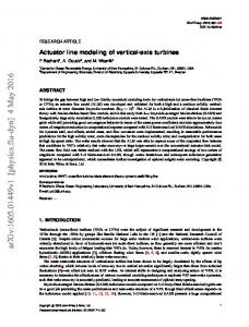
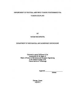
PARTICIPANTS AND METHODS We included 112 participants, mean age 70.0 (SD=5.7) years, who were recruited January 2013 through June 2015 from local senior centers and outpatient medical clinics at two large urban medical centers (a university-based center and an integrated managed-care center), and enrolled in the Specialized Center of Research (SCOR) Kyphosis Project, a randomized controlled trial to investigate gender differences in response to a targeted kyphosis exercise intervention. All participants were screened for kyphometer-derived kyphosis ≥40 degrees and age ≥60 years as inclusion criteria for the SCOR Kyphosis Project. Participants were excluded for inability to straighten the thoracic spine at least five degrees, cognitive impairment13), inability to pass safety tests in the screening examination, or any disorder or disease likely to interfere with safe participation in a group-based exercise class14). Protocols were approved by the University of California San Francisco (IRB No. 12–09348) and Kaiser Permanente Northern California (IRB No. CN-13AGlad-01-H) IRBs and written informed consent was collected for each participant. Cobb angle measurements of kyphosis from lateral spine radiographs acquired at the baseline enrollment visit were used. Standing lateral spine radiographs were acquired using a standardized sagittal view scoliosis protocol from T3 to S1, with arms at a 90-degree angle to the body to prevent superimposition of the vertebrae, knees straight, and upon full inspiration. The degree of kyphosis was calculated by an experienced musculoskeletal radiologist (BF) using digitized Cobb angle derived from the radiographs scans centered at T8 (T4–T12). The Cobb method was used to determine the superior and inferior margins for line placement. Using a translucent digitizer (GTCO, Rockville, MD, USA) and cursor, the reader marked points corresponding to the four corners of the vertebral body at T4 and T12. From the superior surface of T4 and the inferior surface of T12, along the endplates of the vertebrae, a digitization program erected perpendicular lines, the intersection of which is Cobb angle (Fig. 1). The intra-rater reliability ICC for repeated digitized Cobb angle readings was previously reported ICC=0.9915). SVA measurements of sagittal balance were acquired from the same standing lateral spine radiographs by an experienced musculoskeletal radiologist (BF). SVA was identified as the location of the head with respect to the normal center of gravity by a plumb line dropped from the center of the C7 vertebral body to the posterior superior corner of the sacral end plate16). A vertical line drawn from the vertebral body of C7 normally intersects the superior endplate of S1. The horizontal distance of displaced SVA from a reference point on the sacral end plate was determined (Fig. 2). Repeat measurements were performed on a random sample of 15% of the scans by a second observer (JM) to determine the intraobserver and interobserver reliability for the SVA measurement. Additional measurements of thoracic kyphosis and lumbar lordosis were acquired using kyphometer-derived thoracic kyphosis and lumbar lordosis measurements, and radiographic centroid measurement of thoracic kyphosis. The kyphometer angle of kyphosis was measured as the angle formed by the 2 arms of a Debrunner kyphometer (Proteck AG, Berne, Switzerland) protractor-device placed over the spine. The base of the top arm was aligned over the T2–3 interspace, and the base of the lower arm was aligned over the T11–T12 interspace. Similarly, for lumbar lordosis, the kyphometer angle of lordosis was measured as the angle formed by the arms of the device placed over the T11–T12 interspace and the L4–L5 interspace. All kyphometer measures were acquired by a trained exercise physiologist at the UCSF Clinical Research Center. For the radiographic centroid angle method, we used intersecting diagonals between two vertebral corners at both ends of the thoracic spine curve. The centroid curvature angle was defined as the angle between the straight lines drawn perpendicular through the two top and two bottom vertebrae (Fig. 3). Reliability of these methods has been previously established8, 15). Other study covariates: Age and gender were determined at enrollment. Statistical analysis: Characteristics including age, gender, and all spinal variables were summarized by descriptive statisJ. Phys. Ther. Sci. Vol. 30, No. 12, 2018
1418
Fig. 1. Cobb angle of kyphosis measured from standing lateral radiograph. Line a is drawn from the superior endplate of T4; line b is drawn from the inferior endplate of T12; lines c and d are perpendicular lines drawn from lines a and b. Cobb angle of kyphosis is where lines c and d intersect.
Fig. 2. The sagittal vertical axis is measured as the horizontal distance between a plumb line drawn from center of C7 (a) and a line drawn from center of C7 to posterior superior corner of S1 (b).
Fig. 3. Centroid angle was determined by measuring the intersection angle of two lines, which passed through T4– T5 (a) centroid points and T8–T9 centroid points (b).
tics and compared between males and females using t-test or Wilcoxon nonparametric test for continuous variables and the Fisher exact or χ2 test for categorical variables. Nonparametric counterparts were used for variables that were not normally distributed, based upon testing for kurtosis value +217). We used intra-class correlation coefficients (ICC) with 95 percent confidence intervals (95%CI) to compare the relative intra- and inter-rater reliability of SVA measurement. Two readers (BF, JM) blinded to each other’s results, read a random sample of 15 films, and each reader read 15 films twice after a month interval. To evaluate the strength, of the correlation, we used a method outlined by Munro (0.9–1.0=“very high”; 0.7–0.89= “high”; 0.5–0.69= “moderate”; 0.26–0.49= “low”; 0.0–0.25=“little if any”)18). We assessed the absolute reliability of the SVA measurements and calculated the standard error of the measurement [(SEM=standard deviation of sagittal plane measurement*square root of (1−ICC)] and minimal detectable change [MDC=1.96*SEM*square root of (2)]19). We used Spearman correlation coefficients to determine the association between Cobb angle, SVA and other measures of hyperkyphosis, and considered correlations ranging from 0.00 to 0.25 to indicate little or no relationship; 0.25 to 0.5 a fair relationship; 0.5 to 0.75 moderate to good; and values above 0.75 good to excellent20). We used Wilcoxon nonparametric tests for continuous variables to compare SVA in both genders. Kyphometer-measured kyphosis, and lumbar lordosis were included to investigate concurrent validity of the radiographic and clinical measures of hyperkyphosis. Centroid measure of kyphosis
1419
was included to account for endplate tilt. We also categorized SVA as quartiles and assessed the distribution of quartiles of SVA between genders. All analyses were performed with SAS 9.4 (Cary, NC, USA).
RESULTS Characteristics of our study participants, 45 males and 67 females, are presented in Table 1. Age ranged from 60 to 92, with an average age of 70.0 (SD=5.7) years old. Mean Cobb angle was 55.6 (SD=12.1) degrees, with no difference between genders. SVA measurements were acquired on all but 2 of the scans where visualization of C7 vertebral body superiorly was not possible. The intra-rater reliability of the SVA measurement was ICC=0.95 (95%CI: 0.88, 0.98). The inter-rater reliability was ICC=0.93 (95%CI: 0.83, 0.97). The within and between subject SEM was 7.7 and 9.15 mm, and MDC was 21.4 and 25.4 mm, respectively. SVA measurement was skewed in males, although the median SVA in males compared to females was not statistically different, p=0.35 (Table 1). The kurtosis value was 1.7 overall. When we used 5 cm or more of SVA as a threshold for abnormal21), more males than females, n=12 (26.7%) versus n=5 (7.5%), were classified with abnormal SVA, and the difference was significant, p=0.006. There were no differences in centroid angle in males and females, but males had greater kyphometer-derived kyphosis, 53.3 (SD=7.2) vs. 51.3 (SD=7.5) degrees, p=0.06. Males also had less lordosis than females, 23.1 (SD=11.1) vs. 34.9 (SD=10.3) degrees, p