Neuropsychologia 46 (2008) 1161–1169
Repetition of distractor sets improves visual search performance in hemispatial neglect ´ Styrmir Saevarsson a,b,∗ , Sigr´unsif J´oelsd´ottir c , Haukur Hjaltason d,e , Arni Kristj´ansson c,f a
b
Department of Psychology, Neuropsychology, University of Freiburg, Germany Psychological Laboratory, Helmholtz Institute, Utrecht University, The Netherlands c Department of Psychology, University of Iceland, Iceland d Department of Neurology, Landsp´ıtali-University Hospital, Iceland e Department of Medicine, Neurology, University of Iceland, Iceland f Institute of Cognitive Neuroscience, University College London, United Kingdom
Received 4 June 2007; received in revised form 18 October 2007; accepted 19 October 2007 Available online 6 November 2007
Abstract Priming from repeated distractor sets, or search context, in conjunctive visual search was examined in four patients with hemispatial neglect. In the first experiment overall context was either changed or repeated while the target was always the same to control for any modulatory effect of target priming. Considerable priming was seen from repeated context. In the second experiment the context was either repeated on the left side, on the right side, on both sides, or the context was new. Priming from repeated context was found to arise from the left visual field, as well as the right visual field, as well as when overall context was repeated. Brief masked displays were used in experiment 3, the results again showing strong priming from repeated overall context. The results of the three experiments suggest that visual grouping, or perceptual organization, of distractor sets is relatively intact in the affected hemifield of parietal neglect patients. Furthermore, repetition of context may even temporarily ameliorate neglect symptoms in search. These findings are consistent with claims that grouping is distinct from attentional processing and that it operates at lower levels of the perceptual hierarchy. © 2007 Elsevier Ltd. All rights reserved. Keywords: Context priming; Priming; Attention; Visual search; Grouping; Neglect
1. Introduction Hemispatial neglect is a multimodal neuropsychological disorder where patients typically fail to notice, respond to, or orient to novel or important stimuli in their left visual field, even though primary sensory and/or motor processes are unimpaired (Driver & Mattingley, 1998; Heilman, Bowers, Valenstein, & Watson, 1987). A neglect patient may, for instance, read only the right sides of words (McManus, 2001) or repeatedly look for a particular item in his right visual field, seldom orienting to his left (Husain et al., 2001). Neglect is typically caused by brain damage following an acute cerebrovascular accident in areas in the inferior parietal ∗ Corresponding author at: Department of Psychology, Neuropsychology, University of Freiburg, Germany. Tel.: +49 761 203 9442; fax: +49 761 203 9438. E-mail address:
[email protected] (S. Saevarsson).
0028-3932/$ – see front matter © 2007 Elsevier Ltd. All rights reserved. doi:10.1016/j.neuropsychologia.2007.10.020
lobule and in the medial temporoparietal junction (e.g. Mort et al., 2003; Vallar & Perani, 1986), or in the superior temporal gyrus (Karnath, Berger, K¨uker, & Rorden, 2004; Karnath, Ferber, & Himmelbach, 2001), usually in the right hemisphere. No precise compromise regarding neuroanatomical correlates has yet been arrived at, and an exact critical locus for neglect to occur may not exist, but the disorder may rather reflect disruption of the activity of a network for attentional orienting that involves the activity of a number of regions (e.g. Danckert & Ferber, 2006; Kerkhoff, 2001). Disrupted visual search is a common symptom in hemispatial neglect (e.g. Behrmann, Ebert, & Black, 2004; Husain et al., 2001; Mort & Kennard, 2003). However, results from a large number of functional neuroimaging studies show activation in the superior parietal lobule and intraparietal sulcus during visual search in healthy observers. These areas have been found to be more dorsal than lesions that are typically involved in visual neglect (e.g. Corbetta & Shulman, 2002; Geng et al.,
1162
S. Saevarsson et al. / Neuropsychologia 46 (2008) 1161–1169
2006; Kristj´ansson, Vuilleumier, Schwartz, Macaluso, & Driver, 2007). Free exploratory eye movements in visual search have subsequently been found to elicit brain activations that relate more closely to the locations of infarcts that are typical for neglect (Himmelbach, Erb, & Karnath, 2006). Behavioral and physiological evidence indicates, however, that considerable processing of left visual field (LVF) stimuli occurs despite nonawareness of the stimuli on the part of the subject (see e.g. Driver & Vuilleumier, 2001 for review). As an example, Halsband, Gruhn, and Ettlinger (1985) showed that neglect patients were more likely to notice fearsome and sexually arousing pictures compared with neutral pictures when presented to the neglected contralesional hemifield. Neglect symptoms can also be ameliorated by illusory surface completion by stimuli that the patient is unaware of (Mattingley, Davis, & Driver, 1997). Pavlovskaya, Ring, Groswasser, and Hochstein (2002) found reduced neglect of gabor patches in the left hemifield by presenting collinear gabor patches in the unaffected right hemifield. Furthermore, Vuilleumier and Rafal (1999) found that hemispatial neglect disrupted the localization of contralesional stimuli, but the patients were able to count the same stimuli quickly and correctly. Other studies have shown that there is considerable neural response to emotional faces, with or without awareness of those stimuli for neglect patients (Vuilleumier et al., 2002). This occurs in the occipital and ventral stream areas which are usually intact in neglect (see also Driver, Vuilleumier, Eimer, & Rees, 2001; Vuilleumier, Schwartz, Husain, Clarke, & Driver, 2001). 1.1. Priming in visual search tasks Studies of how the normal observer scans the visual field (e.g. Cavanagh & Chase, 1971; Egeth, Jonides, & Wall, 1972; Neisser, 1963; Treisman & Gelade, 1980) have mainly focused on how we search our environment in the here and now, but later studies have indicated that what occurs on a previous trial has a strong effect on how a following scene is processed (e.g. Kristj´ansson, 2006a; Kristj´ansson, Wang, & Nakayama, 2002; Maljkovic & Nakayama, 1994; Olivers & Meeter, 2006; Treisman, 1992; see e.g. Kristj´ansson, 2006b for a review). These studies have revealed that our attention tends to be drawn to those features and items in our visual field that we have recently processed, stimuli that have recently been important to us for one reason or another, or the locations of those stimuli (Maljkovic & Nakayama, 1996). In light of this, Kristj´ansson (2006b) argued that the repetition of the target in visual search led to facilitated attentional processing (see also Kristj´ansson & Nakayama, 2003). Subsequent studies have then shown that context repetition, i.e. repetition of distractor sets in visual search will speed search in a similar manner – independently of any target priming (Geyer, M¨uller, & Krummenacher, 2006; Kristj´ansson & Driver, 2005; Kristj´ansson et al., 2002; Wang, Kristj´ansson, & Nakayama, 2005). With the aim of investigating claims that priming in visual search reflects facilitated attention deployments to recent features of interest, Kristj´ansson, Vuilleumier, Malhotra, Husain, and Driver (2005) studied priming for neglect patients with infe-
rior parietal cortex lesions. The observers performed a visual search task where they had to locate a target diamond of a unique color in the display. Intact location and color priming was found for the patients. Kristj´ansson et al. (2005) speculated that such priming might help patients overcome the attentional deficits in neglect. In addition, they found that location priming requires conscious processing of the preceding target in the LVF when brief displays were used, unlike color priming which was not dependent on the patients’ awareness of the preceding target in the LVF in hemispatial neglect, suggesting a dissociation between priming of color and position. 1.2. Current questions The aim of the current study was to investigate possible priming benefits of context or distractor sets repetition for neglect patients performing a visual search task. Given the amount of evidence for residual processing of stimuli that go unnoticed in neglect the aim was to see whether the benefits of context repetition would aid search. This could address some important aspects of functional damage in neglect – as an example whether or not perceptual organization of vision is intact in the affected hemifield. Some studies have suggested that attention is not necessary for grouping of distractor sets to occur (Moore & Egeth, 1997; Russell & Driver, 2005), which is in line with proposals that surface analysis and grouping, often thought to reflect ‘mid-level’ visual processing (Marr, 1980; Nakayama, He, & Shimojo, 1995) precede object-based processing and attentional processing (Duncan & Humphreys, 1989; Torralba, 2003; Treisman, 1982). If grouping does not require the operation of attention we might expect to see relatively intact priming of context for neglect patients. In fact Sasaki (2007) has, in a recent review argued that grouping processes involve, for the most part, striate and extrastriate areas which are unaffected in neglect. 2. Methods 2.1. Subjects Four patients with chronic hemispatial neglect participated in the study. In experiments 1 and 2, two male patients (N1 and N2) participated while two female patients (N3 and N4) participated in experiment 3. Patient N1 was 55 years old, N2 was 54, N3 was 56 and N4 was 62 at the time of testing. CT scans of patients N1 and N2 revealed a similar infarct in the right artery cerebri media territory resulting in damage in frontal, temporal and parietal cortex and in the centrum semiovale area, including inferior frontal and parietal cortex. The CT scans of patients N1 and N2 showed, on the other hand, intact visual cortices (V1) and frontal eye fields. Patient N3 had similar damage to N1 and N2 as well as an additional infarcted area in the right side of the mesencephalon. Patient N4 suffered from a subarachnoidal hemorrhage localized in the right temporoparietal lobules. A MRI scan 8 months post-stroke showed brain tissue damage including right frontal, temporal and parietal, and the centrum semiovale areas as well as the right side of the mesencephalon and the left frontal area. All patients had intact visual cortices and thus intact visual fields as measured with traditional tasks. The four patients underwent traditional tests of visual neglect before they enrolled in the study. For a 180 mm line bisection task (Wilson, Cockburn, & Halligan, 1987) patients N1 and N2 both showed a 5 mm deviation. On letter cancellation tasks (Mesulam, 1981), patient N1 omitted 2 out of 30 targets on
S. Saevarsson et al. / Neuropsychologia 46 (2008) 1161–1169
1163
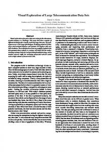
Fig. 1. Stimuli for experiments 1 and 3. There were two possible contexts on any given trial (trial N), either the same context as on the last trial (trial N − 1) or a different context. The two possible contexts were red disks and green donuts, or blue disks and green donuts. the right side and all 30 targets on the left side, while patient N2 omitted no letters on the right side but 7 of 30 letters on the left side. In figure copying (Gainotti, D’Erme, Monteleone, & Silvery, 1986) and line cancellation (Albert, 1973) tasks, patient N1 missed the left side of figure copying but performed line cancellation without omissions while patient N2 performed figure copying and line cancellation more or less without errors. Patient N3 showed a 16 mm right deviation on an 18 mm bisection task and patient N4 showed a 60 mm right deviation for a 140 mm line. On letter cancellation tasks patient N3 omitted 15 out of 30 targets on the right side and 25 targets on the left side, while patient N4 omitted 14 letters on the right side and all 30 letters on the left side. In figure copying and line cancellation tasks, patient N3 missed some objects on the left side during figure copying but performed line cancellation without omissions. Patient N4 missed all objects on the left side in figure copying and missed all targets on the left side but none on the right side on a line cancellation task. In sum, all the patients showed clear signs of hemispatial neglect and had intact visual fields. In experiments 1 and 2 two male control observers (C1 and C2) participated while in experiment 3 two female controls (C3 and C4) participated. The control observers were approximately matched in age to the four patients. Control participants C1 and C2 were both 53 years old. Control participant C3 was 51 years old and control participant C4 was 65. All controls were neurologically healthy and had no history of visual neglect or related problems.
2.2. Stimuli Three different versions of a visual search task were used in the three experiments. In experiment 1 and 2 the visual search array was present on the screen until response, so that the main dependent measure was response time. In experiment 3 the display was presented for 400 ms followed by a mask, and the dependent measure was in this case accuracy. The mask was a field of random dots, either green, red or blue. The size of each dot in the random dot array was 13 arcmin. Two versions of the response time task were used, with regard to context repetition, one in which context across the whole visual field was repeated or not (experiment 1 and 3), and a second where context was repeated on the left or on the right or in both hemifields (so that overall context was repeated) or the whole context was different from the last trial (experiment 2). Context repetition
is defined by whether the distractor sets were the same, partially same or different from the last trial (see Figs. 1 and 2). Observers searched for a green disk and indicated by key press whether they thought that the green disk was present on the screen or not. The target differed in the particular combination of color and shape from the rest of the stimuli on the screen (this is in other words a variation of the well known conjunction search task, since the target was only defined by being unique on two features and shared a feature with each distractor set). The two possible distractor sets were red disks and green donuts, and blue disks and green donuts (see Figs. 1 and 2). The diameter of the discs and donuts was 1.3 arc degree. On any given trial 48 items (a target and 47 distractors on target trials or 48 distractors on the blank trials) appeared on the screen. The target was present on 60% of the trials (30% on left and 30% on right). In an attempt to increase the distinction between hemifields a 4.5◦ gap separated stimuli on the left and right of fixation (as indicated in Figs. 1 and 2). A small fixation cross was present at the center of the screen throughout. The locations of the distractors and target (if present) were determined completely randomly from one trial to the next. The intertrial interval for all three experiments was 1200 to 1700 ms.
2.3. Apparatus An Apple G3 laptop with a 14 in. LCD screen was used to run the experimental tasks. Participants responded by keypress on an external keypad where two response buttons (“4” and “6”) were colored green for target trials and white for non-target trials. Viewing distance was kept as close to 67 cm as possible, at which the area on the screen that stimuli could appear within subtended 17 arc degree (horizontally) by 14 arc degree (vertically). The experimental tasks were all prepared in the C programming language using the Vision Shell function library (Comtois, 2003).
2.4. Procedure Participants were instructed to respond as quickly and as accurately as possible by pressing the green button if they found the green target stimulus or the white button if no unique target was present on the screen. The participants were also told to maintain fixation on the cross. In experiment 3, the patients
1164
S. Saevarsson et al. / Neuropsychologia 46 (2008) 1161–1169
Fig. 2. The experimental display in experiment 2. The figure shows examples of four possible context repetition/non-repetition conditions for trial N following trial N − 1 (counting from the top): Same context as on the last trial; same context on the left; same context on the right; or different context across the whole display. were strongly encouraged to maintain gaze on the centre of the display and the experimenter monitored their eye gaze prompting them to refixate the central fixation marker if their gaze strayed away from screen centre. Ethical approval for the study was provided by the Ethics and Science committee of Landsp´ıtali-University Hospital, and from the National committee of personal rights in Iceland.
3. Results and discussion 3.1. Experiment 1 The aim of experiment 1 was to investigate priming from repeated context across the whole display, by measuring reaction time dependent on whether overall context was repeated or not.
Patient N1 finished 676 trials in experiment 1 (correct responses on 98% of the trials) while patient N2 finished 600 trials (89% correct). Control participants C1 and C2 finished 676 trials (98% and 100% of trials correct, respectively). Fig. 3a shows the effects of repetition of context on response times in the visual search task, for the trials where a target was present. A clear repetition effect was seen for targets in both hemifields (data are shown as a function of the visual field of the target). A clear speed-up when context was repeated was seen both for controls and for the patients for the target present trials. A 2 × 2 ANOVA performed on the individual present trials for each subject, with the factors context repetition and hemifield difference, supported this conclusion. The effect of context repetition was significant for all four participants (N1: F(1,397) = 8.38,
S. Saevarsson et al. / Neuropsychologia 46 (2008) 1161–1169
1165
quite strong, consistent with previous reports (Geyer et al., 2006; Kristj´ansson & Driver, 2005; Kristj´ansson et al., 2002) and are strong both for target and blank trials. 3.2. Experiment 2
Fig. 3. The results from experiment 1. Panel A shows the results for the target present trials as a function of whether context was repeated or not and where the target appeared (in the LVF or RVF, respectively). Panel B shows the results for the target absent trials. The response times for the patients were split into two groups along the median, slow and fast response times (see text for details).
p = 0.004; N2; F(1,319) = 4.96, p = 0.046; C1: F(1,397) = 4.73, p = 0.043; C2: F(1,406) = 5.33, p = 0.027) The effect of hemifield was significant for both patients (N1: F(1,397) = 33.9, p < 0.001; N2: F(1,319) = 37.7, p < 0.01) but unsurprisingly there was no difference between hemifields for the control observers (p > 0.4). There was a rather large number of slow reaction times on the target absent trials for the patients, and our first analysis did not reveal a significant difference between repetition and no repetition for the absent trials. When we split the response times along the median into “slow” and “fast” response times, however, a clear priming effect was found for the faster reaction times (see Fig. 3b). This was confirmed with F-tests. The priming effect was significant for the faster trials but not the slower for both patients (N1, fast trials: F(1,264) = 6.37, p = 0.019, slow trials: F(1,264) = 1.1, n.s.; N2, fast trials F(1,213) = 5.95, p = 0.017; slow trials, F(1,213) = 2.07, n.s.). The context repetition effect was significant for both controls for the absent trials, even without the median split into fast and slow response times as was done for the patients (C1: F(1,265) = 6.98, p = 0.008; C2: F(1,270) = 5.33, p = 0.022). We can conclude from this first experiment that context repetition in visual search speeds search on the following trial considerably both for patients and controls. This indicates that priming of perceptual groups (see e.g. Wang et al., 2005; Kristj´ansson, 2006b) occurs despite neglect and also that perceptual organization is relatively intact in patients despite their lesions. The facilitation effects following context repetition are
A skeptic might argue that faster grouping through context priming from the intact right hemifield simply speeds search on the left, rather than the results reflecting any context priming occurring on the left. Because of this possibility we investigated context priming in neglect further in experiment 2 by asking whether repetition of context in the LVF only or the right visual field (RVF) only would facilitate target detection (see Section 2 for details). In experiment 2, patient N1 finished 1037 trials (correct responses on 98% of trials). Patient N2 finished 867 trials (96% correct). Control participants C1 and C2 finished 1037 trials (correct responses on 98% and 99% of trials, respectively). The mean response times as a function of repetition type are shown in Fig. 4. Clear priming was seen for all participants on present trials as well as absent trials and for both right and left visual hemifields. The largest context priming effect was seen when overall context was repeated. But more importantly, for the present purposes, context repetition on the left, on its own, resulted in priming as well as context repetition on the right, ruling out the possibility that the speed-up of search with overall context repetition seen in experiment 1 was simply due to context priming from the right hemifield only. A one-way ANOVA on target trial response times for each participant as a function of the 4 trial types (context repeated in both RVF and LVF, in RVF only, in LVF only, or not repeated at all) was performed. Since the overall F-test is hard to interpret in terms of differences between the different repetition types, we had to rely on Tukey HSD post hoc tests to determine the significance of measured differences between the different conditions. For the target trials for both patients there was a significant difference between repetition of context in LVF and no context repetition; between RVF repetition only and no repetition; between overall context repetition and no repetition as well as between LVF context repetition only and overall repetition and RVF repetition and overall repetition (all p-values < 0.01). For control participant C1 there was only a significant difference between overall repetition and no repetition; LVF repetition and overall repetition and no repetition and RVF repetition. For control participant C2 the only significant difference was between overall repetition and no repetition (all p-values < 0.01). A similar statistical procedure for the absent trials revealed that again there was, for both patients, a significant difference between repetition of context in LVF and no context repetition; between RVF repetition and no repetition; between overall context repetition and no repetition as well as between LVF repetition only and overall repetition and RVF repetition and overall repetition (all p-values < 0.01). For both controls there was only a significant difference between when overall context was repeated and when overall context changed (all p-values < 0.01). The reason why we do not consistently get significant differences when context is repeated on one side only, for the controls, is probably that this experimental procedure is not particularly sensitive to
1166
S. Saevarsson et al. / Neuropsychologia 46 (2008) 1161–1169
Fig. 4. Response times for patients N1 and N2 and controls C1 and C2 in experiment 2. The first two panels show the response times on the target present trials (target on left in the leftmost panel, target on the right in the middle panel). Trials where the target was absent are shown in the rightmost panel.
context priming effects when response times are brief as is the case for our healthy control observers here. This may, in other words, be a floor effect. We can, at this point, conclude that context repetition leads to facilitated grouping of distractor sets even when attentional processing is disrupted as in neglect. Priming of context is thus unlikely to be solely due to the attentional processing networks that are damaged in neglect, but is likely to reflect facilitated processing at earlier stages in the perceptual hierarchy. We have previously conjectured that context priming operates by allowing faster rejection of distractor sets as not containing a target (Kristj´ansson et al., 2002; Wang et al., 2005) and the present results argue that this process is facilitated when context is repeated. For the neglect patients here, this results in considerable speeding of target detection. Similar facilitation was observed for the control participants. The results from experiment 2 show that the results of experiment 1 cannot be explained by context repetition on the right side only. Repeating context on the left on its own results in priming as does repeating context on the right only, compared with when the context is completely new. When repeated context on right is compared to repeated context on the left, there is very little difference between the two repetition effects. There is, however a large effect on response times of target side for the patients as
expected from their neglect. The results further strengthen our conclusions from experiment 1 that context repetition strongly speeds search even for the affected hemifield of neglect patients, indicating that there is considerable residual processing of stimuli that do not reach awareness consistent with previous reports (see e.g. Driver & Vuilleumier, 2001 for review), in this case grouping of display items for faster rejection as non targets. 3.3. Experiment 3 One might conceivably criticize the results of experiments 1 and 2 on the grounds that observers could more-or-less freely move their eyes around the screen and had unlimited time to view the display. We did not, for example, have much control over where the observers oriented their gaze. Their gaze might, for instance, have lingered at the locus of the last target, on a given trial. Although this would not have aided their search overall since target position was completely unpredictable, we conducted a third experiment where any potential problems due to this issue of eye gaze could be avoided. The patients and controls in this experiment thus had only limited time to search the briefly presented display (400 ms at maximum) and the dependent measure was accuracy (see Section 2 for details). Patient N3 finished 786 trials in experiment 3 (70.4% of the trials correct)
S. Saevarsson et al. / Neuropsychologia 46 (2008) 1161–1169
1167
almost always correct on the target absent trials. There was thus little room for improvement with context repetition since performance was close to ceiling (but the repetition effect for absent trials was nevertheless significant for patient N3). This does not change our conclusion that both LVF and RVF targets were found much more often when context was repeated than when it was not. 4. General discussion
Fig. 5. The results from experiment 3 for the two patients, N3 and N4 as a function of target side as well as for the trials where no target was presented (see text above for discussion of results for the aged matched controls).
while patient N4 finished 899 trials (71.5% correct). Control participants C3 and C4 finished 1000 trials (98% of trials correct for both). A large effect of context repetition was seen for the target present trials (see Fig. 5). When context was repeated, noticing rates of left sided targets went up from less than 10% for both patients, up to 40% for patient N3 and 60% for patient N4. A large difference was also seen on the right when context was repeated. The control participants were at ceiling on the task so it was not possible to assess any repetition priming for them. Chi-square tests were performed on the data for the patients. For patient N3 a significant context repetition effect was found for the LVF (χ2 = 57.42, p < 0.001), for the RVF (χ2 = 8.96, p = 0.002) as well as for the absent trials (χ2 = 11.73, p < 0.001). For patient N4, a significant context repetition effect was found for the LVF (χ2 = 67.54, p < 0.001) as well as for the RVF (χ2 = 15.2, p < 0.001) but χ2 is equal to zero for the absent trials for patient N4 since the percent correct was completely the same for repetition versus no repetition. The patients seem to have adopted a very strict criterion for deciding on target presence which means that they were
In the three experiments presented here we have shown that repetition of context in visual search is relatively intact for patients suffering from hemispatial neglect. One might even say that context repetition can temporarily ameliorate neglect symptoms in such a task. The results suggest that grouping of distractor sets is facilitated if they remain constant from one trial to the next. This allows faster rejection of distractors as nontargets and the target is found faster in consequence. The current findings add to the growing evidence for the importance of what has occurred on previous trials for visual search, in general (see e.g. Kristj´ansson, 2006b), as well as increasing evidence that such repetition can speed search and improve accuracy on search tasks for neglect patients (see e.g. Kristj´ansson et al., 2005). That study only showed this for target repetition, and when the layout of the visual search stimuli was constant across trials, however. The repetition benefit here is, on the other hand, not bound to any particular layout since the positions of target and distractors were decided randomly for each trial. Kristj´ansson and Nakayama (2003; see also Kristj´ansson, 2006b; Nakayama, Maljkovic, & Kristj´ansson, 2004) claimed that visual attention is guided to a surprisingly large extent by what has occurred recently, for example on previous trials in a visual search experiment. They argued for the existence of a primitive memory system for the deployment of visual attention that aids us in orienting our attention to the task at hand at any given moment (see also discussion in Maljkovic & Nakayama, 1994). Such benefits apply to situations where a target is repeated as well as when distractor sets, or context, is repeated (Geyer et al., 2006; Kristj´ansson & Driver, 2005; Kristj´ansson et al., 2002; Wang et al., 2005). Apart from the speeded search (in experiments 1 and 2, for both target and blank trials), the most noticeable result is, perhaps, that noticing rates of a briefly presented target in the affected hemifield can increase from close to zero to approximately 40 to 50% (experiment 3). This shows how dramatic the context repetition effects can be on how we organize a visual scene, seemingly ameliorating neglect symptoms in a visual search task, making it more likely that the patients will orient their attention into the neglected hemifield. 4.1. Neural mechanisms The human neuronal mechanisms of priming following target repetition have been investigated with functional brain imaging techniques such as fMRI. Kristj´ansson et al. (2007; see also Yoshida, Tsubomi, Osaka, & Osaka, 2003; and Geng et al., 2006) found that priming seems to involve well known atten-
1168
S. Saevarsson et al. / Neuropsychologia 46 (2008) 1161–1169
tional mechanisms in frontal and parietal areas (see e.g. Corbetta & Shulman, 2002). There were also neural effects correlated with color priming in color specific areas of ventral cortex (V4) and effects specific to repetition of target position in inferior frontal and inferior parietal cortex. These findings on healthy participants are consistent with findings on hemispatial neglect patients that have inferior parietal cortex damage and show intact position and color priming (Kristj´ansson et al., 2005). We are not aware of any functional neuroimaging studies of context priming in visual search. Durston, Thomas, Worden, Yang, and Casey (2002) have, however, showed that repetition of context in a modified go no-go task affects activity patterns in ventrolateral prefrontal cortex, anterior cingulate gyrus and superior parietal cortex in healthy subjects (see also e.g. Casey, Martinez, Thomas, Worden, & Durston, 2001). Finally, Sasaki (2007) has recently argued that grouping involves the operation of striate and extrastriate areas that are generally intact in neglect patients and indeed in the patients that were tested here. In sum, these studies suggest that priming from repeated context may reflect changes in neural activity at multiple sites of the brain, as has been found for target priming.
results add to the growing evidence for cognitive functions that are preserved in neglect and show that patients’ visual search can greatly benefit from repetition of context, in this case the repetition of distractor identity in visual search.
4.2. Grouping and mid-level vision
References
Marr (1980) argued that surface assignment and grouping preceded object analysis; a point supported by the results of Nakayama and colleagues (summarized in Nakayama et al., 1995; see also discussion in Kristj´ansson, 2006c) who have showed how surface analysis influences higher level motion perception, visual search, figure ground analysis and the perception of apparent motion, and precedes the operation of attention. Sasaki (2007) argued that perceptual organization in vision involves the operation of a number of regions of the visual cortices in the occipital lobes, depending on the nature of the grouping cues. Occipital cortices were intact in all our observers so despite their neglect, lower level perceptual organization could very well be unaffected in the patients. As stated before, Kristj´ansson et al. (2005) found that location priming requires conscious processing of the preceding target in the LVF unlike color priming in the LVF. An interesting question for future research would, for instance, be to explore if such a dissociation can be found between color and position for context priming. Also, any interactions between repetition priming of distractor sets and repetition of spatial layout of distractors (see e.g. Chun & Jiang, 1998) would be of great interest in future studies. 5. Conclusions The current results, combined with previous results, show that intertrial priming in visual search is not largely affected in neglect. This applies both to target and context repetition effects. Contextual priming was found even when the context was only repeated in the affected hemifield of neglect patients. The results suggest that grouping processes of mid-level vision are relatively intact in neglect (consistent with previous proposals; see e.g. Driver & Vuilleumier, 2001 for review), and that neglect affects higher level vision and attentional processing most strongly. The
Acknowledgements ´ S.S., H.H. and A.K. were supported by a grant from the Science fund of the University Hospital of Iceland, number 2005-39. S.S. was supported by a grant from the German Academic Exchange Service (Deutscher Akademischer Austausch ´ Dienst). A.K. was supported by a grant from the University of Iceland research fund and the Human Frontiers Science Program. Albert Postma is thanked for comments at various stages of this study. Chris Olivers and Thomas Geyer are thanked for helpful comments on the manuscript. Experiments 1, 2 and 3 were part of a project towards partial fulfillment of S.S.’ M.Sc. degree at Utrecht University and Ph.D. degree at University of Freiburg. Experiment 3 was also performed in partial fulfillment of S.J.’s M.A. degree at the University of Iceland.
Albert, M. L. (1973). A simple test of visual neglect. Neurology, 23, 658–664. Behrmann, M., Ebert, P., & Black, S. E. (2004). Hemispatial neglect and visual search: A large scale analysis. Cortex, 40, 247–263. Casey, B. J., Martinez, A., Thomas, K., Worden, M., & Durston, S. (2001). A developmental fMRI study of attentional conflict. NeuroImage, 13, S306. Cavanagh, J. P., & Chase, W. G. (1971). The equivalence of target and nontarget processing in visual search. Perception & Psychophysics, 9, 493–495. Comtois, R. (2003). Vision shell PPC [Software libraries]. Cambridge, MA: Raynald Comtois. Corbetta, M., & Shulman, G. L. (2002). Control of goal-directed and stimulusdriven attention in the brain. Nature Reviews Neuroscience, 3, 201–215. Chun, M. M., & Jiang, Y. (1998). Contextual cueing: Implicit learning and memory of visual context guides spatial attention. Cognitive Psychology, 36, 28–71. Danckert, J., & Ferber, S. (2006). Revisiting unilateral neglect. Neuropsychologia, 44, 987–1006. Driver, J., & Mattingley, J. B. (1998). Parietal neglect and visual awareness. Nature Neuroscience, 1, 17–22. Driver, J., & Vuilleumier, P. (2001). Perceptual awareness and its loss in unilateral neglect and extinction. Cognition, 79, 39–88. Driver, J., Vuilleumier, P., Eimer, M., & Rees, G. (2001). Functional magnetic resonance imaging and evoked potential correlates of conscious and unconscious vision in parietal extinction patients. NeuroImage, 14, S68–S75. Duncan, J., & Humphreys, G. W. (1989). Visual search and stimulus similarity. Psychological Review, 96, 433–458. Durston, S., Thomas, K. M., Worden, M. S., Yang, Y., & Casey, B. J. (2002). The effect of preceding context on inhibition: An event-related fMRI study. NeuroImage, 16, 449–543. Egeth, H., Jonides, J., & Wall, S. (1972). Parallel processing of multielement displays. Cognitive Psychology, 3, 674–698. Gainotti, G., D’Erme, P., Monteleone, D., & Silveri, M. C. (1986). Mechanisms of unilateral spatial neglect in relation to laterality of cerebral lesions. Brain, 109, 599–612. Geyer, T., M¨uller, H. J., & Krummenacher, J. (2006). Cross-trial priming in visual search for singleton conjunction targets: Role of repeated target and distractor features. Perception & Psychophysics, 68, 736–749. ´ Rothsthein, P., & Driver, J. Geng, J. J., Eger, E., Ruff, C., Kristj´ansson, A., (2006). On- line attentional selection from competing stimuli in opposite visual fields: Effects on human visual cortex and control processes. Journal of Neurophysiology, 96, 2601–2612.
S. Saevarsson et al. / Neuropsychologia 46 (2008) 1161–1169 Halsband, U., Gruhn, S., & Ettlinger, G. (1985). Unilateral spatial neglect and defective performance in one half of space. International Journal of Neuroscience, 28, 173–195. Heilman, K. M., Bowers, D., Valenstein, E., & Watson, R. T. (1987). Hemispace and hemispatial neglect. In M. Jeannerod (Ed.), Neuropsychological and neuropsychological aspects of spatial neglect (pp. 115–150). New York: Elsevier Science Publishers Company. Himmelbach, M., Erb, M., & Karnath, H.-O. (2006). Exploring the visual world: The neural substrate of spatial orienting. NeuroImage, 32, 1747– 1759. Husain, M., Mannan, S., Hodgson, T., Wojciulik, E., Driver, J., & Kennard, C. (2001). Impaired spatial working memory across saccades contributes to abnormal search in parietal neglect. Brain, 124, 941–952. Karnath, H.-O., Ferber, S., & Himmelbach, M. (2001). Spatial awareness is a function of the temporal not the posterior parietal lobe. Nature, 411, 950–953. Karnath, H.-O., Berger, M. F., K¨uker, W., & Rorden, C. (2004). The anatomy of spatial neglect based on voxelwise statistical analysis: A study of 140 patients. Cerebral Cortex, 14, 1164–1172. Kerkhoff, G. (2001). Spatial hemineglect in humans. Progress in Neurobiology, 63, 1–27. ´ (2006a). Simultaneous priming along multiple dimensions in Kristj´ansson, A. visual search task. Vision Research, 46, 2554–2570. ´ (2006b). Rapid learning in attention shifts—A review. Visual Kristj´ansson, A. Cognition, 13, 324–362. ´ (2006c). Surface assignment modulates object-formation for Kristj´ansson, A. visual short-term memory. Perception, 35, 865–881. ´ & Driver, J. (2005). Priming in vision: Target repetition effects, Kristj´ansson, A., context effects and role reversal effects. Perception, 34(Suppl.), 40c. ´ & Nakayama, K. (2003). A primitive memory system for Kristj´ansson, A., the deployment of transient attention. Perception and Psychophysics, 65, 711–724. ´ Vuilleumier, P., Malhotra, P., Husain, M., & Driver, J. (2005). Kristj´ansson, A., Priming of color and position during visual search in unilateral spatial neglect. Journal of Cognitive Neuroscience, 17, 859–873. ´ Vuilleumier, P., Schwartz, S., Macaluso, E., & Driver, J. (2007). Kristj´ansson, A., Neural basis for priming of pop-out during visual search revealed with fMRI. Cerebral Cortex, 17, 1612–1624. ´ Wang, D., & Nakayama, K. (2002). The role of priming in Kristj´ansson, A., conjunctive visual search. Cognition, 85, 37–52. Maljkovic, V., & Nakayama, K. (1994). Priming of pop-out: I. Role of features. Memory & Cognition, 22, 657–672. Maljkovic, V., & Nakayama, K. (1996). Priming of pop-out: II. Role of position. Perception & Psychophysics, 58, 977–991. Marr, D. (1980). Vision. San Francisco: W.H. Freeman. Mattingley, J. B., Davis, G., & Driver, J. (1997). Preattentive filling-in of visual surfaces in parietal extinction. Science, 275, 671–674. McManus, I. C. (2001). Charles Dickens: A neglected diagnosis. Lancet, 358, 2158–2168. Mesulam, M. M. (1981). A cortical network for directed attention and unilateral neglect. Annals of Neurology, 10, 309–325. Mort, J. M., Malhotra, P., Mannan, S. K., Rorden, C., Pambakian, A., Kennard, C., et al. (2003). The anatomy of visual neglect. Brain, 126, 1986– 1997.
1169
Mort, D. J., & Kennard, C. (2003). Visual search and its disorders. Current Opinion in Neurology, 16, 51–57. Moore, C. M., & Egeth, H. E. (1997). Perception without attention: Evidence for grouping under conditions of inattention. Journal of Experimental Psychology: Human perception & Performance, 23, 339–352. Nakayama, K., He, J., & Shimojo, S. (1995). Visual surface representation: A critical link between lower-level and higher-level vision. In S. M. Kosslyn & D. N. Osherson (Eds.), Visual cognition: An invitation to cognitive science (second ed., vol. 2, pp. 1–70). Cambridge, MA: MIT Press. ´ (2004). Short term memNakayama, K., Maljkovic, V., & Kristj´ansson, A. ory for the rapid deployment of visual attention. In M. Gazzaniga (Ed.), The cognitive neurosciences (third ed., pp. 397–408). Cambridge, MA: MIT Press. Neisser, U. (1963). Decision time without reaction time: Experiments in visual scanning. American Journal of Psychology, 76, 376–385. Olivers, C. N. L., & Meeter, M. (2006). On the dissociation between compound and present/absent tasks in visual search: Intertrial priming is ambiguitydriven. Visual Cognition, 13, 202–222. Pavlovskaya, M., Ring, H., Groswasser, Z., & Hochstein, S. (2002). Searching with unilateral neglect. Journal of Cognitive Neuroscience, 14, 745–756. Russell, C., & Driver, J. (2005). New indirect measures of “inattentive” visual grouping in a change detection task. Perception & Psychophysics, 67, 606–623. Sasaki, Y. (2007). Processing local signals into global patterns. Current Opinion in Neurobiology, 17, 1–8. Treisman, A. (1982). Perceptual grouping and attention in visual search for features and for objects. Journal of Experimental Psychology: Human Perception and Performance, 8, 194–214. Treisman, A. (1992). Perceiving and re-perceiving objects. American Psychologist, 47, 862–875. Treisman, A., & Gelade, G. (1980). A feature-integration theory of attention. Cognitive Psychology, 12, 97–136. Torralba, A. (2003). Contextual priming for object detection. International Journal of Computer Vision, 53, 169–191. Vallar, G., & Perani, D. (1986). The anatomy of unilateral neglect after righthemisphere stroke lesions. A clinical/CT-scan correlation study in man. Neuropsychologia, 24, 609–622. Vuilleumier, P., Armony, J. L., Clarke, K., Husain, M., Driver, J., & Dolan, R. J. (2002). Neural response to emotional faces with and without awareness: Event-related fMRI in parietal patient with visual extinction and spatial neglect. Neuropsychologia, 40, 2156–2166. Vuilleumier, P., & Rafal, R. (1999). “Both” means more than “two”: Localizing and counting in patients with visuospatial neglect. Nature Neuroscience, 2, 783–784. Vuilleumier, P., Schwartz, S., Husain, M., Clarke, M., & Driver, J. (2001). Implicit processing and learning of visual stimuli in parietal extinction and neglect. Cortex, 37, 741–744. ´ & Nakayama, K. (2005). Efficient visual search Wang, D., Kristj´ansson, A., without top-down or bottom-up guidance. Perception & Psychophysics, 67, 239–253. Wilson, B., Cockburn, J., & Halligan, P. W. (1987). Behavioral inattention test. Bury St. Edmunds: Thomas Valley. Yoshida, T., Tsubomi, H., Osaka, M., & Osaka, N. (2003). Priming of a popout—An fMRI study. Perception, 32, 93c.