INTERNATIONAL JOURNAL OF ONCOLOGY 42: 1743-1753, 2013
Repression of exogenous gene expression by the retinoic acid target gene G0S2 TIAN MA1, JESSICA P. DONG4, DAVID J. SEKULA1, DENNIS LIANG FEI1, WILLIAM W. LAMPH5, MICHAEL HENDERSON1, YUN LU1, STEVEN BLUMEN1, SARAH J. FREEMANTLE1 and ETHAN DMITROVSKY1-3 Departments of 1Pharmacology and Toxicology, and 2Medicine, Geisel School of Medicine at Dartmouth, Hanover, NH 03755; 3Norris Cotton Cancer Center, Dartmouth-Hitchcock Medical Center, Lebanon, NH 03756; 4Dartmouth College, Hanover, NH 03755; 5Ligand Pharmaceutical, San Diego, CA 92121, USA Received January 6, 2013; Accepted February 18, 2013 DOI: 10.3892/ijo.2013.1876 Abstract. The G0/G1 switch gene 2 (G0S2) is rapidly induced by all-trans-retinoic acid (RA)-treatment of acute promyelocytic leukemia (APL) and other cells. G0S2 regulates lipolysis via inhibition of adipose triglyceride lipase (ATGL). This study found that retinoic acid receptor (RAR), but not retinoid X receptor (RXR) agonists induced G0S2 expression in APL cells. Novel G0S2 functions were uncovered that included repression of exogenous gene expression and transcriptional activity. Transient G0S2 transfection repressed the activities of multiple reporter constructs (including the retinoid-regulated species RARβ, UBE1L and G0S2); this occurred in diverse cell contexts. This inhibition was antagonized by siRNA-mediated G0S2 knockdown. To determine the inhibitory effects were not due to transient G0S2 expression, G0S2 was stably overexpressed in cells without appreciable basal G0S2 expression. As expected, this repressed transcriptional activities. Intriguingly, transfection of G0S2 did not affect endogenous RARβ, UBE1L or G0S2 expression. Hence, only exogenously expressed genes were affected by G0S2. The domain responsible for this repression was localized to the G0S2 hydrophobic domain (HD). This was the same region responsible for the ability of G0S2 to inhibit ATGL activity. Whether an interaction with ATGL accounted for this new G0S2 activity was studied. Mimicking the inhibition of ATGL by oleic acid treatment that increased lipid droplet size or ATGL siRNA knockdown did not recapitulate G0S2 repressive effects. Engineered gain of ATGL expression did not rescue G0S2 transcriptional repression either. Thus, transcriptional repression by G0S2 did not depend on the ability of G0S2 to inhibit ATGL. Subcellular localization studies revealed
Correspondence to: Dr Ethan Dmitrovsky, Department of Pharmacology and Toxicology, Geisel School of Medicine, HB 7650, Hanover, NH 03755, USA E-mail:
[email protected]
Key words: retinoic acid, G0/G1 switch gene 2, transcriptional repression
that endogenous and exogenously-expressed G0S2 proteins were localized to the cytoplasm, particularly in the perinuclear region. Expression of a mutant G0S2 species that lacked the HD domain altered cytosolic G0S2 localization. This linked G0S2 subcellular localization to G0S2 transcriptional repression. The potential mechanisms responsible for this G0S2 repression are examined. Introduction All-trans retinoic acid (RA) is a derivative of vitamin A, which is required for development, vision and immune function (1-3). As a signaling molecule, RA affects target gene transcription through retinoid receptor-mediated mechanisms (4). There are two families of nuclear retinoid receptors: retinoic acid receptors (RARs) and retinoid X receptors (RXRs). RXR/RAR heterodimers and RXR homodimers exist; these respective complexes bind to defined retinoic acid response elements (RAREs) in the promoter regions of retinoid target genes, as reviewed (4). In the absence of RA-treatment, these receptors basally associate with an inhibitory co-repressor complex and upon RA-treatment a stimulatory co-activator complex is recruited that leads to chromatin remodeling and retinoid target gene transcription, as reviewed (4). In addition to its physiological roles, RA is also used as therapy for acute promyelocytic leukemia (APL) (4,5). RA-treatment of APL is a successful example of differentiation therapy. The majority of clinical APL cases exhibit a balanced chromosomal translocation t(15;17), resulting in a fusion protein between the promyelocytic leukemia (PML) and the retinoic acid receptor-α (RARα) gene products (6,7). This fusion protein retains the ability to bind to an RARE, but also has a strong association with its co-repressor complex, as reviewed (7). Physiological retinoid levels are not able to dissociate the co-repressor complex, resulting in transcriptional repression of retinoid target genes. Since these target genes are critical for induced cellular differentiation, basal repression of transcription of these species can block maturation of immature promyelocytes, leading to APL (7). In contrast, pharmacological concentrations of RA can overcome the inhibitory association between the co-repressor
1744
MA et al: G0S2 REPRESSION OF EXOGENOUS GENE EXPRESSION
complex and the PML/RARα fusion protein and can recruit a stimulatory co-activator complex that leads to the transcription of retinoid target genes. One consequence is retinoid-induced degradation of the PML/RARα fusion protein, as reviewed (8). Together, these pathways contribute to the maturation of APL cells and clinical remission of APL patients. Clinical use of retinoids is limited by toxicity and resistance (4,7). In a search for retinoid target genes that could serve as candidate therapeutic targets in APL, the G0/G1 switch gene 2 (G0S2) was found. G0S2 is one of the most rapid and prominently-induced RA target genes in APL (9,10). G0S2 is a small basic protein with 103 amino acids (11). It does not have apparent homology to other proteins and its functions are under intensive study. The G0S2 gene was discovered in a screen to identify species regulated in the lectin-induced G0 to G1 cell cycle change of human peripheral blood mononuclear cells (11). However, its precise role in cell cycle regulation has been elusive (11). G0S2 is expressed in white and brown adipose tissue; it is highly expressed in the liver, heart and skeletal muscle (12,13). G0S2 is a regulator of lipolysis (13). It is a target of the peroxisome-proliferator-activated receptor γ (PPARγ) in adipocytes and G0S2 is upregulated in adipogenesis (12). G0S2 is also known to regulate adipose lipolysis through its inhibition of adipose triglyceride lipase (ATGL) activity (13). In settings of high metabolic demand, ATGL mediates hydrolysis of triglyceride (TAG) stored in lipid droplets of adipocytes to diglyceride (DAG) and free fatty acid (FFA) for subsequent energy use. It is through its hydrophobic domain (HD) that G0S2 binds to ATGL, which can inhibit lipolysis (13). As expected, G0S2 knockdown was found to enhance lipolysis in adipocytes, whereas G0S2 overexpression reduced lipolysis; this resulted in TAG accumulation and an increase in lipid droplet size (13). G0S2 is involved in diverse cellular activities. For example, G0S2 is upregulated after treatment with the lymphocyte mitogen lectin and downregulated in peripheral blood mononuclear cells by treatment with the immunosuppressive agent cyclosporine (11,14). G0S2 is also upregulated in peripheral blood or bone marrow-derived mononuclear cells isolated from patients with different autoimmune diseases, including psoriasis, rheumatoid arthritis, vasculitis and lupus (15-17). Although engineered G0S2 transgenic mice did not exhibit evidence for an autoimmune disease, these mice did have autoimmunityrelated antibodies in their serum (17). Together, these findings implicated a role for G0S2 in immune regulation. G0S2 was proposed to act as a tumor suppressor. This hypothesis came about from evidence for hypermethylation of the G0S2 promoter that conferred its silencing in head and neck squamous cell carcinomas (18,19) and squamous cell lung carcinomas (20,21). G0S2 overexpression also augmented apoptosis in lung and colon cancer cells by interacting with Bcl-2, which in turn antagonized the formation of anti‑apoptotic Bcl-2/Bax heterodimers (22). These studies were consistent with a tumor suppressive role for G0S2. In this study, G0S2 was shown to be induced in APL cells after treatment with RAR, but not RXR agonists. A previously unrecognized function of G0S2 was uncovered. G0S2 was found to repress exogenous gene expression and reporter activity. Yet, G0S2 did not affect endogenous expression of the examined species. These inhibitory effects were not restricted to APL cells, but were also detected in diverse cellular contexts,
including those that were retinoid differentiation-responsive or not. The studies reported here indicate that these inhibitory G0S2 effects were mediated through an overlapping domain that conferred ATGL repression and altered G0S2 subcellular localization. Yet, this new G0S2 function was not rescued by gain of ATGL expression or mimicked by antagonizing ATGL activity. Thus, these findings revealed that these G0S2 effects are independent of its previously recognized role in regulating ATGL activity. The biological implications of this G0S2 repression are discussed. Materials and methods Cell culture and reagents. Cell lines were cultured in their respective media supplemented with penicillin (100 U/ml) and streptomycin (100 µg/ml) (Mediatech, Manassas, VA) in a humidified incubator at 37˚C with 5% CO2. The NB4 human APL cell line (9) was cultured in advanced RPMI‑1640 media supplemented with 2% fetal bovine serum (FBS) and 4 mM L-glutamine. BEAS-2B immortalized human bronchial epithelial cells were cultured in LHC-9 media, as before (23). The human 293T embryonic kidney cell line (ATCC, Manassas, VA) was cultured in DMEM media supplemented with 10% FBS. The multipotent NTERA-2 clone D1 (NT2/D1) human embryonal carcinoma cells were cultured in DMEM media supplemented with 10% FBS and 2 mM L-glutamine (24). Murine ED-1 (25) and the human A549 (ATCC) lung cancer cell lines were each cultured in RPMI‑1640 media supplemented with 10% FBS. To engineer cell lines with stable G0S2 expression, ED-1 cells were transduced with a G0S2 lentivirus (Addgene, Cambridge, MA) (designated as ED-1-G0S2) and comparisons were made to an insertless control lentivirus (ED-1-vector). Cells were then selected in media supplemented with blasticidin S HCl (17.43 µM, Invitrogen, Grand Island, NY) in RPMI-1640 media supplemented with 10% FBS. Independent retinoid and rexinoid effects on G0S2 expression. NB4 APL cells were individually treated for 2 days with the RAR (RA, 1 µM) or RXR (LG268, 1 µM; Ligand Pharmaceutical, La Jolla, CA) agonists. The proteasome inhibitors MG132 (Calbiochem) and ALLN (Calbiochem), protease inhibitors PMSF (Sigma, 1 mM) and EDTA (Sigma, 1 mM) and the lysosomal inhibitor NH4Cl (Sigma, 2 mM) were each purchased. Five hours after transfection, the original transfection medium was removed and replenished with fresh media supplemented individually with each of these inhibitors, except for the proteasome inhibitors (MG132, 10 µM and ALLN, 50 µM), which were each added 44 h after transfection. Luciferase assays were performed 48 h after transfection. Plasmids and siRNAs. For luciferase assay experiments, pRL-TK (Promega, Madison, WI), pGL3E (Promega), βRARE-TK-luc (26), pGL3-UBE1L-TK-luc (27) and pGL3G0S2-FL-luc (9) were respectively used as reporter constructs. For G0S2 gain of expression experiments with the CMV-myc-G0S2 (myc-G0S2) vector (9), the results were compared to its empty vector (CMV-myc ∆ HD) as a control (9). The ∆ HD G0S2 mutation of CMV-myc-G0S2 [myc-G0S2 was generated by polymerase chain reaction (PCR) assays with deletions accomplished using primers that flanked the region to
INTERNATIONAL JOURNAL OF ONCOLOGY 42: 1743-1753, 2013
be deleted in the full length CMV-myc-G0S2 vector. Primer sequences were: forward primer 5'-GATGGTGAAGCTG ATGGAGACTGTGTGCAGC-3' and reverse primer 5'-CACA GTCTCCATCAGCTTCACCATCTTCCC-3'. For ATGL engineered overexpression experiments, the pCMV-SPORT6ATGL vector (Thermo Fisher, Rockford, IL) was used to overexpress murine ATGL. An empty vector pCMV-SPORT6 served as a control vector. Target sequence for G0S2 siRNA (Thermo Fisher) was: 5'-AGATGGTGAAGCTGTACGT-3'. The target sequences for ATGL siRNAs (Thermo Fisher) were: human ATGL siRNA1: 5'-GTAAAGATCATCCGC AGTT-3' and human ATGL siRNA2: 5'-GGGCGAGAGTGAC ATCTGT-3'; and for murine ATGL siRNA1: 5'-GAAATTGG GTGACCATCTG-3'; and murine ATGL siRNA2: 5'-GGAGA GAACGTCATCATAT-3'. A non-targeting RISC-free siRNA (Thermo Fisher) was used as a control. Transient transfection and luciferase assays. NB4 APL cells were transiently co-transfected with myc-G0S2 or a corresponding empty vector control with the indicated luciferase construct using the AMAXA cell line Nucleofector kit V (Lonza, Basel, Switzerland) according to the manufacturer's protocol. Following transfection, cells were plated at 2x10⁶ cells/ml in individual wells of a 12-well tissue culture plate and treated with RA (1 µM) or dimethyl sulfoxide (DMSO) as vehicle control for 6 h. Cells were then harvested in Passive Lysis Buffer as part of the Dual-Luciferase Reporter Assay System kit (Promega). Analyses for luciferase activities were performed according to the manufacturer's recommended protocol and luciferase activity was measured with a TD-20/20 Luminometer (Promega). Renilla luciferase activity was also measured. To normalize for total protein, total protein concentrations within studied cell lysates were measured using the BCA protein assay kit (Thermo Fisher). Luciferase activities were normalized to the respective cellular protein concentrations and activities were subsequently normalized to the vehicle-treated insertless vector experimental arm. Similar transfection efficiencies were confirmed by co-transfecting fluorescein-labeled siRNA or GFP in desired cells and then by measuring fluorescein or GFP-positive cells using flow cytometry (Becton Dickinson FACScan cytometer, Franklin Lakes, NJ or MACSQuant VYB, Miltenyi Biotec, Bergisch Gladbach, Germany). Cell lysates were harvested in radioimmunoprecipitation assay (RIPA) buffer (Thermo Fisher) supplemented with protease arrest (GBioscience, St. Louis, MO) for immunoblot analysis to confirm that G0S2 knockdown or engineered overexpression was achieved in the desired cells. BEAS-2B, NT2/D1, ED-1, A549, ED-1-G0S2 and ED-1vector cells were individually plated at densities of 2x10⁵, 2x10⁵, 3.5x10⁴ to 1x10⁵, 6x10⁵, 2x10⁵ and 2x10⁵ cells/well in 6-well tissue culture plates, respectively. BEAS-2B cells were transiently transfected the next day with indicated constructs using Fugene 6 (Roche, Indianapolis, IN). ED-1, NT2/D1 and A549 cells were individually transfected with Lipofectamine 2000 (Invitrogen). ED-1-G0S2 and ED-1-vector cells were each transfected with TransIT-LT1 transfection reagent (Mirus, Madison, WI) using the respective manufacturer's protocol. Twenty-four hours after transient transfection, the medium was replaced with fresh medium supplemented respectively with RA or DMSO as a vehicle for NT2/D1 and BEAS-2B cells, and with fresh media for ED-1 and A549 cells. For oleic acid
1745
treatment experiments, varying concentrations of oleic acid (Sigma, St. Louis, MO) were added 24 h after transfection; cell lysates for the NT2/D1, BEAS-2B, ED-1 and A549 cell lines were individually harvested 48 h after transfection for luciferase activity analyses. Cell lysates for stably transfected ED-1-G0S2 and ED-1-vector cells were harvested 24 h after transfection to measure luciferase activity and also placed in RIPA buffer for immunoblot analyses. Real-time PCR assays. To evaluate effects of G0S2 transient transfection on endogenous gene expression, ED-1 cells were transfected with the myc-G0S2 vector. RA or vehicle (DMSO) was added 24 h after transfection. Total RNA was isolated 48 h after transfection using TRIzol reagent (Invitrogen). Reverse transcription was performed using the High Capacity cDNA Reverse Transcription Kit (Life Technologies, Carlsbad, CA) with a Peltier Thermal Cycler (GMI, Ramsey, MN). Real‑time PCR assays were performed using SYBR‑Green PCR master mix (Life Technology) with the 7500 fast Real‑time PCR system (Life Technology). Primer sequences were as follows: murine RARβ forward primer: 5'-CAGTGAGCTGGCCACCAAGT-3'; reverse primer: 5'-GCGATGGTCAGACCTGTGAA-3'; murine UBE1L forward primer: 5'-CTACGAGCGACTCCATAT ACCT-3'; reverse primer: 5'-TACACACAGGGTAGGGA GCAT-3'; murine G0S2 forward primer: 5'-AGTGCTGCCTCT CTTCCCAC-3'; reverse primer: 5'-TTTCCATCTGAGCTCT GGGC-3'; murine GAPDH forward primer: 5'-AGGTCGGTG TGAACGGATTTG-3' and reverse primer: 5'-TGTAGACCAT GTAGTTGAGGTCA-3'. Subcellular localization and immunoblot analysis. For subcellular localization of endogenous G0S2, NB4 cells were plated at 105/ml and treated with RA (1 µM) for 48 h. Cells were then harvested and fractionated using the Subcellular Protein Fractionation Kit according to manufacturer's protocol (Thermo Fisher, Rockford, IL). To confirm that the ∆ HD G0S2 mutant protein was of the expected size and to establish that respective gain or loss of G0S2 or ATGL expression was achieved, myc-G0S2 and myc-G0S2 ∆ HD plasmids were individually transfected into 293T cells. Human ATGL siRNAs were individually transfected into A549 cells, and murine ATGL siRNA and the pCMV-SPORT6-ATGL vector were each transfected into ED-1 cells. Cell lysates were harvested 48 h after transfection in RIPA buffer (Thermo Fisher) with protease arrest (GBioscience) added. Protein concentrations were determined using the BCA Protein Assay Kit (Thermo Fisher). Samples were run on SDS-PAGE gels and transferred to nitrocellulose membrane, as before (9). Membranes were individually probed with antibody recognizing G0S2 (9) to detect endogenous or stably overexpressed G0S2 proteins with antibody recognizing myc (Covance, Princeton, NJ) to individually detect myc-G0S2 and myc-G0S2 ∆ HD, with antibody recognizing ATGL (Cell Signaling, Danvers, MA) to detect both human and mouse ATGL species, with antibody recognizing transglutaminase II (TGase II) (Thermo Fisher), or with respective antibodies that recognized UBE1L (9) or RARβ (Santa Cruz Biotechnology, Santa Cruz, CA) proteins. Antibodies that recognized actin, COX-4 or nucleoporin (all from Santa Cruz Biotechnology) were used to confirm similar protein loadings were achieved for the desired subcellular immunoblot analyses.
1746
MA et al: G0S2 REPRESSION OF EXOGENOUS GENE EXPRESSION
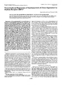
Figure 1. Transient G0S2 transfection reduced activity of different reporter constructs containing a retinoid responsive element. (A) G0S2 transient transfection independently decreased reporter plasmid activity of βRARE-Tk-Luc, pGL3-UBE1L-Luc or pGL3-G0S2-FL-Luc reporter constructs in NB4 APL cells, both in the presence and absence of RA (1 µM)-treatment. (B) G0S2 knockdown was achieved using an siRNA that repressed G0S2 mRNA expression relative to a control siRNA. G0S2 knockdown partially rescued this transcriptional repression by G0S2 transfection. Immunoblot analyses confirmed the expected decline of G0S2 protein in these APL cells, as shown in the insert. (C) G0S2 transient transfection did not affect transfection efficiency of NB4 cells as compared to vehicle control, as indicated by scoring the percentage of cells transfected with fluorescein-linked siRNA by flow analysis. (D) Schematic of studied G0S2 constructs that respectively included wild-type (WT) G0S2, myc-tagged wild-type G0S2 (Myc-G0S2), and myc-tagged G0S2 with the hydrophobic domain (HD) deleted (myc‑G0S2 ∆ HD). (E) The immunoblot displayed in this panel indicates relative to vehicle (DMSO) control that G0S2 protein is induced by the RAR agonist RA (1 µM), but not by the RXR agonist LG268 (1 µM) in NB4 APL cells after 2 days of treatment. Representative results are shown from three independent experiments (each performed in triplicate) with error bars representing standard deviations. *P