Review
Reprogramming and transdifferentiation for cardiovascular development and regenerative medicine: where do we stand? Antje D Ebert1,2,3, Sebastian Diecke4,5, Ian Y Chen1,2,3 & Joseph C Wu1,2,3,*
Abstract Heart disease remains a leading cause of mortality and a major worldwide healthcare burden. Recent advances in stem cell biology have made it feasible to derive large quantities of cardiomyocytes for disease modeling, drug development, and regenerative medicine. The discoveries of reprogramming and transdifferentiation as novel biological processes have significantly contributed to this paradigm. This review surveys the means by which reprogramming and transdifferentiation can be employed to generate induced pluripotent stem cell-derived cardiomyocytes (iPSC-CMs) and induced cardiomyocytes (iCMs). The application of these patient-specific cardiomyocytes for both in vitro disease modeling and in vivo therapies for various cardiovascular diseases will also be discussed. We propose that, with additional refinement, human disease-specific cardiomyocytes will allow us to significantly advance the understanding of cardiovascular disease mechanisms and accelerate the development of novel therapeutic options. Keywords cardiomyocytes; disease modeling; genome editing; human induced pluripotent stem cells; tissue engineering DOI 10.15252/emmm.201504395 | Received 22 March 2015 | Revised 7 June 2015 | Accepted 15 June 2015 | Published online 16 July 2015 EMBO Mol Med (2015) 7: 1090–1103 See the Glossary for abbreviations used in this article.
Introduction Despite advances in medical therapy, cardiovascular disease (CVD) remains a leading cause of morbidity and mortality worldwide. Concerted efforts in fundamental and translational research are required to provide novel diagnostic tools and effective therapeutic approaches for CVD. Mechanistic modeling of CVD as well as preclinical validation of therapeutic strategies will assist in the
1 2 3 4 5
1090
development of next-generation medical therapies that incorporate recent discoveries in stem cell biology.
Human induced pluripotent stem cell-derived cardiomyocytes as a novel platform Although animal models have provided indispensable insights into systemic whole-organ function in vivo as well as in vitro disease mechanisms (Fiedler et al, 2014; Houser et al, 2012; Duncker et al, 2015), not all findings from research on rodent cardiomyocytes can be translated to human cardiomyocytes at the cellular and molecular levels. Human cardiomyocytes, on the other hand, are a limited resource and cannot be indefinitely maintained in culture. These facts emphasize the need for novel human cellular and physiological models of CVD. Over the past decade, rapid technological advances have combined medical and basic sciences in the development and evaluation of novel therapeutics. One exciting advance has been the ability to generate patient-specific induced pluripotent stem cells (iPSCs; Takahashi & Yamanaka, 2006). Human iPSCs resemble human embryonic stem cells (ESCs), the “gold standard” for pluripotency, in their biological properties but without the ethical and political concerns associated with the use of human embryos. Therefore, iPSCs and their differentiated cardiomyocytes (iPSC-CMs) are considered a viable new and ethically less problematic, alternative platform for studying mechanisms of CVD and evaluating novel therapeutic avenues (Fig 1). In addition, human iPSCs present the unprecedented opportunity to study disease-specific differences in a patient-specific manner, taking into account individual drug responses within a patient population. The validity of this approach is exemplified by the successful application of human iPSCs to model LEOPARD syndrome (Carvajal-Vergara et al, 2010), Timothy syndrome (Yazawa et al, 2011), long QT syndrome (Moretti et al, 2010; Itzhaki et al, 2011; Wang et al, 2014), arrhythmogenic right ventricular dysplasia (ARVD) (Kim et al, 2013; Asimaki et al, 2014), familial dilated cardiomyopathy (DCM; Sun et al, 2012), familial hypertrophic cardiomyopathy (HCM; Lan et al,
Stanford Cardiovascular Institute, Stanford University School of Medicine, Stanford, CA, USA Department of Medicine, Division of Cardiology, Stanford University School of Medicine, Stanford, CA, USA Institute of Stem Cell Biology and Regenerative Medicine, Stanford University School of Medicine, Stanford, CA, USA Max Delbrück Center, Berlin, Germany Berlin Institute of Health, Berlin, Germany *Corresponding author. Tel: +1 650 736 2246; E-mail:
[email protected]
EMBO Molecular Medicine Vol 7 | No 9 | 2015
ª 2015 The Authors. Published under the terms of the CC BY 4.0 license
Antje D Ebert et al
Stem cells in cardiovascular development and therapy
EMBO Molecular Medicine
Glossary Cell therapy Transplantation of therapeutic cell populations (e.g., adult stem cells or PSC-CMs) to ameliorate damage in the infarcted heart and improve cardiac function Disease modeling Understanding the functional and molecular causes of diseases by recapitulating their respective phenotypes in organisms (e.g., mouse, zebrafish) or tissue-specific cell models (e.g., human iPSC-CMs) Drug discovery Identifying new therapeutics by employing a wide range of scientific methods and model systems Genomewide association study (GWAS) A whole-genome association study that assesses common genetic variants (e.g., single nucleotide polymorphisms) in defined populations of individuals Genome editing Modifying the DNA of a given genome in specific sites with genetically engineered nucleases
Investigational new drug (IND) application Filing admission of research on a new drug, treatment, or patient population in human subjects with a regulatory agency (e.g., the US Food and Drug Administration) Non-integrating reprogramming De-differentiation of somatic cells to induced pluripotent stem cells (iPSCs) using reprogramming factors that do not integrate into the host cell genome Paracrine mechanisms Chemical signals secreted from one cell (e.g., growth factors or chemokines) to induce functional changes in nearby cells Tissue engineering A strategy to replace damaged myocardium via generation of engineered heart muscle (EHM), which is a functional tissue-like structure constructed in vitro from adult stem cells or PSC derivatives Transdifferentiation Direct somatic cell reprogramming from one germ layer of origin to another
2013), viral cardiomyopathy (Sharma et al, 2014) and aldehyde dehydrogenase 2 genetic polymorphism (Ebert et al, 2014). These accomplishments demonstrate the tremendous power and versatility of iPSC-CMs in helping to develop novel therapeutic approaches for CVD and paving innovative avenues for precision medicine in the future.
De Los Angeles et al, 2012). Naı¨ve human iPSCs can be derived by reversion of primed iPSCs into a state that resembles naı¨ve mouse ESCs (Gafni et al, 2013; Theunissen et al, 2014). Currently, it is unknown whether these naı¨ve human ESCs and iPSCs represent distinct intermediates in embryonic development. Further research is required to elucidate whether human naı¨ve iPSCs may be more amenable to introduction of genomic modifications (Gafni et al, 2013) or may differentiate more efficiently into somatic tissues (Honda et al, 2013; Rais et al, 2013; Theunissen et al, 2014). It has been acknowledged that reprogrammed iPSCs can retain specific DNA methylation profiles associated with their parental source cell type (Bar-Nur et al, 2011; Kim et al, 2011; Lister et al, 2011). Variations in these signatures also appear to account for intraline variability among different clones originating from the same iPSC line (Kim et al, 2011; Lister et al, 2011). The long-term effect of epigenetic pattern retention, such as methylation profiles from the originating somatic cell type, is not yet fully understood. However, the somatic source cell type is known to affect differentiation efficiency into iPSC-CMs. For example, cardiac progenitor cell-derived iPSC lines have shown an enhanced ability to differentiate into cardiomyocytes compared to fibroblast-derived iPSC lines (SanchezFreire et al, 2014). Prolonged propagation of iPSCs through many passages reduces these effects, suggesting that residual epigenetic memory is attenuated in the course of long-term culture (Ohi et al, 2011; Sanchez-Freire et al, 2014). These studies demonstrate that epigenetic memory is a key determinant of iPSC differentiation into lineages that are distinct from the parental cell type. Pluripotent reprogramming and transdifferentiation of cells from one germ layer to another (Ladewig et al, 2013) have altered the concept of cell fate as determined by unidirectional progression (Takahashi & Yamanaka, 2006; Ieda et al, 2010; Vierbuchen et al, 2010; Huang et al, 2011) and illustrate the plasticity of differentiation and lineage specification. Epigenetic roadblocks repressing chromatin in its inactive state occur during iPSC reprogramming (Kim et al, 2010; Carey et al, 2011; Theunissen & Jaenisch, 2014), and a similar role for chromatin remodeling complexes also exists during cardiovascular development (Chang & Bruneau, 2012; Bevilacqua et al, 2014). Epigenetic modulators can alter DNA methylation and
Reprogramming of somatic cells to iPSCs The initial proof-of-concept studies on generation of ESC-like cells were performed using retroviral transduction of mouse fibroblasts with the transcription factors Oct4, Sox2, Klf4, and c-Myc (Takahashi & Yamanaka, 2006). These first-generation iPSCs featured unlimited self-renewal, differentiation into tissues of all germ layers, and the ability to generate an entire organism. However, these earlier approaches involved random insertion of reprogramming factors into the cellular genome, with consequent risk of oncogenic transformation. Subsequently, newer and safer non-integrating approaches employing Sendai virus (Ban et al, 2011), adenovirus (Fusaki et al, 2009; Zhou & Freed, 2009), episomal plasmids (Okita et al, 2008; Yu et al, 2009), minicircle (Jia et al, 2010) or co-MIP (Diecke et al, 2015), mRNA (Warren et al, 2010) or microRNAs (Lin et al, 2011), and direct protein delivery (Kim et al, 2009) have been developed (Fig 1). Based on initial observations in mouse ESCs, two states of pluripotency were defined, an earlier one occurring in normal embryonal development termed “naı¨ve” versus a “primed” state (Nichols & Smith, 2009). Naı¨vety is the ground state of pluripotency. Naı¨ve pluripotent stem cells (PSCs) can be maintained in vitro by supplying leukocyte inhibitory factor (LIF) combined with inhibition of MAPK/ERK kinase (MEK) and glycogen synthase kinase 3 (GSK3) signaling and are characterized by two active X chromosomes in female lines. Primed PSCs are dependent on fibroblast growth factor 2 (FGF2) signaling and transforming growth factor-b (TGFb) signaling and display inactivation of one X chromosome (Nichols & Smith, 2009; Hirai et al, 2012). Human ESCs and iPSCs are considered to share some properties of naı¨ve mouse ESCs, but mainly resemble primed murine epiblast stem cells (Nichols & Smith, 2009;
ª 2015 The Authors
EMBO Molecular Medicine Vol 7 | No 9 | 2015
1091
EMBO Molecular Medicine
Stem cells in cardiovascular development and therapy
iPSC REPROGRAMMING
Antje D Ebert et al
DIRECT REPROGRAMMING Somatic cells Fibroblasts
Fibroblasts
PBMCs Oct4 Sox2 Klf4 cMyc Lin28 Nanog
EPIGENETIC REGULATION
Gata4 Mef2c Tbx5 Hand2 Mesp1 Myocardin ESRRG
iPSCs Bmp4 FGF2 ActivinA Dkk1 VEGF
CHROMATIN REMODELING
miR-1 miR-133
CHIR99021 IWR1
iPSC-CMs
DISEASE MECHANISMS
iCMs
CARDIOVASCULAR DISEASE THERAPY
DRUG DEVELOPMENT TOXICITY TESTING
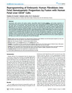
Figure 1. Generation and applications of patient-specific cardiomyocytes. From isolated patient-specific source cells such as dermal fibroblasts or peripheral blood mononuclear cells (PBMCs), cardiomyocytes can be generated via iPSC reprogramming and subsequent differentiation to iPSC-CMs, or by transdifferentiation into iCMs. Both strategies employ a set of defined factors that cause drastic modulatory changes in the cellular epigenome. Disease-specific mutations within iPSCs can be corrected via genome editing approaches and can be employed for studying disease mechanisms, drug discovery, and regenerative medicine. While in vivo applications of iCMs are already being evaluated, the suitability of iCMs for other purposes such as disease mechanism and drug development studies remains to be ascertained.
histone acetylation profiles, thereby opening or repressing chromatin in target gene loci that direct lineage fate. Clearly, epigenetic checkpoint regulators of development and lineage differentiation (Takeuchi & Bruneau, 2009; Wang, 2012) are candidates for targeted modulation during iPSC reprogramming and cardiac differentiation. In this context, small molecule compounds are a highly promising resource for further improving the efficiency of cardiovascular lineage differentiation (Efe et al, 2011).
Cardiac differentiation of iPSCs Today, efficient differentiation protocols allow the generation of large quantities of highly enriched cardiomyocyte populations. These achievements have been made possible by pivotal work utilizing spontaneous aggregation of iPSCs in suspension as embryoid bodies (EBs) in combination with stage-defined growth factors
1092
EMBO Molecular Medicine Vol 7 | No 9 | 2015
(Kehat et al, 2001; Mummery et al, 2007; Burrdige et al, 2011; Kattman et al, 2011). Subsequently, these protocols led to the establishment of monolayer systems that stimulate the superfamily of TGFb receptors via growth factors such as activin A and bone morphogenetic protein 4 (BMP4; Paige et al, 2010; Zhang et al, 2012), resulting in activation and repression of the canonical Wnt signaling pathway, respectively. Most recently, specific small molecules have been employed to replace growth factors as modulators of these signaling pathways (Fig 1; Lian et al, 2012). The relative immaturity of iPSC-CMs remains a challenge because it limits their use for disease modeling, drug discovery, and regenerative medicine purposes (Karakikes et al, 2015). Attempts to bypass this limitation have demonstrated that long-term culture enhances the appearance of more mature sarcomeric structural organization in iPSC-CMs (Kamakura et al, 2013). In addition, external cues such as electrical stimulation and mechanical cyclic stretching have been reported to aid in obtaining functionally
ª 2015 The Authors
Antje D Ebert et al
Stem cells in cardiovascular development and therapy
mature iPSC-CMs (Lieu et al, 2013; Hirt et al, 2014a). Improvements in iPSC-CM maturation were also achieved via novel 3D culture methods (Nunes et al, 2013; Rao et al, 2013) and genetic overexpression of distinct factors (Fu et al, 2011; Bett et al, 2013; Lieu et al, 2013), and these approaches are currently subjects of intense research. Maturation affects specification of cardiomyocyte subtypes and is vital for accurate recapitulation of disease phenotypes (Lan et al, 2013), including fundamental functional features such as more mature ion channel currents, densities, and kinetics (Sartiani et al, 2007; Yang et al, 2014). The early developmental stage produced by state-of-the-art iPSC-CM differentiation protocols is sufficient for analysis of certain hereditary channelopathies that cause ventricular tachyarrhythmias (Sallam et al, 2015). However, depending on the exact stage of development, there are numerous differences in electrical conduction and coupling as well as the contractile rate and force, compared to human adult cardiomyocytes (Karakikes et al, 2015). Furthermore, insufficient maturation of iPSC-CMs compared to adult human cardiomyocytes has also hindered a broader application of these cells for primary drug discovery and validation. Nevertheless, their use in exploratory studies and for examination of drug toxicity is clearly justified. A related concern regarding iPSC-CM differentiation is the varying degree of heterogeneity achieved in the generated cardiomyocyte population. Current iPSC or ESC differentiation strategies yield a heterogeneous mixture of atrial-like and ventricular-like lineages, as well as pacemaker-like lineages such as atrioventricular node-like, sinoatrial node-like, and Purkinje fiber-like cells (Burridge et al, 2012). A deeper understanding of directed lineage differentiation, followed by its modulation, would facilitate subtype-specific cardiac differentiation. This can include direct manipulations at the epigenetic level or by achieving mRNA-based delivery of lineage-specific factors (Ong et al, 2015). The most immediate need, however, is to achieve defined culture conditions and standardized protocols that address the issue of iPSC-CM maturation. In a broader sense, reproducibility and standardization throughout the scientific community will be a key to ensuring comparable datasets, as well as strides toward a broader applicability of iPSC-CMs for disease modeling and drug development. Although still at its infancy, the field has already made significant progress toward the defined derivation and propagation of human iPSCs and iPSC-CMs (Chen et al, 2011; Burridge et al, 2014; Ribeiro et al, 2015).
Genetic engineering and personalized medicine To understand the molecular and genetic determinants of CVD, advanced genome editing techniques are required to study genotype/phenotype relationships and to allow for the correction of patient-specific mutations in human iPSCs (Wang et al, 1995; Chen et al, 1998; Schwartz et al, 2000; Benson et al, 2003; Fig 1). Initial pioneering work was performed using zinc-finger nucleases (ZFNs), a widely used technology for genomic correction that relies on the fusion of the FokI restriction endonuclease with zinc-finger proteins. These nucleases induce target site-specific double-stranded breaks, which stimulate endogenous DNA repair pathways. Due to the complexity of the required engineering steps, ZFNs have been
ª 2015 The Authors
EMBO Molecular Medicine
largely supplanted by transcription activator-like effector nucleases (TALENs), and more recently by the clustered regulatory interspaced short palindromic repeats (CRISPRs)/Cas9 nuclease system. TALENs display enhanced specificity as well as reduced off-target action compared to ZFNs. Importantly, single-base pair recognition by TALENs or CRISPRs can correct single nucleotide exchange mutations (Hockemeyer et al, 2011; Ding et al, 2013; Lin et al, 2014). CRISPRs are the most accessible means to facilitate and optimize genetic engineering. Their specificity and off-target effects are currently being evaluated, as these nucleases have the potential to bind and cut sites other than the primary target site (Hendel et al, 2015). Nevertheless, in a relatively short time, CRISPRs have been demonstrated to be a cost-effective and time-efficient approach for genomic correction or introduction of site-specific mutations (Sander & Joung, 2014). Genome-corrected and disease-introduced isogenic cell lines are particularly valuable, as they share a common genotype with the exception of the disease-causing mutation, thereby eliminating confounding effects from genetic heterogeneity. Genomic modification to directly correct disease-specific point mutations in vitro is also valuable for exploring drug development in patient-specific cardiomyocytes. Human iPSC-CMs are currently being utilized as a system to evaluate novel and existing medications and to test patient-specific drug responses (Liang et al, 2013; Navarrete et al, 2013; Wang et al, 2014). For instance, iPSC-CMs from patients carrying long QT syndrome mutations (e.g., KCNQ1 G269S) and genome-edited iPSC-CMs with these disease-causing mutations were both shown to display long QT phenotypes (Liang et al, 2013; Wang et al, 2014). Furthermore, both cell types revealed comparable disease-specific responses following drug treatment (e.g., nifedipine) to rescue prolongation of action potential duration (APD) (Liang et al, 2013; Matsa et al, 2014; Wang et al, 2014). Overall, these examples illustrate the potential for using genome editing to generate accurate, reliable, and less expensive in vitro human models for understanding CVD and for accelerating drug discovery (Fig 1; Ebert et al, 2012). Moreover, genome editing may accelerate the future clinical application of integration-free cellbased gene therapy, including the autologous transplantation of patient-specific, genome-corrected iPSC-CMs. The complexity of genotype/phenotype relationships is further magnified by genetic background variation and variability among iPSC lines (Table 1). Genome-wide association studies (GWAS) and subsequent data mining identify signaling pathways governing the control of disease-relevant targets. Large numbers of critical gene loci and related mutations have been described by GWAS and linked to pathogenic phenotypes. Variants occurring in these regions can influence the regulation of disease-relevant gene expression (Fig 1). Moreover, late-onset or incomplete penetration of the disease phenotype can complicate further readout and genotypic correlation. In those cases, response profiling of well-established compounds and drugs in CVD might provide further insight. However, the presence of line-to-line and genetic background variation implies that additional layers of control are required to confirm genotype/phenotype relationships. Rescue of pathogenic functional features following genomic correction of the disease-related locus via TALENs or CRISPRs, the use of isogenic controls, and sufficiently powered studies are means to address these limitations. Given our ability to introduce specific disease-causing mutations into both iPSCs and ESCs, it is likely that instead of isolating
EMBO Molecular Medicine Vol 7 | No 9 | 2015
1093
EMBO Molecular Medicine
Stem cells in cardiovascular development and therapy
Antje D Ebert et al
Table 1. Challenges and opportunities of de novo generated cardiomyocytes for disease modeling, drug discovery, and regenerative therapies. CM generation strategy Parameters
iPSC reprogramming and differentiation
Direct reprogramming
Human ESC differentiation
Mechanism
De-differentiation to iPSCs followed by specific differentiation to CMs
Transdifferentiation
Specific differentiation to CMs
Timeline
2–3 months
2–3 weeks
2–3 weeks
Efficiencies (% cTnT)
90–95%
9–13%
90–95%
Genome editing, isogenic controls
Yes
No
Yes
Genetic variation
Yes
Not yet determined
No
Disease modeling, drug development
Yes
Current efficiencies too low
Yes
Patient-specific assessment of phenotypes and drug function
Yes
Currently undergoing investigation
No
In vivo preclinical evaluation of regenerative therapies
Yes
Yes
Yes
Clinical safety and efficacy
Not yet determined
Not yet determined
Currently undergoing investigation
Ethical concerns
No
No
Yes
primary cells from affected patients and generating disease-specific iPSCs and iPSC-CMs, the field will evolve toward standardized procedures based on introducing mutations of interest into fully sequenced and characterized reference stem cell lines (iPSCs and/or ESCs) to assess disease-specific genotypic and phenotypic relationships (Sallam et al, 2015).
Direct conversion to induced cardiomyocytes (iCMs) There are both advantages and disadvantages in reprogramming of somatic cells to iPSCs. The intrinsic properties of iPSCs enable the use of tools such as genome editing to facilitate our understanding of basic disease mechanisms, as well as to evaluate precision medicine approaches (Wilson & Wu, 2015). Nevertheless, despite methodological advances, the entire process of generating patient-specific iPSC-CMs still requires several months and presents a potential risk of teratoma formation for regenerative medicine, given that the presence of residual pluripotent cells in the final product cannot be completely excluded (Lee et al, 2013). As a result, other approaches that eliminate the need for pluripotent stem cell generation are being explored. In recent years, proof-of-concept studies have shown that somatic cells can be directly converted to cardiomyocytes (Fig 1; Ieda et al, 2010; Efe et al, 2011; Qian et al, 2012). Transgenic expression of three cardiac-specific transcription factors (Gata4, Mef2c, and Tbx5) resulted in the transdifferentiation of murine fibroblasts into contracting cardiomyocytes referred to as induced cardiomyocytes (iCMs; Ieda et al, 2010). Intriguingly, the same outcome has been observed during an epigenetic activation phase in the early stage of reprogramming. This approach employed ectopic expression of transcription factors together with growth factors (Efe et al, 2011), demonstrating elegantly how cellular complexity can be harnessed for the understanding of specific molecular processes. Other reports have shown that direct reprogramming of somatic cells to iCMs is also feasible using various
1094
EMBO Molecular Medicine Vol 7 | No 9 | 2015
small molecules and miRNAs (Jayawardena et al, 2012, 2014; Protze et al, 2012). Recently, direct reprogramming of human fibroblasts has also been achieved (Islas et al, 2012). Several studies showed that the murine direct reprogramming factors Gata4, Mef2c, and Tbx5 (GMT), or GMT plus Hand2 (GHMT), were insufficient to transform human fibroblasts into iCMs (Nam et al, 2013; Wada et al, 2013), indicating that the differences between mouse and human cardiovascular development need to be considered for optimal transdifferentiation to human iCMs. A combination of Gata4, Hand2, Tbx5, myocardin, miR-1, and miR-133 could convert human adult dermal fibroblasts into induced cardiomyocyte-like cells (iCMLs). The transduction of these factors promoted substantial cardiac troponin T expression in at least 9% of the source population (Nam et al, 2013). Shortly afterward, introduction of GMT plus Mesp1 and myocardin (GMTMM) was also shown to successfully convert human fibroblasts to iCMLs (Wada et al, 2013). Since then, alternative approaches have succeeded in generating human iCMs with gene expression profiles and functional characteristics similar to those detected in ESC-CMs (Fu et al, 2013).
Current limitations and the routes toward therapeutic application Direct reprogramming as an alternative to deriving human iPSCs offers the advantage of a 2-week timeline, compared to 2–3 months (Table 1). However, current methods for producing iCMs suffer from low efficiencies compared to iPSC differentiation (Chen et al, 2012). Depending on the combination of transcription factors used, human fibroblasts convert into iCMs with different efficiencies, ranging from 5% (GMTMM; Ieda et al, 2010) to 13% (Gata4, Hand2, Tbx5, Myocd, miRNA 1, and miRNA 133; Fu et al, 2013) based on troponin T-positive cells. Recently, polycistronic vectors have been used to express the GMT factors in appropriate stoichiometry as a single mRNA, which has significantly increased the efficiency of mouse
ª 2015 The Authors
Antje D Ebert et al
Stem cells in cardiovascular development and therapy
fibroblast conversion in vitro up to 25% (Inagawa et al, 2012; Wang et al, 2015). However, these results remain to be replicated in human fibroblasts. In addition, successful generation of pure iCM populations has not yet been reported. Indeed, direct transdifferentiation has so far generated heterogeneous populations of cardiomyocyte-like cells representing various early developmental stages, which rarely display spontaneous beating and produce only sporadic action potentials (Fu et al, 2013). In general, the overall low transdifferentiation efficiency of iCMs into bona fide cardiomyocytes is the main obstacle for the required scale-up of cell production. Like iPSC-CMs, iCMs must undergo additional maturation before they can serve as true models of adult cardiomyocytes (Bedada et al, 2014; Yang et al, 2014; Jayawardena et al, 2015). The accurate determination of the differences between these cell types requires the direct comparison of iCMs with both iPSC-CMs and human adult cardiomyocytes (Protze et al, 2012; Wada et al, 2013). While similarity of iCMs to ESC-CMs (Fu et al, 2013) has been reported, including subtype specification as a feature of mature cardiomyocyte populations (Nam et al, 2014), other studies have indicated that human iCMs generated in vitro may be even more immature than human iPSC-CMs (Wada et al, 2013). These findings imply that iCMs may reflect the early fetal stage of embryonic cardiomyocytes, and hence, co-stimulation with appropriate factors may drive maturation of iCMs in vitro. Potential approaches include mechanical stimuli or secreted molecules related to the normal myocardial environment, such as cardiac matrix scaffolds and secreted paracrine factors. Exposure to stretching forces is also thought to accelerate iCM generation and maturation (Qian & Srivastava, 2013). Thus, current limitations of efficient generation and maturation of iCMs in vitro may be addressed by advancing in vivo reprogramming instead. miRNAs have been demonstrated to be sufficient for direct reprogramming to iCMs without addition of any transcription factors (Jayawardena et al, 2012). Current research has thus far focused on their use as powerful drivers of lineage fates (Cordes & Srivastava, 2009; Boon & Dimmeler, 2015). It is likely that miRNAs promote cardiac induction by suppressing fibroblast signatures, as for example, miRNA133-mediated inhibition of Snai1-controlled expression pathways (Muraoka et al, 2014). Therefore, an optimized cocktail to promote the generation of more mature iCMs may include specific miRNAs, as well as specific chemical epigenetic modulators. Should such strategies result in higher efficiency of iCM reprogramming and improved maturation, safety and efficacy would need to be assessed in studies similar to those required for human iPSCCMs. For now, a direct comparison of iCMs with iPSC-CMs regarding their functional properties remains to be performed. Initial comparative evaluations should focus on functional parameters such as electrophysiology and calcium handling, and also gene and protein expression patterns. Subsequently, the beneficial effects of either cell type on improving cardiac function in preclinical models of CVD will need to be demonstrated. Likewise, the long-term stability of iPSC-CM and iCM phenotypes must be assessed to address safety and efficacy issues.
Heart disease and novel therapeutic approaches Two of the main classes of genetically inherited heart diseases include channelopathies and cardiomyopathies. Channelopathies or
ª 2015 The Authors
EMBO Molecular Medicine
arrhythmic cardiac disorders are caused by mutations in genes encoding ion channels, such as SCN5A that encodes the cardiac Na+ channel a-subunit. The functional characteristics include voltage gating and/or protein trafficking defects, which can result in gain or loss of function in the Na+ channel and subsequent ventricular arrhythmias, leading to diseases such as long QT syndromes (Lehnart et al, 2007). By contrast, cardiomyopathies, or defects in heart muscle contraction, are most frequently caused by mutations in cytoskeletal or contractile proteins (McNally et al, 2013). The relevant pathogenic features are transversely isotropic, consisting of irreversible ventricular dilatation and systolic dysfunction that cause severely impaired ventricular contraction. Both channelopathies and cardiomyopathies can also be caused by non-hereditary, acquired determinants such as chronic or acute ischemia (Fig 2), and by drugs or autoimmune events (Kass, 2005). Human models of iPSC-CMs have successfully recapitulated numerous genetically determined CVD, including long QT, DCM, HCM, and ARVD (Moretti et al, 2010; Yazawa et al, 2011; Davis et al, 2012; Sun et al, 2012; Caspi et al, 2013; Lan et al, 2013). These studies have demonstrated that iPSC-CMs display phenotypic disease features such as abnormal sarcomere alignment and striation, as well as critical functional properties such as propagation of calcium transients in amplitude, time to peak, duration, and decay (Moretti et al, 2010; Yazawa et al, 2011; Davis et al, 2012; Sun et al, 2012; Caspi et al, 2013; Lan et al, 2013). Importantly, iPSCCMs allow for quantitative analysis of electrical properties regarding the action potential’s upstroke velocity, time to peak, and duration. For example, abnormally prolonged APD and decreased repolarization velocity are detected in long QT patient-derived iPSC-CMs (Yazawa et al, 2011; Table 1).
Pathogenesis of myocardial infarction and regeneration of the heart The early phase during and after an infarction is characterized by inflammatory, necrotic, and apoptotic cellular responses. The ensuing late or chronic phase includes an expansion of the infarcted region in the myocardial wall, recruitment of myeloid cells, tissue necrosis, and degradation of the extracellular matrix (ECM). Subsequent neoangiogenesis and remodeling of the left ventricle (LV) entails scar formation, hypertrophic expansion of cardiomyocytes, and fibrosis. The resulting dilation of the LV is accompanied by increased frequency of arrhythmias, myocardial dysfunction, and eventually heart failure. Conventional therapy of fibrosis and LV dilatation by angiotensin converting enzyme (ACE) inhibitors and angiotensin II receptor blockers (ARBs) partially counteracts these deleterious consequences and attenuates adverse remodeling (Dorn, 2009). Nevertheless, viable cardiomyocytes are lost to a large extent in the area of myocardial infarction (MI). Recent studies suggest that the heart is capable of limited endogenous regeneration (Bergmann et al, 2009; Parmacek & Epstein, 2009; Porrello et al, 2011; van Berlo et al, 2014). While proliferation of the heart may occur to a minor extent throughout the lifetime of an organism, active cell division of cardiomyocytes is limited to the embryonic stage (Bergmann et al, 2009; Porrello et al, 2011). A different source of endogenous heart regeneration is the resident adult stem cell population, known as cardiac progenitor cells
EMBO Molecular Medicine Vol 7 | No 9 | 2015
1095
EMBO Molecular Medicine
Stem cells in cardiovascular development and therapy
Antje D Ebert et al
iCMs Gata4 Mef2c Tbx5
Adult stem and progenitor cells
Hand2 Mesp1 Myocardin miR-1 miR-133
EHMs
IN SITU REPROGRAMMING iPSC-CMs
REGENERATED MYOCARDIUM
Figure 2. Cell therapy and tissue engineering approaches for cardiovascular disease therapy. Heart failure due to ischemic heart disease or genetic disorders remains a major healthcare burden. Potential novel treatment options include transplantation of iPSC-CMs or ESC-CMs, as well as direct in vivo reprogramming of cardiac fibroblasts in the scar region to iCMs. The regenerative capacity of adult stem and progenitor cell populations is also being evaluated. Tissue engineering is a new method that aims to re-muscularize damaged myocardium via transplantation of in vitro engineered heart muscle made from iPSC-CMs or ESC-CMs.
(CPCs), reported to be capable of differentiating and proliferating to replenish apoptotic cardiomyocytes (Fig 2; Dimmeler et al, 2005; Leri et al, 2011). The full regenerative capacities of these cells remain controversial and are discussed in detail elsewhere (Laflamme & Murry, 2011; Anversa et al, 2013; Maillet et al, 2013, van Berlo et al, 2014). Overall, the endogenous proliferation and repair abilities of the heart are not sufficient to allow the repopulation of damaged myocardial areas with new cardiomyocytes following MI.
Stem cell-derived cardiomyocytes for heart disease therapy Previous clinical trials have employed various adult stem cell and progenitor cell populations to test their efficacy for therapeutic applications (Fig 2; Assmus et al, 2002; Schachinger et al, 2004; Losordo et al, 2007; Chugh et al, 2012; Hare et al, 2012; Makkar et al, 2012; Traverse et al, 2012; Vrtovec et al, 2013; Karantalis et al, 2014). Safety and feasibility of these cells have been demonstrated in these scenarios, and extensive efforts have been spent on exploring the therapeutic potential of these cells. Overall, the results have shown varying degrees of clinical benefit in MI patients (Sanganalmath & Bolli, 2013). Currently, additional approaches are being explored, including transplantation of new cell types (e.g.,
1096
EMBO Molecular Medicine Vol 7 | No 9 | 2015
human ESC-cardiac progenitor cells (Menasche et al, 2015) or iPSCCMs), or application of alternative delivery approaches such as implantation of in vitro constructed cell sheets of engineered heart muscles (EHMs) (Fig 2; Zimmermann, 2013; Emmert et al, 2014; Hirt et al, 2014b). One mechanism by which cell therapy (e.g., ESC-CMs or iPSC-CMs) may improve outcomes is via engraftment of transplanted cells within the host environment, which in theory would lead to the replacement of damaged cardiomyocytes and fibrotic tissue, and restore structural support of the ventricular walls. Extensive studies have focused on grafts within the non-infarcted versus infarcted myocardium in small and large animal models (Laflamme et al, 2007; van Laake et al, 2008). Human grafts express cardiac markers and displayed sarcomere alignment as well as integration with the host’s vasculature. In this context, coupling of transplanted cardiomyocytes with the host myocardium is considered a vital factor contributing to improved cardiac function. Stem cell-derived cardiomyocytes couple to a limited extent with the host myocardial cells in small animal models (Kehat et al, 2004). Large animal models such as pigs and non-human primates are more valuable due to the greater resemblance of their heart rates with the beating frequency of transplanted cardiomyocytes (Chong et al, 2014). However, to a large extent, functional improvement in left ventricular ejection fraction (LVEF) in some of these transplantation models has been suggested to result through the release of paracrine factors
ª 2015 The Authors
Antje D Ebert et al
(Gnecchi et al, 2005; Gu et al, 2012; Huber et al, 2013). Transplanted cells may secrete signaling molecules that exert beneficial functions directly or by altering gene expression patterns in the surrounding myocardium. Such paracrine mechanisms have been frequently proposed to contribute to the recovery of cardiac function (Gnecchi et al, 2005). Well-studied factors such as vascular endothelial growth factor (VEFG; Zangi et al, 2013) and thymosin b-4 (TB4; Smart et al, 2010, 2011) have been selectively characterized in murine infarct models for their capacity to mediate cardiac repair. Recently, targeted approaches to identify specific paracrine factors revealed novel paracrine-acting proteins that could improve tissue and heart function following MI (Korf-Klingebiel et al, 2015). In the future, cell-based therapies may benefit from these findings by integrating delivery of specific factors into the transplanted therapeutic composite. Moreover, complementing the cell mixture with iPSCderived endothelial cells may lead to beneficial effects from the developed vasculature (Ye et al, 2014). Together, these synergistic approaches may help promote engraftment, vascularization, and structural integrity of the ventricular walls. To date, three fundamental issues have slowed the clinical translation of iPSC-CMs or ESC-CMs: the risk of tumor formation, poor survival of transplanted cells, and the need for immunosuppression for allogeneic ESC and iPSC derivatives. The first obstacle includes both tumors potentially arising from random insertion of integrating reprogramming vectors, and the risk of teratomas arising from residual undifferentiated stem cells (Lee et al, 2009, 2013). Novel non-integrating iPSC reprogramming strategies may decrease risk, while increasing the efficiency of differentiation or the purity of the final cell product could minimize teratoma risk (Tang et al, 2011). The second major obstacle is acute donor cell death due to hypoxia, anoikis, and inflammation, as well as lack of blood supply (Li et al, 2009a,b; Liu et al, 2012; Nguyen et al, 2014). Hence, the majority of current cardiac cell therapies (both basic and clinical) appear to achieve beneficial effects without long-term persistence of the cells, presumably through the release of paracrine factors to the host heart prior to transplanted cell loss as described earlier (Gnecchi et al, 2005). Relating practical issues include generation of sufficient infarct sizes in the chosen species to induce a measureable decline in heart function without killing the animal. In some cases (e.g., guinea pigs and dogs), the collateral circulation is so high that meaningful infarcts cannot be generated by coronary artery ligation (Verdouw et al, 1998). These concerns are critical for investigational new drug (IND)-enabling large animal studies in the evaluation of the safety and efficacy of stem cell-derived therapies. The third obstacle toward clinical applications of iPSC-CMs or ESC-CMs is the need for effective immunosuppression to reduce rejection in allogeneic settings, which can be daunting (Pearl et al, 2011, 2012). The use of more sophisticated immunosuppressive or tolerance induction strategies (Huber et al, 2013) as well as combinations of iPSC-CMs with potentially immunotolerant iPSC-derived mesenchymal stem cells (iPSC-MSCs) is being investigated (Lian et al, 2010). An alternative approach is the creation of human leukocyte antigen (immunosuppression HLA)-matched cell banks from healthy donors that contain selected iPSC lines with maximized HLA genotype overlap, which may minimize the need for immunosuppression (Taylor et al, 2012; Neofytou et al, 2015). However, an important caveat to this approach was recently found
ª 2015 The Authors
EMBO Molecular Medicine
Stem cells in cardiovascular development and therapy
in the heterogeneity of human mitochondria and, specifically, mismatched mitochondrial antigens, which by themselves can trigger rejection in transplant models (Deuse et al, 2015).
In vivo applications of iCMs Direct application of iCM reprogramming in vivo may promote patient-specific precision therapy by reducing the accompanying costs and efforts, which are considerable with in vitro generation of patient-specific iPSC-CMs. Induced cardiac regeneration in vivo via iCMs might circumvent current unresolved issues in iPSC-CM therapy, such as poor survival and engraftment of transplanted cells. However, the degree of functional cardiac improvement resulting from in situ transdifferentiated iCMs is unknown, as is the extent of their coupling and integration within the host myocardium (Table 1). Safety and potential off-target effects of iCM reprogramming cocktails have yet to be studied in detail, and the consequences of in vivo transfection of “off-target” cells such as endothelial, smooth muscle, or cardiac cells in the heart are also unknown and can be problematic. Finally, another consideration is the reproducibility of iCM generation using viral delivery approaches, which can lead to host immune response, as compared to non-viral or small molecule approaches, which may have poor pharmacokinetics in vivo (Chen et al, 2012). In summary, many challenges remain to be resolved before therapeutic application of iCMs in the clinic can even begin.
Tissue engineering Currently, heart transplantation is the only viable therapy for end-stage heart failure but remains problematic due to a chronic shortage of organ supply, as well as the persistent risk of immune rejection. An alternative strategy for regeneration of damaged myocardium is to exploit therapeutic cells such as iPSC-CMs for the construction of 3D structures in vitro, and subsequent transplantation of these engineered cardiac patches (Caspi et al, 2007; Tulloch et al, 2011; Kawamura et al, 2012). This technology is known as “tissue engineering” or generation of engineered heart muscle (EHM). Transplantation of a tissue patch/EHM ensures increased precision of delivery onto damaged myocardial areas, as well as full retention of transplanted material. EHM transplants may also allow direct substitution of scar tissue in the infarcted area with new, healthy cardiac muscle, minimizing long-term damage resulting from scar growth and ultimately reducing adverse remodeling and improving cardiac function (Fig 2). Moreover, it is expected that 3D cardiac tissues may mature into more adult-like structures compared to single cardiomyocytes, which is considered essential for optimal integration into the host environment. Several key features of maturation, such as alignment, orientation, and binucleation of cardiomyocytes (particularly their sarcomeric structural organization), were found to be improved in engineered tissues (Zimmermann et al, 2002; Tiburcy et al, 2011; Zhang et al, 2013). The beneficial outcomes of tissue engineering-based therapy have been extensively demonstrated in small animal models (Naito et al, 2006; Zimmermann et al, 2006; Sekine et al, 2008; Tulloch et al, 2011) and are currently being tested in large animal models. Eventually, EHMs
EMBO Molecular Medicine Vol 7 | No 9 | 2015
1097
EMBO Molecular Medicine
Stem cells in cardiovascular development and therapy
Pending issues Maturation of pluripotent stem cell-derived cardiomyocytes (e.g., iPSCCMs or ESC-CMs) and iCMs, as well as defined populations of cardiomyocyte subtypes (e.g., atrial-, nodal-, and ventricular-like cells). Standardization of efficient, reproducible, and defined culture conditions to generate comparable data. Improving the conclusiveness of patient-specific iPSC-CM models with approaches to address line-to-line variability and genetic background variations via CRISPR-generated isogenic controls, or fully sequenced and characterized reference stem cell lines (either iPSCs or ESCs). Optimizing efficiencies in direct reprogramming to iCMs in vitro and in vivo. Assessing efficacy of ESC-CM-, iPSC-CM-, iCM-, and EHM-based therapies.
Antje D Ebert et al
Conflict of interest JCW is a cofounder of Stem Cell Theranostics. The other authors declare that they have no conflict of interest.
References Anversa P, Kajstura J, Rota M, Leri A (2013) Regenerating new heart with stem cells. J Clin Invest 123: 62 – 70 Asimaki A, Kapoor S, Plovie E, Karin Arndt A, Adams E, Liu Z, James CA, Judge DP, Calkins H, Churko J et al (2014) Identification of a new modulator of the intercalated disc in a zebrafish model of arrhythmogenic cardiomyopathy. Sci Transl Med 6: 240ra274 Assmus B, Schachinger V, Teupe C, Britten M, Lehmann R, Dobert N, Grunwald F, Aicher A, Urbich C, Martin H et al (2002) Transplantation of
may facilitate patient-specific organ transplantation via in vitro generation of partial or whole-organ structures. Significant potential problems, such as poor survival of transplanted EHMs and their problematic integration into the host myocardium, need to be resolved before human trials can commence. A recent study using mouse cardiac sliced tissue as a benchmark to validate and model tissue-engineered patches showed poor survival with > 400 lm thickness (Riegler et al, 2014). Detailed understanding of the complex molecular mechanisms that determine the engraftment of transplanted EHMs within the host will lead to better strategies to address these issues. Possible solutions include co-delivery of supporting scaffold matrices, pro-survival cocktails, and stimulation of host myocardium via specific chemical molecules. Standardization of protocols for EHM generation, performance, and maturation will be a crucial step before moving forward to clinical trials. The substantial progress made during the past decade holds promise for a future clinical translation of EHM technology (Tee et al, 2012; Sekine et al, 2013; Ye et al, 2013; Hirt et al, 2014b).
progenitor cells and regeneration enhancement in acute myocardial infarction (TOPCARE-AMI). Circulation 106: 3009 – 3017 Ban H, Nishishita N, Fusaki N, Tabata T, Saeki K, Shikamura M, Takada N, Inoue M, Hasegawa M, Kawamata S et al (2011) Efficient generation of transgene-free human induced pluripotent stem cells (iPSCs) by temperature-sensitive Sendai virus vectors. Proc Natl Acad Sci USA 108: 14234 – 14239 Bar-Nur O, Russ HA, Efrat S, Benvenisty N (2011) Epigenetic memory and preferential lineage-specific differentiation in induced pluripotent stem cells derived from human pancreatic islet beta cells. Cell Stem Cell 9: 17 – 23 Bedada FB, Chan SS, Metzger SK, Zhang L, Zhang J, Garry DJ, Kamp TJ, Kyba M, Metzger JM (2014) Acquisition of a quantitative, stoichiometrically conserved ratiometric marker of maturation status in stem cell-derived cardiac myocytes. Stem Cell Reports 3: 594 – 605 van Berlo JH, Molkentin JD (2014) An emerging consensus on cardiac regeneration. Nat Med 20: 1386 – 1393 Benson DW, Wang DW, Dyment M, Knilans TK, Fish FA, Strieper MJ, Rhodes TH, George AL Jr (2003) Congenital sick sinus syndrome caused by recessive mutations in the cardiac sodium channel gene (SCN5A). J Clin
Conclusions
Invest 112: 1019 – 1028 Bergmann O, Bhardwaj RD, Bernard S, Zdunek S, Barnabe-Heider F, Walsh S,
Pluripotent stem cell-derived cardiomyocytes, induced cardiomyocytes, and engineered heart muscle present exciting new opportunities for the development of novel CVD treatments. While iCM production is currently being optimized, iPSC-CMs provide a stateof-the-art patient-specific model system to study disease mechanisms and develop new drugs. Future studies will have to ascertain whether ESC-CM-, iPSC-CM-, iCM-, or EHM-based transplantation can achieve sustained improvement of cardiac function. These synergistic, multidisciplinary approaches should improve understanding of the mechanisms governing cardiovascular health and disease at the molecular, cellular, and organ levels. Transformation of this knowledge into therapeutic strategies is the key to achieve the full potential of regenerative medicine and open a new era of advances in cardiovascular therapy.
Zupicich J, Alkass K, Buchholz BA, Druid H et al (2009) Evidence for cardiomyocyte renewal in humans. Science 324: 98 – 102 Bett GC, Kaplan AD, Lis A, Cimato TR, Tzanakakis ES, Zhou Q, Morales MJ, Rasmusson RL (2013) Electronic “expression” of the inward rectifier in cardiocytes derived from human-induced pluripotent stem cells. Heart Rhythm 10: 1903 – 1910 Bevilacqua A, Willis MS, Bultman SJ (2014) SWI/SNF chromatin-remodeling complexes in cardiovascular development and disease. Cardiovasc Pathol 23: 85 – 91 Boon RA, Dimmeler S (2015) MicroRNAs in myocardial infarction. Nat Rev Cardiol 12: 135 – 142 Burridge PW, Keller G, Gold JD, Wu JC (2012) Production of de novo cardiomyocytes: human pluripotent stem cell differentiation and direct reprogramming. Cell Stem Cell 10: 16 – 28 Burridge PW, Matsa E, Shukla P, Lin ZC, Churko JM, Ebert AD, Lan F, Diecke S,
Acknowledgements We thank Joseph Gold and Blake Wu for the critical reading of the manuscript. This work is supported by research grants from the National Institute of
1098
Huber B, Mordwinkin NM et al (2014) Chemically defined generation of human cardiomyocytes. Nat Methods 11: 855 – 860 Carey BW, Markoulaki S, Hanna JH, Faddah DA, Buganim Y, Kim J, Ganz K,
Health T32 training grant (IYC), American Heart Association 13EIA14420025,
Steine EJ, Cassady JP, Creyghton MP et al (2011) Reprogramming factor
NIH R01 HL123968, and NIH R01 HL126527, CIRM DR2A-05394, and CIRM
stoichiometry influences the epigenetic state and biological properties of
TR3-05556 (JCW).
induced pluripotent stem cells. Cell Stem Cell 9: 588 – 598
EMBO Molecular Medicine Vol 7 | No 9 | 2015
ª 2015 The Authors
Antje D Ebert et al
EMBO Molecular Medicine
Stem cells in cardiovascular development and therapy
Carvajal-Vergara X, Sevilla A, D’Souza SL, Ang YS, Schaniel C, Lee DF, Yang L, Kaplan AD, Adler ED, Rozov R et al (2010) Patient-specific induced pluripotent stem-cell-derived models of LEOPARD syndrome. Nature 465: 808 – 812 Caspi O, Huber I, Gepstein A, Arbel G, Maizels L, Boulos M, Gepstein L (2013) Modeling of arrhythmogenic right ventricular cardiomyopathy with
Dorn GW 2nd (2009) Novel pharmacotherapies to abrogate postinfarction ventricular remodeling. Nat Rev Cardiol 6: 283 – 291 Duncker DJ, Bakkers J, Brundel BJ, Robbins J, Tardiff JC, Carrier L (2015) Animal and in silico models for the study of sarcomeric cardiomyopathies. Cardiovasc Res 105: 439 – 448 Ebert AD, Kodo K, Liang P, Wu H, Huber BC, Riegler J, Churko J, Lee J,
human induced pluripotent stem cells. Circ Cardiovasc Genet 6:
de Almeida P, Lan F et al (2014) Characterization of the molecular
557 – 568
mechanisms underlying increased ischemic damage in the aldehyde
Caspi O, Huber I, Kehat I, Habib M, Arbel G, Gepstein A, Yankelson L, Aronson D, Beyar R, Gepstein L (2007) Transplantation of human embryonic stem cell-derived cardiomyocytes improves myocardial performance in infarcted rat hearts. J Am Coll Cardiol 50: 1884 – 1893 Chang CP, Bruneau BG (2012) Epigenetics and cardiovascular development. Annu Rev Physiol 74: 41 – 68 Chen G, Gulbranson DR, Hou Z, Bolin JM, Ruotti V, Probasco MD, Smuga-Otto K, Howden SE, Diol NR, Propson NE et al (2011) Chemically defined conditions for human iPSC derivation and culture. Nat Methods 8: 424 – 429 Chen JX, Krane M, Deutsch MA, Wang L, Rav-Acha M, Gregoire S, Engels MC, Rajarajan K, Karra R, Abel ED et al (2012) Inefficient reprogramming of fibroblasts into cardiomyocytes using Gata4, Mef2c, and Tbx5. Circ Res 111: 50 – 55 Chen Q, Kirsch GE, Zhang D, Brugada R, Brugada J, Brugada P, Potenza D,
dehydrogenase 2 genetic polymorphism using a human induced pluripotent stem cell model system. Sci Transl Med 6: 255ra130 Ebert AD, Liang P, Wu JC (2012) Induced pluripotent stem cells as a disease modeling and drug screening platform. J Cardiovasc Pharmacol 60: 408 – 416 Efe JA, Hilcove S, Kim J, Zhou H, Ouyang K, Wang G, Chen J, Ding S (2011) Conversion of mouse fibroblasts into cardiomyocytes using a direct reprogramming strategy. Nat Cell Biol 13: 215 – 222 Emmert MY, Hitchcock RW, Hoerstrup SP (2014) Cell therapy, 3D culture systems and tissue engineering for cardiac regeneration. Adv Drug Deliv Rev 69–70: 254 – 269 Fiedler LR, Maifoshie E, Schneider MD (2014) Mouse models of heart failure: cell signaling and cell survival. Curr Top Dev Biol 109: 171 – 247 Fu JD, Rushing SN, Lieu DK, Chan CW, Kong CW, Geng L, Wilson KD, Chiamvimonvat N, Boheler KR, Wu JC et al (2011) Distinct roles of
Moya A, Borggrefe M, Breithardt G et al (1998) Genetic basis and
microRNA-1 and -499 in ventricular specification and functional
molecular mechanism for idiopathic ventricular fibrillation. Nature 392:
maturation of human embryonic stem cell-derived cardiomyocytes. PLoS
293 – 296 Chong JJ, Yang X, Don CW, Minami E, Liu YW, Weyers JJ, Mahoney WM, Van
ONE 6: e27417 Fu JD, Stone NR, Liu L, Spencer CI, Qian L, Hayashi Y, Delgado-Olguin P, Ding S,
Biber B, Cook SM, Palpant NJ et al (2014) Human embryonic-stem-cell-
Bruneau BG, Srivastava D (2013) Direct reprogramming of human fibroblasts
derived cardiomyocytes regenerate non-human primate hearts. Nature
toward a cardiomyocyte-like state. Stem Cell Reports 1: 235 – 247
510: 273 – 277 Chugh AR, Beache GM, Loughran JH, Mewton N, Elmore JB, Kajstura J, Pappas P, Tatooles A, Stoddard MF, Lima JA et al (2012) Administration of cardiac stem cells in patients with ischemic cardiomyopathy: the SCIPIO trial: surgical aspects and interim analysis of myocardial function and viability by magnetic resonance. Circulation 126: S54 – S64 Cordes KR, Srivastava D (2009) MicroRNA regulation of cardiovascular development. Circ Res 104: 724 – 732 Davis RP, Casini S, van den Berg CW, Hoekstra M, Remme CA, Dambrot C,
Fusaki N, Ban H, Nishiyama A, Saeki K, Hasegawa M (2009) Efficient induction of transgene-free human pluripotent stem cells using a vector based on Sendai virus, an RNA virus that does not integrate into the host genome. Proc Jpn Acad Ser B Phys Biol Sci 85: 348 – 362 Gafni O, Weinberger L, Mansour AA, Manor YS, Chomsky E, Ben-Yosef D, Kalma Y, Viukov S, Maza I, Zviran A et al (2013) Derivation of novel human ground state naive pluripotent stem cells. Nature 504: 282 – 286 Gnecchi M, He H, Liang OD, Melo LG, Morello F, Mu H, Noiseux N, Zhang L,
Salvatori D, Oostwaard DW, Wilde AA, Bezzina CR et al (2012)
Pratt RE, Ingwall JS et al (2005) Paracrine action accounts for marked
Cardiomyocytes derived from pluripotent stem cells recapitulate
protection of ischemic heart by Akt-modified mesenchymal stem cells.
electrophysiological characteristics of an overlap syndrome of cardiac sodium channel disease. Circulation 125: 3079 – 3091 De Los Angeles A, Loh YH, Tesar PJ, Daley GQ (2012) Accessing naive human pluripotency. Curr Opin Genet Dev 22: 272 – 282 Deuse T, Wang D, Stubbendorff M, Itagaki R, Grabosch A, Greaves LC, Alawi M, Grunewald A, Hu X, Hua X et al (2015) SCNT-derived ESCs with
Nat Med 11: 367 – 368 Gu M, Nguyen PK, Lee AS, Xu D, Hu S, Plews JR, Han L, Huber BC, Lee WH, Gong Y et al (2012) Microfluidic single-cell analysis shows that porcine induced pluripotent stem cell-derived endothelial cells improve myocardial function by paracrine activation. Circ Res 111: 882 – 893 Hare JM, Fishman JE, Gerstenblith G, DiFede Velazquez DL, Zambrano JP,
mismatched mitochondria trigger an immune response in allogeneic
Suncion VY, Tracy M, Ghersin E, Johnston PV, Brinker JA et al (2012)
hosts. Cell Stem Cell 16: 33 – 38
Comparison of allogeneic vs autologous bone marrow-derived
Diecke S, Lu J, Lee J, Termglinchan V, Kooreman NG, Burridge PW, Ebert AD, Churko JM, Sharma A, Kay MA et al (2015) Novel codon-optimized miniintronic plasmid for efficient, inexpensive, and xeno-free induction of pluripotency. Sci Rep 5: 8081 Dimmeler S, Zeiher AM, Schneider MD (2005) Unchain my heart: the scientific foundations of cardiac repair. J Clin Invest 115: 572 – 583 Ding Q, Lee YK, Schaefer EA, Peters DT, Veres A, Kim K, Kuperwasser N, Motola DL, Meissner TB, Hendriks WT et al (2013) A TALEN genomeediting system for generating human stem cell-based disease models. Cell Stem Cell 12: 238 – 251
ª 2015 The Authors
mesenchymal stem cells delivered by transendocardial injection in patients with ischemic cardiomyopathy: the POSEIDON randomized trial. JAMA 308: 2369 – 2379 Hendel A, Fine EJ, Bao G, Porteus MH (2015) Quantifying on- and off-target genome editing. Trends Biotechnol 33: 132 – 140 Hirai H, Firpo M, Kikyo N (2012) Establishment of LIF-dependent human iPS cells closely related to basic FGF-dependent authentic iPS cells. PLoS ONE 7: e39022 Hirt MN, Boeddinghaus J, Mitchell A, Schaaf S, Bornchen C, Muller C, Schulz H, Hubner N, Stenzig J, Stoehr A et al (2014a) Functional improvement
EMBO Molecular Medicine Vol 7 | No 9 | 2015
1099
EMBO Molecular Medicine
Stem cells in cardiovascular development and therapy
and maturation of rat and human engineered heart tissue by chronic electrical stimulation. J Mol Cell Cardiol 74: 151 – 161 Hirt MN, Hansen A, Eschenhagen T (2014b) Cardiac tissue engineering: state of the art. Circ Res 114: 354 – 367 Hockemeyer D, Wang H, Kiani S, Lai CS, Gao Q, Cassady JP, Cost GJ, Zhang L,
Karantalis V, DiFede DL, Gerstenblith G, Pham S, Symes J, Zambrano JP, Fishman J, Pattany P, McNiece I, Conte J et al (2014) Autologous mesenchymal stem cells produce concordant improvements in regional function, tissue perfusion, and fibrotic burden when administered to patients undergoing coronary artery bypass grafting: the Prospective
Santiago Y, Miller JC et al (2011) Genetic engineering of human
Randomized Study of Mesenchymal Stem Cell Therapy in Patients
pluripotent cells using TALE nucleases. Nat Biotechnol 29: 731 – 734
Undergoing Cardiac Surgery (PROMETHEUS) trial. Circ Res 114:
Honda A, Hatori M, Hirose M, Honda C, Izu H, Inoue K, Hirasawa R, Matoba S, Togayachi S, Miyoshi H et al (2013) Naive-like conversion overcomes the limited differentiation capacity of induced pluripotent stem cells. J Biol Chem 288: 26157 – 26166 Houser SR, Margulies KB, Murphy AM, Spinale FG, Francis GS, Prabhu SD, Rockman HA, Kass DA, Molkentin JD, Sussman MA et al (2012) Animal models of heart failure: a scientific statement from the American Heart Association. Circ Res 111: 131 – 150 Huang P, He Z, Ji S, Sun H, Xiang D, Liu C, Hu Y, Wang X, Hui L (2011)
1302 – 1310 Kass RS (2005) The channelopathies: novel insights into molecular and genetic mechanisms of human disease. J Clin Invest 115: 1986 – 1989 Kattman SJ, Witty AD, Gagliardi M, Dubois NC, Niapour M, Hotta A, Ellis J, Keller G (2011) Stage-specific optimization of activin/nodal and BMP signaling promotes cardiac differentiation of mouse and human pluripotent stem cell lines. Cell Stem Cell 8: 228 – 240 Kawamura M, Miyagawa S, Miki K, Saito A, Fukushima S, Higuchi T, Kawamura T, Kuratani T, Daimon T, Shimizu T et al (2012) Feasibility,
Induction of functional hepatocyte-like cells from mouse fibroblasts by
safety, and therapeutic efficacy of human induced pluripotent stem cell-
defined factors. Nature 475: 386 – 389
derived cardiomyocyte sheets in a porcine ischemic cardiomyopathy
Huber BC, Ransohoff JD, Ransohoff KJ, Riegler J, Ebert A, Kodo K, Gong Y, Sanchez-Freire V, Dey D, Kooreman NG et al (2013) Costimulation-adhesion
model. Circulation 126: S29 – S37 Kehat I, Kenyagin-Karsenti D, Snir M, Segev H, Amit M, Gepstein A, Livne E,
blockade is superior to cyclosporine A and prednisone immunosuppressive
Binah O, Itskovitz-Eldor J, Gepstein L (2001) Human embryonic stem cells
therapy for preventing rejection of differentiated human embryonic stem
can differentiate into myocytes with structural and functional properties
cells following transplantation. Stem Cells 31: 2354 – 2363 Ieda M, Fu JD, Delgado-Olguin P, Vedantham V, Hayashi Y, Bruneau BG,
of cardiomyocytes. J Clin Invest 108: 407 – 414 Kehat I, Khimovich L, Caspi O, Gepstein A, Shofti R, Arbel G, Huber I, Satin J,
Srivastava D (2010) Direct reprogramming of fibroblasts into functional
Itskovitz-Eldor J, Gepstein L (2004) Electromechanical integration of
cardiomyocytes by defined factors. Cell 142: 375 – 386
cardiomyocytes derived from human embryonic stem cells. Nat Biotechnol
Inagawa K, Miyamoto K, Yamakawa H, Muraoka N, Sadahiro T, Umei T, Wada R, Katsumata Y, Kaneda R, Nakade K et al (2012) Induction of
22: 1282 – 1289 Kim C, Wong J, Wen J, Wang S, Wang C, Spiering S, Kan NG, Forcales S, Puri
cardiomyocyte-like cells in infarct hearts by gene transfer of Gata4, Mef2c,
PL, Leone TC et al (2013) Studying arrhythmogenic right ventricular
and Tbx5. Circ Res 111: 1147 – 1156
dysplasia with patient-specific iPSCs. Nature 494: 105 – 110
Islas JF, Liu Y, Weng KC, Robertson MJ, Zhang S, Prejusa A, Harger J,
Kim D, Kim CH, Moon JI, Chung YG, Chang MY, Han BS, Ko S, Yang E,
Tikhomirova D, Chopra M, Iyer D et al (2012) Transcription factors ETS2
Cha KY, Lanza R et al (2009) Generation of human induced pluripotent
and MESP1 transdifferentiate human dermal fibroblasts into cardiac
stem cells by direct delivery of reprogramming proteins. Cell Stem Cell
progenitors. Proc Natl Acad Sci USA 109: 13016 – 13021 Itzhaki I, Maizels L, Huber I, Zwi-Dantsis L, Caspi O, Winterstern A, Feldman O,
4: 472 – 476 Kim K, Doi A, Wen B, Ng K, Zhao R, Cahan P, Kim J, Aryee MJ, Ji H, Ehrlich LI
Gepstein A, Arbel G, Hammerman H et al (2011) Modelling the long QT
et al (2010) Epigenetic memory in induced pluripotent stem cells. Nature
syndrome with induced pluripotent stem cells. Nature 471: 225 – 229
467: 285 – 290
Jayawardena T, Mirotsou M, Dzau VJ (2014) Direct reprogramming of cardiac
Kim K, Zhao R, Doi A, Ng K, Unternaehrer J, Cahan P, Huo H, Loh YH, Aryee
fibroblasts to cardiomyocytes using microRNAs. Methods Mol Biol 1150:
MJ, Lensch MW et al (2011) Donor cell type can influence the epigenome
263 – 272
and differentiation potential of human induced pluripotent stem cells. Nat
Jayawardena TM, Egemnazarov B, Finch EA, Zhang L, Payne JA, Pandya K, Zhang Z, Rosenberg P, Mirotsou M, Dzau VJ (2012) MicroRNA-mediated
Biotechnol 29: 1117 – 1119 Korf-Klingebiel M, Reboll MR, Klede S, Brod T, Pich A, Polten F, Napp LC,
in vitro and in vivo direct reprogramming of cardiac fibroblasts to
Bauersachs J, Ganser A, Brinkmann E et al (2015) Myeloid-derived growth
cardiomyocytes. Circ Res 110: 1465 – 1473
factor (C19orf10) mediates cardiac repair following myocardial infarction.
Jayawardena TM, Finch EA, Zhang L, Zhang H, Hodgkinson CP, Pratt RE, Rosenberg PB, Mirotsou M, Dzau VJ (2015) MicroRNA induced cardiac reprogramming in vivo: evidence for mature cardiac myocytes and improved cardiac function. Circ Res 116: 418 – 424 Jia F, Wilson KD, Sun N, Gupta DM, Huang M, Li Z, Panetta NJ, Chen ZY, Robbins RC, Kay MA et al (2010) A nonviral minicircle vector for deriving human iPS cells. Nat Methods 7: 197 – 199 Kamakura T, Makiyama T, Sasaki K, Yoshida Y, Wuriyanghai Y, Chen J, Hattori
Nat Med 21: 140 – 149 van Laake LW, Passier R, Doevendans PA, Mummery CL (2008) Human embryonic stem cell-derived cardiomyocytes and cardiac repair in rodents. Circ Res 102: 1008 – 1010 Ladewig J, Koch P, Brustle O (2013) Leveling Waddington: the emergence of direct programming and the loss of cell fate hierarchies. Nat Rev Mol Cell Biol 14: 225 – 236 Laflamme MA, Chen KY, Naumova AV, Muskheli V, Fugate JA, Dupras SK,
T, Ohno S, Kita T, Horie M et al (2013) Ultrastructural maturation of
Reinecke H, Xu C, Hassanipour M, Police S et al (2007) Cardiomyocytes
human-induced pluripotent stem cell-derived cardiomyocytes in a long-
derived from human embryonic stem cells in pro-survival factors enhance
term culture. Circ J 77: 1307 – 1314 Karakikes I, Ameen M, Termglinchan V, Wu JC (2015) Human induced pluripotent stem cell-derived cardiomyocytes: insights into molecular, cellular, and functional phenotypes. Circ Res 117: 80 – 88
1100
Antje D Ebert et al
EMBO Molecular Medicine Vol 7 | No 9 | 2015
function of infarcted rat hearts. Nat Biotechnol 25: 1015 – 1024 Laflamme MA, Murry CE (2011) Heart regeneration. Nature 473: 326 – 335 Lan F, Lee AS, Liang P, Sanchez-Freire V, Nguyen PK, Wang L, Han L, Yen M, Wang Y, Sun N et al (2013) Abnormal calcium handling properties
ª 2015 The Authors
Antje D Ebert et al
EMBO Molecular Medicine
Stem cells in cardiovascular development and therapy
underlie familial hypertrophic cardiomyopathy pathology in patientspecific induced pluripotent stem cells. Cell Stem Cell 12: 101 – 113 Lee AS, Tang C, Cao F, Xie X, van der Bogt K, Hwang A, Connolly AJ, Robbins RC, Wu JC (2009) Effects of cell number on teratoma formation by human embryonic stem cells. Cell Cycle 8: 2608 – 2612 Lee AS, Tang C, Rao MS, Weissman IL, Wu JC (2013) Tumorigenicity as a clinical hurdle for pluripotent stem cell therapies. Nat Med 19: 998 – 1004 Lehnart SE, Ackerman MJ, Benson DW Jr, Brugada R, Clancy CE, Donahue JK, George AL Jr, Grant AO, Groft SC, January CT et al (2007) Inherited arrhythmias: a National Heart, Lung, and Blood Institute and Office of Rare Diseases workshop consensus report about the diagnosis, phenotyping, molecular mechanisms, and therapeutic approaches for primary cardiomyopathies of gene mutations affecting ion channel function. Circulation 116: 2325 – 2345 Leri A, Kajstura J, Anversa P (2011) Role of cardiac stem cells in cardiac pathophysiology: a paradigm shift in human myocardial biology. Circ Res 109: 941 – 961 Li Z, Han Z, Wu JC (2009a) Transplantation of human embryonic stem cell-
Maillet M, van Berlo JH, Molkentin JD (2013) Molecular basis of physiological heart growth: fundamental concepts and new players. Nat Rev Mol Cell Biol 14: 38 – 48 Makkar RR, Smith RR, Cheng K, Malliaras K, Thomson LE, Berman D, Czer LS, Marban L, Mendizabal A, Johnston PV et al (2012) Intracoronary cardiosphere-derived cells for heart regeneration after myocardial infarction (CADUCEUS): a prospective, randomised phase 1 trial. Lancet 379: 895 – 904 Matsa E, Burridge PW, Wu JC (2014) Human stem cells for modeling heart disease and for drug discovery. Sci Transl Med 6: 239 ps236 McNally EM, Golbus JR, Puckelwartz MJ (2013) Genetic mutations and mechanisms in dilated cardiomyopathy. J Clin Invest 123: 19 – 26 Menasche P, Vanneaux V, Hagege A, Bel A, Cholley B, Cacciapuoti I, Parouchev A, Benhamouda N, Tachdjian G, Tosca L et al (2015) Human embryonic stem cell-derived cardiac progenitors for severe heart failure treatment: first clinical case report. Eur Heart J 36: 2011 – 2017 Moretti A, Bellin M, Welling A, Jung CB, Lam JT, Bott-Flugel L, Dorn T,
derived endothelial cells for vascular diseases. J Cell Biochem 106:
Goedel A, Hohnke C, Hofmann F et al (2010) Patient-specific induced
194 – 199
pluripotent stem-cell models for long-QT syndrome. N Engl J Med 363:
Li Z, Lee A, Huang M, Chun H, Chung J, Chu P, Hoyt G, Yang P, Rosenberg J, Robbins RC et al (2009b) Imaging survival and function of transplanted cardiac resident stem cells. J Am Coll Cardiol 53: 1229 – 1240 Lian Q, Zhang Y, Zhang J, Zhang HK, Wu X, Zhang Y, Lam FF, Kang S, Xia JC, Lai WH et al (2010) Functional mesenchymal stem cells derived from human induced pluripotent stem cells attenuate limb ischemia in mice. Circulation 121: 1113 – 1123 Lian X, Hsiao C, Wilson G, Zhu K, Hazeltine LB, Azarin SM, Raval KK, Zhang J, Kamp TJ, Palecek SP (2012) Robust cardiomyocyte differentiation from human pluripotent stem cells via temporal modulation of canonical Wnt signaling. Proc Natl Acad Sci USA 109: E1848 – E1857 Liang P, Lan F, Lee AS, Gong T, Sanchez-Freire V, Wang Y, Diecke S, Sallam K, Knowles JW, Wang PJ et al (2013) Drug screening using a library of
1397 – 1409 Mummery C, van der Heyden MA, de Boer TP, Passier R, Ward D, van den Brink S, van Rooijen M, van de Stolpe A (2007) Cardiomyocytes from human and mouse embryonic stem cells. Methods Mol Med 140: 249 – 272 Muraoka N, Yamakawa H, Miyamoto K, Sadahiro T, Umei T, Isomi M, Nakashima H, Akiyama M, Wada R, Inagawa K et al (2014) MiR-133 promotes cardiac reprogramming by directly repressing Snai1 and silencing fibroblast signatures. EMBO J 33: 1565 – 1581 Naito H, Melnychenko I, Didie M, Schneiderbanger K, Schubert P, Rosenkranz S, Eschenhagen T, Zimmermann WH (2006) Optimizing engineered heart tissue for therapeutic applications as surrogate heart muscle. Circulation 114: I72 – I78
human induced pluripotent stem cell-derived cardiomyocytes reveals
Nam YJ, Lubczyk C, Bhakta M, Zang T, Fernandez-Perez A, McAnally J, Bassel-
disease-specific patterns of cardiotoxicity. Circulation 127: 1677 – 1691
Duby R, Olson EN, Munshi NV (2014) Induction of diverse cardiac cell
Lieu DK, Fu JD, Chiamvimonvat N, Tung KC, McNerney GP, Huser T, Keller G,
types by reprogramming fibroblasts with cardiac transcription factors.
Kong CW, Li RA (2013) Mechanism-based facilitated maturation of human pluripotent stem cell-derived cardiomyocytes. Circ Arrhythm Electrophysiol 6: 191 – 201 Lin SL, Chang DC, Lin CH, Ying SY, Leu D, Wu DT (2011) Regulation of somatic cell reprogramming through inducible mir-302 expression. Nucleic Acids Res 39: 1054 – 1065 Lin Y, Fine EJ, Zheng Z, Antico CJ, Voit RA, Porteus MH, Cradick TJ, Bao G
Development 141: 4267 – 4278 Nam YJ, Song K, Luo X, Daniel E, Lambeth K, West K, Hill JA, DiMaio JM, Baker LA, Bassel-Duby R et al (2013) Reprogramming of human fibroblasts toward a cardiac fate. Proc Natl Acad Sci USA 110: 5588 – 5593 Navarrete EG, Liang P, Lan F, Sanchez-Freire V, Simmons C, Gong T, Sharma A, Burridge PW, Patlolla B, Lee AS et al (2013) Screening drug-induced arrhythmia using human induced pluripotent stem cell-derived
(2014) SAPTA: a new design tool for improving TALE nuclease activity.
cardiomyocytes and low-impedance microelectrode arrays. Circulation 128:
Nucleic Acids Res 42: e47
S3 – S13
Lister R, Pelizzola M, Kida YS, Hawkins RD, Nery JR, Hon G, Antosiewicz-
Neofytou E, O’Brien CG, Couture LA, Wu JC (2015) Hurdles to clinical
Bourget J, O’Malley R, Castanon R, Klugman S et al (2011) Hotspots of
translation of human induced pluripotent stem cells. J Clin Invest 125:
aberrant epigenomic reprogramming in human induced pluripotent stem
2551 – 2557
cells. Nature 471: 68 – 73 Liu J, Narsinh KH, Lan F, Wang L, Nguyen PK, Hu S, Lee A, Han L, Gong Y, Huang M et al (2012) Early stem cell engraftment predicts late cardiac functional recovery: preclinical insights from molecular imaging. Circ Cardiovasc Imaging 5: 481 – 490 Losordo DW, Schatz RA, White CJ, Udelson JE, Veereshwarayya V, Durgin M, Poh KK, Weinstein R, Kearney M, Chaudhry M et al (2007) Intramyocardial transplantation of autologous CD34+ stem cells for intractable angina: a phase I/IIa double-blind, randomized controlled trial. Circulation 115: 3165 – 3172
ª 2015 The Authors
Nguyen PK, Riegler J, Wu JC (2014) Stem cell imaging: from bench to bedside. Cell Stem Cell 14: 431 – 444 Nichols J, Smith A (2009) Naive and primed pluripotent states. Cell Stem Cell 4: 487 – 492 Nunes SS, Miklas JW, Liu J, Aschar-Sobbi R, Xiao Y, Zhang B, Jiang J, Masse S, Gagliardi M, Hsieh A et al (2013) Biowire: a platform for maturation of human pluripotent stem cell-derived cardiomyocytes. Nat Methods 10: 781 – 787 Ohi Y, Qin H, Hong C, Blouin L, Polo JM, Guo T, Qi Z, Downey SL, Manos PD, Rossi DJ et al (2011) Incomplete DNA methylation underlies a
EMBO Molecular Medicine Vol 7 | No 9 | 2015
1101
EMBO Molecular Medicine
Stem cells in cardiovascular development and therapy
transcriptional memory of somatic cells in human iPS cells. Nat Cell Biol 13: 541 – 549 Okita K, Nakagawa M, Hyenjong H, Ichisaka T, Yamanaka S (2008) Generation of mouse induced pluripotent stem cells without viral vectors. Science 322: 949 – 953 Ong SG, Lee WH, Kodo K, Wu JC (2015) MicroRNA-mediated regulation of
Antje D Ebert et al
Sartiani L, Bettiol E, Stillitano F, Mugelli A, Cerbai E, Jaconi ME (2007) Developmental changes in cardiomyocytes differentiated from human embryonic stem cells: a molecular and electrophysiological approach. Stem Cells 25: 1136 – 1144 Schachinger V, Assmus B, Britten MB, Honold J, Lehmann R, Teupe C, Abolmaali ND, Vogl TJ, Hofmann WK, Martin H et al (2004)
differentiation and trans-differentiation in stem cells. Adv Drug Deliv Rev
Transplantation of progenitor cells and regeneration enhancement in
88: 3 – 15
acute myocardial infarction: final one-year results of the TOPCARE-AMI
Paige SL, Osugi T, Afanasiev OK, Pabon L, Reinecke H, Murry CE (2010) Endogenous Wnt/beta-catenin signaling is required for cardiac differentiation in human embryonic stem cells. PLoS ONE 5: e11134 Parmacek MS, Epstein JA (2009) Cardiomyocyte renewal. N Engl J Med 361: 86 – 88 Pearl JI, Kean LS, Davis MM, Wu JC (2012) Pluripotent stem cells: immune to the immune system? Sci Transl Med 4: 164ps125 Pearl JI, Lee AS, Leveson-Gower DB, Sun N, Ghosh Z, Lan F, Ransohoff J, Negrin RS, Davis MM, Wu JC (2011) Short-term immunosuppression promotes engraftment of embryonic and induced pluripotent stem cells. Cell Stem Cell 8: 309 – 317 Porrello ER, Mahmoud AI, Simpson E, Hill JA, Richardson JA, Olson EN, Sadek HA (2011) Transient regenerative potential of the neonatal mouse heart. Science 331: 1078 – 1080 Protze S, Khattak S, Poulet C, Lindemann D, Tanaka EM, Ravens U (2012) A new
Trial. J Am Coll Cardiol 44: 1690 – 1699 Schwartz PJ, Priori SG, Dumaine R, Napolitano C, Antzelevitch C, StrambaBadiale M, Richard TA, Berti MR, Bloise R (2000) A molecular link between the sudden infant death syndrome and the long-QT syndrome. N Engl J Med 343: 262 – 267 Sekine H, Shimizu T, Hobo K, Sekiya S, Yang J, Yamato M, Kurosawa H, Kobayashi E, Okano T (2008) Endothelial cell coculture within tissueengineered cardiomyocyte sheets enhances neovascularization and improves cardiac function of ischemic hearts. Circulation 118: S145 – S152 Sekine H, Shimizu T, Sakaguchi K, Dobashi I, Wada M, Yamato M, Kobayashi E, Umezu M, Okano T (2013) In vitro fabrication of functional threedimensional tissues with perfusable blood vessels. Nat Commun 4: 1399 Sharma A, Marceau C, Hamaguchi R, Burridge PW, Rajarajan K, Churko JM, Wu H, Sallam KI, Matsa E, Sturzu AC et al (2014) Human induced
approach to transcription factor screening for reprogramming of fibroblasts
pluripotent stem cell-derived cardiomyocytes as an in vitro model for
to cardiomyocyte-like cells. J Mol Cell Cardiol 53: 323 – 332
coxsackievirus B3-induced myocarditis and antiviral drug screening
Qian L, Huang Y, Spencer CI, Foley A, Vedantham V, Liu L, Conway SJ, Fu JD, Srivastava D (2012) In vivo reprogramming of murine cardiac fibroblasts into induced cardiomyocytes. Nature 485: 593 – 598 Qian L, Srivastava D (2013) Direct cardiac reprogramming: from developmental biology to cardiac regeneration. Circ Res 113: 915 – 921
platform. Circ Res 115: 556 – 566 Smart N, Bollini S, Dube KN, Vieira JM, Zhou B, Davidson S, Yellon D, Riegler J, Price AN, Lythgoe MF et al (2011) De novo cardiomyocytes from within the activated adult heart after injury. Nature 474: 640 – 644 Smart N, Dube KN, Riley PR (2010) Identification of Thymosin beta4 as an
Rais Y, Zviran A, Geula S, Gafni O, Chomsky E, Viukov S, Mansour AA, Caspi I,
effector of Hand1-mediated vascular development. Nat Commun 1: 46
Krupalnik V, Zerbib M et al (2013) Deterministic direct reprogramming of
Sun N, Yazawa M, Liu J, Han L, Sanchez-Freire V, Abilez OJ, Navarrete EG,
somatic cells to pluripotency. Nature 502: 65 – 70 Rao C, Prodromakis T, Kolker L, Chaudhry UA, Trantidou T, Sridhar A, Weekes C, Camelliti P, Harding SE, Darzi A et al (2013) The effect of microgrooved culture substrates on calcium cycling of cardiac myocytes derived from human induced pluripotent stem cells. Biomaterials 34: 2399 – 2411 Ribeiro MC, Tertoolen LG, Guadix JA, Bellin M, Kosmidis G, D’Aniello C, Monshouwer-Kloots J, Goumans MJ, Wang YL, Feinberg AW et al (2015) Functional maturation of human pluripotent stem cell derived
cells as a model for familial dilated cardiomyopathy. Sci Transl Med 4: 130ra147 Takahashi K, Yamanaka S (2006) Induction of pluripotent stem cells from mouse embryonic and adult fibroblast cultures by defined factors. Cell 126: 663 – 676 Takeuchi JK, Bruneau BG (2009) Directed transdifferentiation of mouse mesoderm to heart tissue by defined factors. Nature 459: 708 – 711 Tang C, Lee AS, Volkmer JP, Sahoo D, Nag D, Mosley AR, Inlay MA, Ardehali R,
cardiomyocytes in vitro - Correlation between contraction force and
Chavez SL, Pera RR et al (2011) An antibody against SSEA-5 glycan on
electrophysiology. Biomaterials 51: 138 – 150
human pluripotent stem cells enables removal of teratoma-forming cells.
Riegler J, Gillich A, Shen Q, Gold JD, Wu JC (2014) Cardiac tissue slice transplantation as a model to assess tissue-engineered graft thickness, survival, and function. Circulation 130: S77 – S86 Sallam K, Li Y, Sager PT, Houser SR, Wu JC (2015) Finding the rhythm of sudden cardiac death: new opportunities using induced pluripotent stem cell–derived cardiomyocytes. Circ Res 116: 1989 – 2004 Sanchez-Freire V, Lee AS, Hu S, Abilez OJ, Liang P, Lan F, Huber BC, Ong SG, Hong WX, Huang M et al (2014) Effect of human donor cell source on differentiation and function of cardiac induced pluripotent stem cells. J Am Coll Cardiol 64: 436 – 448 Sander JD, Joung JK (2014) CRISPR-Cas systems for editing, regulating and targeting genomes. Nat Biotechnol 32: 347 – 355 Sanganalmath SK, Bolli R (2013) Cell therapy for heart failure: a comprehensive overview of experimental and clinical studies, current challenges, and future directions. Circ Res 113: 810 – 834
1102
Hu S, Wang L, Lee A et al (2012) Patient-specific induced pluripotent stem
EMBO Molecular Medicine Vol 7 | No 9 | 2015
Nat Biotechnol 29: 829 – 834 Taylor CJ, Peacock S, Chaudhry AN, Bradley JA, Bolton EM (2012) Generating an iPSC bank for HLA-matched tissue transplantation based on known donor and recipient HLA types. Cell Stem Cell 11: 147 – 152 Tee R, Morrison WA, Dusting GJ, Liu GS, Choi YS, Hsiao ST, Dilley RJ (2012) Transplantation of engineered cardiac muscle flaps in syngeneic rats. Tissue Eng Part A 18: 1992 – 1999 Theunissen TW, Jaenisch R (2014) Molecular control of induced pluripotency. Cell Stem Cell 14: 720 – 734 Theunissen TW, Powell BE, Wang H, Mitalipova M, Faddah DA, Reddy J, Fan ZP, Maetzel D, Ganz K, Shi L et al (2014) Systematic identification of culture conditions for induction and maintenance of naive human pluripotency. Cell Stem Cell 15: 471 – 487 Tiburcy M, Didie M, Boy O, Christalla P, Doker S, Naito H, Karikkineth BC, El-Armouche A, Grimm M, Nose M et al (2011) Terminal differentiation,
ª 2015 The Authors
Antje D Ebert et al
EMBO Molecular Medicine
Stem cells in cardiovascular development and therapy
advanced organotypic maturation, and modeling of hypertrophic growth in engineered heart tissue. Circ Res 109: 1105 – 1114 Traverse JH, Henry TD, Pepine CJ, Willerson JT, Zhao DX, Ellis SG, Forder JR, Anderson RD, Hatzopoulos AK, Penn MS et al (2012) Effect of the use and
Yang X, Pabon L, Murry CE (2014) Engineering adolescence: maturation of human pluripotent stem cell-derived cardiomyocytes. Circ Res 114: 511 – 523 Yazawa M, Hsueh B, Jia X, Pasca AM, Bernstein JA, Hallmayer J, Dolmetsch RE
timing of bone marrow mononuclear cell delivery on left ventricular
(2011) Using induced pluripotent stem cells to investigate cardiac
function after acute myocardial infarction: the TIME randomized trial.
phenotypes in Timothy syndrome. Nature 471: 230 – 234
JAMA 308: 2380 – 2389
Ye L, Chang YH, Xiong Q, Zhang P, Zhang L, Somasundaram P, Lepley M,
Tulloch NL, Muskheli V, Razumova MV, Korte FS, Regnier M, Hauch KD, Pabon L,
Swingen C, Su L, Wendel JS et al (2014) Cardiac repair in a porcine model
Reinecke H, Murry CE (2011) Growth of engineered human myocardium
of acute myocardial infarction with human induced pluripotent stem cell-
with mechanical loading and vascular coculture. Circ Res 109: 47 – 59 Verdouw PD, van den Doel MA, de Zeeuw S, Duncker DJ (1998) Animal models in the study of myocardial ischaemia and ischaemic syndromes. Cardiovasc Res 39: 121 – 135 Vierbuchen T, Ostermeier A, Pang ZP, Kokubu Y, Sudhof TC, Wernig M (2010) Direct conversion of fibroblasts to functional neurons by defined factors. Nature 463: 1035 – 1041 Vrtovec B, Poglajen G, Lezaic L, Sever M, Socan A, Domanovic D, Cernelc P, Torre-Amione G, Haddad F, Wu JC (2013) Comparison of transendocardial and intracoronary CD34+ cell transplantation in patients with nonischemic dilated cardiomyopathy. Circulation 128: S42 – S49 Wada R, Muraoka N, Inagawa K, Yamakawa H, Miyamoto K, Sadahiro T, Umei T, Kaneda R, Suzuki T, Kamiya K et al (2013) Induction of human cardiomyocyte-like cells from fibroblasts by defined factors. Proc Natl Acad Sci USA 110: 12667 – 12672 Wang L, Liu Z, Yin C, Asfour H, Chen O, Li Y, Bursac N, Liu J, Qian L (2015) Stoichiometry of Gata4, Mef2c, and Tbx5 influences the efficiency and quality of induced cardiac myocyte reprogramming. Circ Res 116: 237 – 244 Wang Q, Shen J, Splawski I, Atkinson D, Li Z, Robinson JL, Moss AJ, Towbin JA, Keating MT (1995) SCN5A mutations associated with an inherited cardiac arrhythmia, long QT syndrome. Cell 80: 805 – 811 Wang QT (2012) Epigenetic regulation of cardiac development and function by polycomb group and trithorax group proteins. Dev Dyn 241: 1021 – 1033 Wang Y, Liang P, Lan F, Wu H, Lisowski L, Gu M, Hu S, Kay MA, Urnov FD, Shinnawi R et al (2014) Genome editing of isogenic human induced
derived cardiovascular cells. Cell Stem Cell 15: 750 – 761 Ye L, Zimmermann WH, Garry DJ, Zhang J (2013) Patching the heart: cardiac repair from within and outside. Circ Res 113: 922 – 932 Yu J, Hu K, Smuga-Otto K, Tian S, Stewart R, Slukvin II, Thomson JA (2009) Human induced pluripotent stem cells free of vector and transgene sequences. Science 324: 797 – 801 Zangi L, Lui KO, von Gise A, Ma Q, Ebina W, Ptaszek LM, Spater D, Xu H, Tabebordbar M, Gorbatov R et al (2013) Modified mRNA directs the fate of heart progenitor cells and induces vascular regeneration after myocardial infarction. Nat Biotechnol 31: 898 – 907 Zhang D, Shadrin IY, Lam J, Xian HQ, Snodgrass HR, Bursac N (2013) Tissueengineered cardiac patch for advanced functional maturation of human ESC-derived cardiomyocytes. Biomaterials 34: 5813 – 5820 Zhang J, Klos M, Wilson GF, Herman AM, Lian X, Raval KK, Barron MR, Hou L, Soerens AG, Yu J et al (2012) Extracellular matrix promotes highly efficient cardiac differentiation of human pluripotent stem cells: the matrix sandwich method. Circ Res 111: 1125 – 1136 Zhou W, Freed CR (2009) Adenoviral gene delivery can reprogram human fibroblasts to induced pluripotent stem cells. Stem Cells 27: 2667 – 2674 Zimmermann WH (2013) Biomechanical regulation of in vitro cardiogenesis for tissue-engineered heart repair. Stem Cell Res Ther 4: 137 Zimmermann WH, Melnychenko I, Wasmeier G, Didie M, Naito H, Nixdorff U, Hess A, Budinsky L, Brune K, Michaelis B et al (2006) Engineered heart tissue grafts improve systolic and diastolic function in infarcted rat hearts. Nat Med 12: 452 – 458 Zimmermann WH, Schneiderbanger K, Schubert P, Didie M, Munzel F, Heubach JF, Kostin S, Neuhuber WL, Eschenhagen T (2002) Tissue
pluripotent stem cells recapitulates long QT phenotype for drug testing. J
engineering of a differentiated cardiac muscle construct. Circ Res 90:
Am Coll Cardiol 64: 451 – 459
223 – 230
Warren L, Manos PD, Ahfeldt T, Loh YH, Li H, Lau F, Ebina W, Mandal PK, Smith ZD, Meissner A et al (2010) Highly efficient reprogramming to
License: This is an open access article under the
pluripotency and directed differentiation of human cells with synthetic
terms of the Creative Commons Attribution 4.0
modified mRNA. Cell Stem Cell 7: 618 – 630
License, which permits use, distribution and reproduc-
Wilson KD, Wu JC (2015) Induced pluripotent stem cells. JAMA 313: 1613 – 1614
ª 2015 The Authors
tion in any medium, provided the original work is properly cited.
EMBO Molecular Medicine Vol 7 | No 9 | 2015
1103