RESEARCH ARTICLE
© 2002 Nature Publishing Group http://biotech.nature.com
Reprogramming fibroblasts to express T-cell functions using cell extracts Anne-Mari Håkelien1,3, Helga B. Landsverk1, James M. Robl3, Bjørn S. Skålhegg2, and Philippe Collas1,3*
We demonstrate here the functional reprogramming of a somatic cell using a nuclear and cytoplasmic extract derived from another somatic cell type. Reprogramming of 293T fibroblasts in an extract from primary human T cells or from a transformed T-cell line is evidenced by nuclear uptake and assembly of transcription factors, induction of activity of a chromatin remodeling complex, histone acetylation, and activation of lymphoid cell–specific genes. Reprogrammed cells express T cell–specific receptors and assemble the interleukin-2 receptor in response to T cell receptor–CD3 (TCR–CD3) complex stimulation. Reprogrammed primary skin fibroblasts also express T cell–specific antigens. After exposure to a neuronal precursor extract, 293T fibroblasts express a neurofilament protein and extend neurite-like outgrowths. In vitro reprogramming of differentiated somatic cells creates possibilities for producing isogenic replacement cells for therapeutic applications.
Results and discussion
Reprogramming the functions of differentiated somatic cells would benefit the production of isogenic replacement cells to treat a variety of diseases. Classic examples of reprogramming include the cloning of animals1–3 and the derivation of embryonic stem (ES) cells4–6 after transplantation of a somatic nucleus into an oocyte. Epigenetic reprogramming also occurs after fusion of somatic cells with ES or embryonic germ (EG) cells7–9. In addition, neural, hematopoietic, or mesenchymal stem cells can give rise to developmentally unrelated tissues in vivo10. Lastly, hormonal stimulation or coculture has been shown to promote differentiation of myoblasts into adipocytes11, pancreatic cells into hepatocytes12, keratocytes into myoblasts13, and endothelial cells into cardiomyocytes14. These successes, however, are hampered by low efficiency and a need for prior knowledge of the regulatory mechanisms controlling cell function. Egg extracts have been used to investigate DNA replication in quiescent nuclei15. Chromatin remodeling and transcriptional regulation using chromatin templates incubated with purified transcription factors or nuclear extracts have also been studied16,17. Thus far, however, there exists no in vitro system capable of altering gene expression in intact nuclei or cells. Here we describe an in vitro reprogramming system based on the incubation of nuclei or cells in a somatic cell extract. The activation of resting T cells by the TCR–CD3 complex and the costimulatory receptor CD28 induces activation of numerous genes, including the interleukin-2 gene (IL2)18, which is regulated by the stimulation-dependent activators NFAT and NFκB, the AP-1 complex, and the constitutive transcription factor Oct-1 (refs 19, 20). We show that exposure of human fibroblasts and fibroblast nuclei to extracts from T cells and from lymphoblastic leukemia JurkatTAg cells activates several T cell–specific genes and functions, including CD3, CD4, TCRαβ chains, and IL-2 receptor synthesis. In addition, we examined in vitro reprogramming of fibroblasts using a neuronal precursor extract.
1Institute
460
Preparation of extracts. We prepared a nuclear and cytoplasmic extract from human peripheral blood T cells 5–10 min after initiation of anti-CD3 stimulation21 (2 h before the onset of IL2 transcription; data not shown). Stimulated T-cell extracts (STEs) did not contain any IL2 mRNA, as shown by reverse transcription–PCR (RT-PCR; Fig. 1A). Extracts from unstimulated T cells (UTE) were also devoid of IL2 mRNA (Fig. 1A). First, we investigated reprogramming of 293T fibroblast nuclei. The nuclear reprogramming reaction consisted of incubating intact nuclei for 30 min at 30°C in STE containing an ATP-generating system. Nuclear uptake and chromatin binding of transcription factors. We examined the assembly of IL2 transcriptional activators in 293T nuclei incubated in STE. STE, but not UTE, supported nuclear uptake of NFAT and NFκB and assembly of the AP-1 transcription complex (Fig. 1B, C). UTE supported nuclear import of BSA–NLS conjugates, indicating that nuclear import of IL2 transcription factors was specific to STE. Active nuclear uptake was demonstrated by inhibition of import after blocking of nuclear pore function with an antibody against nucleoporins (mAb414, Fig. 1C). Oct-1 was detected in 293T input nuclei, consistent with its DNA-binding property in several cell types19. Immunoblotting of chromatin and nuclear matrices of STE-treated nuclei indicated that NFAT, AP-1, NFκB, and Oct-1 cofractionated primarily with chromatin (Fig. 1D). Thus, the extract promoted uptake and assembly of transcriptional regulators of the IL2 gene in fibroblast nuclei. Anchoring and activity of the BAF complex in fibroblast nuclei. Anti-CD3 stimulation of T cells elicited intranuclear anchoring of the nucleosome remodeling complex BAF (ref. 22), as determined by immunoblotting of Triton X-100-soluble and -insoluble nuclear fractions with an antibody against BRG1, a component of the BAF complex (Fig. 2A). Similarly, >80% of BAF was detected in a bound form in STE-treated 293T nuclei, whereas
of Medical Biochemistry, P.O. Box 1112, and 2Institute for Nutrition Research, P.O. Box 1046 Blindern, University of Oslo, Oslo 0317, Norway. 3Nucleotech LLC, 33 Riverside Avenue, Westport, CT 06880. *Corresponding author (
[email protected]). nature biotechnology
•
VOLUME 20
•
MAY 2002
•
http://biotech.nature.com
RESEARCH ARTICLE
© 2002 Nature Publishing Group http://biotech.nature.com
A
C
D
tive of H4 hypoacetylation (Fig. 2D). By contrast, the IL2 promoter was detected exclusively in anti-acH4 bound chromatin of STEtreated nuclei, indicating H4 acetylation of the promoter (Fig. 2D). Similar results were obtained with resting T-cell nuclei incubated B in STE (data not shown). Probing filters with a probe against the β-actin gene (ACTB) showed hyperacetylated H4 at the ACTB locus (Fig. 2D). Therefore, the STE elicits H4 acetylation in the IL2 promoter in fibroblast nuclei, providing evidence for chromatin remodeling. IL2 gene activation. Induction of IL2 transcription in 293T nuclei exposed to STE, but Figure 1. Import and chromatin binding of transcriptional activators of the IL-2 gene in 293T not UTE, was shown by RT-PCR (Fig. 2E). nuclei exposed to STE. (A) Absence of IL2 transcripts in the reprogramming extract. RT-PCR was done with IL2 primers using RNA extracted from UTE, STE, STE pretreated with RNase A, The IL2 transcript was absent from input and STE containing 2 ng exogenously added mRNA from IL2-transcribing T cells (Pos. control nuclei and input STE (Fig. 1A), and from STE) with or without RNase treatment prior to RNA isolation and RT-PCR. (B) Nuclei purified nuclei exposed to STE containing mAb414 or from 293T fibroblasts (0 min) were incubated in STE for 30 min and uptake of NFAT was 50 nM of the RNA polymerase II (Pol II) examined by immunofluorescence. Bar, 10 µm. (C) Nuclear uptake of NFAT, AP-1, NFκB, Oct-1, and BSA-NLS was examined by immunoblotting analysis of input 293T nuclei (Input n.) and inhibitor actinomycin D (Fig. 2E). Thus, the 293T nuclei exposed for 30 min to STE, UTE, or STE with mAb414. Anti-histone H4 antibodies IL2 transcript detected results from tranwere used as loading control. (D) Nuclear matrix (Mtx) and chromatin (Chr) fractions were scription and not from contaminating prepared from STE-treated 293T nuclei and immunoblotted using indicated antibodies. NuMA endogenous RNA. Pretreatment of the STE and RNA Pol IIo were used as matrix and chromatin markers, respectively. Similar results were with 100 µg/ml DNAse I did not affect IL2 obtained in three replicate experiments. transcription, indicating that the gene was not transcribed from contaminating DNA it remained soluble in nuclei exposed to UTE (Fig. 2A). The pres(Fig. 2E). Similar results were obtained with NT2 nuclei, resting Tence of β-actin in the BAF complex22 in stimulated T-cell nuclei cell nuclei, and nuclei from primary human umbilical vein and in 293T nuclei incubated in STE was demonstrated by coimendothelial cells (HUVEC; Fig. 2E). munoprecipitation with BRG1 (Fig. 2B). β-actin, an intrinsic An in vitro cell reprogramming strategy. We investigated the feacomponent of the BAF complex, is required for association of BAF sibility of reprogramming whole cells, as opposed to nuclei, in vitro. with the chromatin and nuclear matrix after lymphocyte activa293T fibroblasts were permeabilized with 200 ng/ml streptolysin tion in the mouse22. O and exposed for 1 h to an extract of stimulated Jurkat cells. ATPase activity of the BAF complex was determined in a Analysis of nucleic acids from the extract showed RNAs but no luciferin–luciferase assay after immunoprecipitation of the comdetectable DNA (data not shown). RNAse A (50 µg/ml) treatment plex with anti-BRG1 antibodies. BRG1 immune precipitates of the extract eliminated RNAs and inhibited RT-PCR amplifica(BRG1-IPs; Fig. 2C, blots) purified from 293T input nuclei or tion of the tubulin β1 (TUBB1) gene transcript from the extract UTE-treated nuclei showed no ATPase activity. However, BRG1-IP (data not shown). The extract was devoid of detectable DNA, as isolated from STE-treated nuclei showed an approximately sixfold PCR amplification of the IL2 promoter from the extract before increase in ATPase activity (Fig. 2C). ATPase activity of BRG1-IPs and after DNAse I digestion was unsuccessful. Fibroblasts exposed was also triggered in resting T-cell nuclei exposed to STE (Fig. 2C), to the extract were resealed for 2 h with 2 mM CaCl2 and expanded in culture. illustrating the relevance of STE-induced ATPase activity of the Alteration of gene expression in reprogrammed cells. To BAF complex in fibroblast nuclei. account for possible variations in gene expression due to fibroblast As β-actin exists in a complex with BRG1, we examined whether handling, we compared the transcription profile of reprogrammed β-actin influenced the ATPase activity of the BAF complex22. We used latrunculin B, a specific inhibitor that sequesters actin cells to that of control fibroblasts (permeabilized, exposed to a monomers22. Addition of 600 µM latrunculin B to BRG1-IPs from 293T extract, resealed, and cultured) 10 and 13 days post reproT-cell nuclei or 293T nuclei exposed to STE reduced ATPase activity gramming reaction (d.p.r.). A cytokine expression array was used by approximately threefold (Fig. 2C). Cytochalasin D, which blocks to monitor changes in gene expression in two independent analyactin filament growth, did not inhibit ATPase activity (Fig. 2C). ses. Expression of >120 genes was altered as a result of reprogramThese observations are consistent with an earlier report that actin is ming (Fig. 3A; only transcripts upregulated or downregulated probably in a monomeric form in the BAF complex22. Therefore, more than twofold are shown, except for housekeeping genes). STE promotes intranuclear anchoring and ATPase activity of the Hematopoietic genes, including IL2, were activated or upregulatBAF nucleosome remodeling complex in fibroblast nuclei. ed, whereas genes for fibroblast growth factors, adhesion moleHistone H4 acetylation of the IL2 promoter. Potential for gene cules, and cytoskeletal proteins were downregulated. Expression of activity generally correlates with hyperacetylation of histone H4 in housekeeping genes was not affected. Expression of the promoter regions23. To monitor changes in H4 acetylation of the hematopoietic cell–specific marker genes IL2, IL7, CD3 (CD3Z), IL2 promoter in STE-treated 293T nuclei, we performed chromatin CD4, and RANTES (also known as SCYA5), and downregulation of immunoprecipitations (ChIPs)23 using an antibody against all the integrin β1 gene (ITGB1), were verified on northern blots after forms of acetylated H4 (acH4). Precipitated DNA was hybridized three reprogramming reactions (Fig. 3B, Rep. 1–3). Activation of with a probe covering the 300 bp proximal IL2 promoter region. In two other hematopoietic genes, cytotoxic T-lymphocyte–associated input 293T nuclei and nuclei exposed to UTE, the IL2 promoter protein 4 (CTLA4) and the interleukin-2 receptor β (IL2RB), was was detected exclusively in anti-acH4 unbound chromatin, indicaalso observed by RT-PCR (data not shown). http://biotech.nature.com
•
MAY 2002
•
VOLUME 20
•
nature biotechnology
461
RESEARCH ARTICLE
© 2002 Nature Publishing Group http://biotech.nature.com
A
B
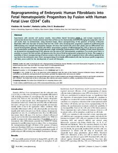
ing also persisted after culture, freezing, and subsequent culture for several months (data not shown). Fibroblasts exposed to a 293T extract did not display hematopoietic markers (Fig. 4A, B). TCRαβ synthesis occurred in cells exposed to extract treated with 50 µg/ml C RNAse A or 500 µg/ml DNAse I before the reprogramming reaction (Fig. 4D), indicating that protein expression resulted from transcription in the reprogrammed cells rather than from nucleic acids carried over from the extract. Reprogramming was also applicD able to primary cells. Over 60% of normal human skin fibroblasts from a 13-year-old male (American Type Culture Collection) exposed to Jurkat extract expressed CD3 and TCRαβ at 5 and 12 d.p.r. (Fig. 4E). Whether fibroblasts of older individuals are E also amenable to reprogramming remains to be tested. Induction of a T cell–specific function. We next investigated whether new intracellular functions were elicited upon expression of immune cell surface receptors in reprogrammed fibroblasts. UnFigure 2. Chromatin remodeling and activation of the IL2 gene in STE. (A) Intranuclear anchoring of the BAF stimulated T cells express the lowcomplex. Left panel: nuclei were isolated from resting (–) or anti-CD3-stimulated (α-CD3) T cells and intranuclear free and bound BAF assessed by immunoblotting of Triton X-100-soluble and insoluble nuclear affinity IL-2 receptor β (IL-2Rβ) fractions using anti-BRG1 antibodies. Right panel: free and bound BAF fractions were visualized as above in chain, whereas the high-affinity 293T nuclei incubated in STE and sedimented through sucrose. Graph: proportions of bound BAF were IL-2R requires induction of the determined by densitometric analysis of duplicate blots. (B) β-actin is part of the BAF complex in 293T nuclei IL-2Rα chain by stimulation of the exposed to STE. Anti-BRG1 immunoprecipitates of stimulated T-cell nuclei (T cell) or 293T nuclei exposed to STE were immunoblotted using anti-BRG1 and anti-β-actin antibodies. IgG, control immunoprecipitation using TCR–CD3 signaling pathway24,25. pre-immune IgGs. (C) ATPase activity of the BAF complex. Following exposure of 293T nuclei (black columns) Reprogrammed 293T cells were or resting T-cell nuclei (white columns) to STE or UTE, BRG1 was immunoprecipitated from nuclear lysates and stimulated with anti-CD3 antibodhydrolysis of 1 nM exogenous ATP (ATP+) by the immune precipitate (BRG1-IP) was determined in a ies and phorbolmyristylacetate luminometric assay. Control precipitations were performed using pre-immune IgGs (Pre-I IgG). Top panel shows an anti-BRG1 blot of the BRG1-IPs. Where indicated, ATPase activity was evaluated after treatment of BRG1(PMA), an act-ivator of the protein IPs with 600 µM latrunculin B or 200 µM cytochalasin D. ATPase activity is expressed as mean (± s.d.) relative kinase C pathway leading to T-cell light units (RLU) in the assay subtracted from the RLU of the control preimmune precipitate (mean RLU = 2,610; activation. Similar proportions not shown). Data are from five measurements taken in three replicates (except latrunculin and cytochalasin (80–90%) of reprogrammed treatments (six measurements in two replicates)). (D) Histone H4 acetylation in the IL2 proximal promoter in fibroblasts and Jurkat cells 293T nuclei exposed to STE. Micrococcal nuclease–soluble chromatin was prepared from input 293T nuclei (Input n.) and 293T nuclei incubated in UTE or STE. Acetylated H4 was immunoprecipitated and DNA was expressed IL-2Rβ, but not IL-2Rα, isolated from anti-acH4 precipitate (Bound) and supernatant (Unbound) fractions. DNA was dot-blotted and in the absence of stimulation hybridized to an IL2 promoter–specific probe or a β-actin (ACTB) probe. DNA (200 ng) was applied onto each (Fig. 5). However, stimulation spot. (E) Transcription of the IL2 gene in purified non-T-cell nuclei in vitro. Nuclei from 293T, NT2, HUVEC, and elicited expression of IL-2Rα in resting T cells were incubated for 2 h in UTE (Nuclei/UTE) or STE (Nuclei/STE). As controls, nuclei were incubated in STE containing mAb414 or 50 nM actinomycin D (ActD). Nuclei were also exposed to STE treated almost all reprogrammed cells that with 100 µg/ml DNAse I prior to the transcription reaction. RNA was isolated from the reaction mix and IL2 expressed IL2-Rβ. Similar observatranscription was examined by RT-PCR. Data from one of three to six replicates are shown. tions were made in Jurkat cells. Stimulation of control 293T fibrobExpression of hematopoietic cell surface receptors. Expression of lasts did not invoke IL-2Rα synthesis. This suggests that complex, T hematopoietic cell–specific surface antigens in reprogrammed cell–specific intracellular signaling pathways are induced in the fibroblasts was evaluated by immunofluorescence. Most fibroblasts reprogrammed cells. reprogrammed in Jurkat extract expressed CD3, CD4, and CD8 at 4 Fibroblasts exposed to NT2 extract express a neurofilament d.p.r. (Fig. 4A) and 11 d.p.r. (Fig. 4B). CD45 was absent at 4 d.p.r. but protein. Our method of in vitro cell reprogramming may be was detected by 11 d.p.r. (Fig. 4A, B). Remarkably, TCRαβ chains extended to other extracts or cell types. In a preliminary experiwere expressed in ∼90% of reprogrammed fibroblasts (Fig. 4A, B). ment, we exposed permeabilized 293T fibroblasts to an extract Expression of these antigens was first seen 24–60 h after reprogramfrom NT2 neuronal precursors. Fifteen days after reprogramming, ming (Fig. 4C), indicating that they had not been carried over from neurofilament protein NF200 (ref. 26) was expressed in ∼18% of the extract but were newly translated molecules. Fluorescence labelthe cells (Fig. 6). NF200 was restricted to polarized outgrowths 462
nature biotechnology
•
VOLUME 20
•
MAY 2002
•
http://biotech.nature.com
RESEARCH ARTICLE B
© 2002 Nature Publishing Group http://biotech.nature.com
A
Figure 3. 293T fibroblasts reprogrammed in Jurkat-TAg extract express hematopoietic cell-specific genes. (A) Relative mRNA levels in 293T fibroblasts incubated in stimulated Jurkat extract or in control 293T extract were compared using a cDNA macroarray. Data show fold increase or decrease in transcription level of indicated genes in Jurkat extract–treated cells, measured as the ratio of reprogrammed cell probe hybridization signal strength to that of control cells. Only genes with a greater than twofold increase or decrease in expression level are shown (except housekeeping genes, all shown). Data from two separate experiments are shown (bars, dots). Colored backgrounds designate distinct gene groups. (B) Northern blot analysis of IL-2, IL-7, CD3, CD4, RANTES, integrin β1, and β-actin expression in 293T cells, Jurkat cells, 293T cells exposed to 293T extract, and reprogrammed cells (three replicates, Rep.1–3). RNAs from Rep. 1 and Rep. 2 were those used in the array shown in A.
Reprogramming is illustrated by chromatin remodeling, activation of lymphoid-specific genes, synthesis of cell surface receptors, and induction of a complex intracellular regulatory pathway. Persistence of lymphoid cell–specific molecules weeks after exposure to extract suggests that the changes elicited are to some extent heritable. Acetylation of the IL2 promoter in the fibroblast nuclei also indicates that aspects of epigenetic reprogramming have taken place. In contrast to cell hybridization studies in which somatic nuclei are continuously exposed to reprogramming cytoplasm7,8, our results suggest that transient exposure of nuclei to a heterologous environment may be sufficient for reprogramming. In vitro reprogramming of differentiated somatic cells from primary cultures should allow repression of the donor cell program for cloning purposes and production of isogenic replacement cells for therapeutic applications.
resembling elongating neurites, which occasionally contacted neighboring cells in culture. Conclusions. Our results demonstrate the functional reprogramming of a somatic cell by exposure to a nuclear and cytoplasmic extract derived from a heterologous somatic cell type. http://biotech.nature.com
•
MAY 2002
•
VOLUME 20
•
nature biotechnology
463
RESEARCH ARTICLE 5–10 × 107 cells/ml in RPMI 1640 (Gibco-BRL, Paisley, UK). The TCR–CD3 complex was stimulated with 5 µg/ml anti-CD3 antibodies and cells were incubated on ice for 30 min. Cells were spun at 400g at 4°C for 7 min, washed, and resuspended to 5 × 10⁷ cells/ml in ice-cold RPMI 1640. Anti-mouse Fab fragments (10 µg/ml) were added as crosslinkers and cells were incubated at 37°C (t = 0 min post stimulation).
© 2002 Nature Publishing Group http://biotech.nature.com
A
Cell extracts. To prepare extracts from stimulated T cells, cells were frozen in liquid nitrogen at 5–10 min post stimulation, thawed, washed in ice-cold lysis buffer27, and sedimented; pellets were resuspended in 2 volumes of lysis buffer. Cells and nuclei were disrupted with a tip sonicator (2 mm diameter) and the lysate was cleared at 15,000g for 15 min at 4°C. Unstimulated T-cell extracts were prepared from mock (H2O)–stimulated T cells. Extracts from Jurkat-TAg cells were prepared as above after costimulation for 2 h with 40 ng/ml anti-CD3 and 0.1 µM PMA. NT2 extract was prepared from confluent NT2 cells. Extracts were used either fresh or frozen for up to several weeks and thawed, without noticeable differences in the results.
C
B
E
D
Nuclei, chromatin, and ChIP. Nuclei were isolated from 293T, NT2, HUVEC, and unstimulated peripheral blood T cells by Dounce homogenization, and were stored frozen27. Soluble chromatin was prepared from purified nuclei by micrococcal nuclease digestion28. Triton X-100-, Figure 4. Fibroblasts reprogrammed in Jurkat extract exhibit hematopoietic cell markers. (A) 293T fibroblasts DNAse-, and RNAse-resistant nuclear exposed to a 293T extract (control) or a stimulated Jurkat extract, and Jurkat cells were analyzed by immunofluorescence using indicated FITC-conjugated antibodies. Labeling shown of CD3, CD4, CD8, and TCRαβ matrices were isolated as described29. was detected at 4 d.p.r. CD45, absent at 4 d.p.r. (not shown), was detected at 11 d.p.r. The catalytic subunit of PKA ChIP was performed after solubiliza(PKA-C) was examined as positive control. DNA was labeled with propidium iodide. (B) Proportions of each cell tion of chromatin with micrococcal type expressing indicated antigens at 11 d.p.r. (mean ± s.d.; n > 300 cells/treatment/marker in >3 replicates). nuclease (0.1 units/µg DNA)28 using (C) Onset of TCRαβ expression in reprogrammed cells. Reprogrammed (Rep.) and control (Cont.) cells were an anti–pan-acetylated histone H4 analyzed as in A at indicated time points after reprogramming reaction. (D) 293T fibroblasts were exposed to antibody. DNA was isolated by pheJurkat extract treated with no nuclease (–), 50 µg/ml RNAse A, or 500 µg/ml DNAse I before reaction. Expression nol–chloroform extraction from of TCRαβ was examined at 5 d.p.r. (E) Human primary skin fibroblasts exposed to Jurkat extract or mockreprogrammed were cultured for five days and analyzed by immunofluorescence using anti-CD3 and anti-TCRαβ antibody-bound and -unbound fracantibodies. Bars, 10 µm. tions, and the IL2 promoter was identified by dot blotting. A fluoresceinated IL2 promoter probe (Gene Images CDP-Star, Amersham, Uppsala, Sweden) was synthesized by ranExperimental protocol Immunological procedures. Anti-CD3 mAb (clone SpvT3d) was a gift of dom priming using a cloned 430 bp insert encompassing the 360 bp of the A.M. Rasmussen (Norwegian Radium Hospital, Montebello, Norway). We promoter region proximal to the start site and the first 70 bp of the IL2 obtained anti-H4 and -acH4 antibodies from Serotec; anti-CD3, -CD4, -CD8, coding sequence. The ACTB probe was synthesized and hybridization was and -CD45 from Diatec; anti-TCRαβ from Pharmingen (Erembodegem, performed as described30. Belgium); anti-IL-2Rα and -IL-2Rβ from R&D Systems (Minneapolis, MN); Nuclear reprogramming. Nuclear reprogramming reactions consisted of 20 and anti-NF200 from Sigma (St. Louis, MO). All other antibodies were µl (or multiples thereof) of STE or UTE, containing 100,000 nuclei and an obtained from Santa Cruz Biotechnology and Transduction Laboratories ATP-generating system (1 mM ATP, 10 mM creatine phosphate, 25 µg/ml (both in Santa Cruz, CA). Immunofluorescence (NFAT, IL-2Rα, and IL-2Rβ) creatine kinase, 100 µM GTP). Reactions were incubated at 30°C for 30 min was performed as described27 or using FITC-conjugated primary antibodies unless indicated otherwise, and nuclei were sedimented through sucrose. (CD3, CD4, CD8, CD45, and TCRαβ). Western blotting27 was performed Alternatively, total RNA was extracted (Qiagen, Valencia, CA) from the using antibody dilutions of 1:500. BRG1 was immunoprecipitated from reaction mix for RT-PCR. micrococcal nuclease–soluble chromatin precleared with rabbit IgGs, using anti-BRG1 antibodies (diluted 1:40) and protein A–Sepharose beads27. Cell reprogramming. 293T fibroblasts were grown on round 16 mm poly-Llysine-coated coverslips in RPMI 1640 medium, to 50,000–100,000 cells per T-cell stimulation. T cells were purified from peripheral blood from healthy coverslip in 12-well plates. Cells were permeabilized with 200 ng/ml strepdonors21. Cells were cultured for 20 h and incubated on ice for 15 min at
464
nature biotechnology
•
VOLUME 20
•
MAY 2002
•
http://biotech.nature.com
RESEARCH ARTICLE
© 2002 Nature Publishing Group http://biotech.nature.com
A
B
Figure 6. Fibroblasts reprogrammed in NT2 extract express neurofilament protein NF200. NT2 or control 293T extract–treated fibroblasts were examined by immunofluorescence using anti-NF200 antibodies at 15 d.p.r. DNA was labeled with Hoechst 33342. Bar, 10 µm. Proportions of cells expressing NF200 are shown.
mRNA expression profiling. mRNA was isolated from cells frozen in liquid nitrogen at 10 and 13 d.p.r. mRNA (1 µg) was used as template for cDNA synthesis and labeling with [α-33P]dCTP (R&D Systems). Probes were hybridized to human cytokine expression arrays (R&D Systems) under recommended conditions, arrays were exposed to a phosphor screen for six days, and hybridization was quantified on a phosphorimager and analyzed using Phoretix Array V.2 (NonLinear Dynamics, Newcastle, UK). For northern analyses, total or poly(A)+ RNA was isolated, fractionated through 1% (wt/vol) agarose and transferred to a nylon membrane. Transcripts were detected with fluoresceinated probes prepared from IL2, IL7, CD3, CD4, RANTES (SCYA5), ITGB1, and ACTB cDNAs. Hybridization, washes, and detection were performed as described elsewhere31.
Figure 5. Induction of a T cell–specific signaling pathway in reprogrammed cells. (A) Jurkat cells, and 293T fibroblasts exposed to a 293T extract or Jurkat extract were stimulated with anti-CD3 antibodies and PMA for 24 h starting at 2 d.p.r. Stimulated and unstimulated cells were analyzed by immunofluorescence using anti-IL-2Rβ (green) and anti-IL-2Rα (red) antibodies. DNA was labeled with Hoechst 33342. Merged images are shown. Bars, 10 µm. (B) Proportions of cells expressing IL-2Rβ and IL-2Rα before and after stimulation as in A (mean ± s.d; n > 300 per treatment).
tolysin O in Ca2+-free Hanks’ balanced salt solution (Gibco-BRL) for 50 min at 37°C. Over 80% of cells were permeabilized, as judged by propidium iodide uptake. Streptolysin O was aspirated, and coverslips were overlaid with 80 µl of 293T, Jurkat-TAg, or NT2 extract and incubated for 1 h at 37°C in air. Extracts contained an ATP-generating system and 1 mM each of ATP, CTP, GTP, and UTP. To reseal plasma membranes, 1.5 ml of RPMI 1640 containing 2 mM CaCl2 was added and cells were incubated for 2 h at 37°C. CaCl2-containing RPMI was replaced by RPMI and cells were expanded for several weeks.
Acknowledgments We are grateful to T. Küntziger, A. Spurkland, and Y. Qi for discussion, primers, probes, and technical assistance. This work was supported by the Research Council of Norway and Nucleotech, LLC. P.C. is a fellow of the Norwegian Cancer Society.
Reverse transcription (RT)–PCR. Total RNA was isolated from cells, nuclei, or reprogramming reactions using the Qiagen RNeasy kit and 15 ng (Figs. 1, 2) or 100 ng (Fig. 3) RNA was used as template for RT-PCR. A 467-bp product was amplified using the human IL2 gene-specific primers 5′-ATGTACAGGATGCAACTCCTGTCTT-3′ and 5′-GTTAGTGTTGAGATGATGCTTTGAC-3′.
Competing interests statement The authors declare competing financial interests: see the Nature Biotechnology website (http://biotech.nature.com) for details.
1. Gurdon, J.B. & Uehlinger, V. “Fertile” intestine nuclei. Nature 210, 1240–1241 (1966). 2. Wilmut, I., Schnieke, A.E., McWhir, J., Kind, A.J. & Campbell, K.H.S. Viable offspring derived from fetal and adult mammalian cells. Nature 385, 810–813 (1997). 3. Cibelli, J.B. et al. Cloned transgenic calves produced from nonquiescent fetal fibroblasts. Science 280, 1256–1258 (1998). 4. Cibelli, J.B. et al. Transgenic bovine chimeric offspring produced from somatic cell–derived stem-like cells. Nat. Biotechnol. 16, 642–646 (1998). 5. Munsie, M.J. et al. Isolation of pluripotent embryonic stem cells from reprogrammed adult mouse somatic cell nuclei. Curr. Biol. 10, 989–992 (2000). 6. Wakayama, T. et al. Differentiation of embryonic stem cell lines generated from adult somatic cells by nuclear transfer. Science 292, 740–743 (2001). 7. Tada, M., Takahama, Y., Abe, K., Nakatsuji, N. & Tada, T. Nuclear reprogramming of somatic cells by in vitro hybridization with ES cells. Curr. Biol. 11, 1553–1558 (2001). 8. Tada, M., Tada T., Lefebvre, L., Barton, S.C. & Surani, M.A. Embryonic germ cells induce epigenetic reprogramming of somatic nucleus in hybrid cells. EMBO J. 16, 6510–6520 (1997). 9. Surani, M.A. Reprogramming of genome function through epigenetic inheritance. Nature 414, 122–128 (2001). 10. Morrison, S.J. Stem cell potential: can anything make anything? Curr. Biol. 11, R7–R9 (2001).
11. Hu, E., Tontonoz, P. & Spiegelman, B.M. Transdifferentiation of myoblasts by the adipogenic transcription factors PPARγ and C/EBPα. Proc. Natl. Acad. Sci. USA 92, 9856–9860 (1995). 12. Shen, C.N., Slack, J.M. & Tosh, D. Molecular basis of transdifferentiation of pancreas to liver. Nat. Cell Biol. 2, 879–887 (2001). 13. Funderburgh, J.L., Funderburgh, M.L., Mann, M.M., Corpuz, L.M. & Roth, M.R. Proteoglycan expression during transforming growth factor β–induced keratocyte–myofibroblast transdifferentiation. J. Biol. Chem. 276, 44173–44178 (2001). 14. Condorelli, G. et al. Cardiomyocytes induce endothelial cells to trans-differentiate into cardiac muscle: implications for myocardium regeneration. Proc. Natl. Acad. Sci. USA 98, 10733–10738 (2001). 15. Wangh, L.J., DeGrace, D., Sanchez, J.A., Golf, A. & Yeghiazarians, Y. Efficient reactivation of Xenopus erythrocyte nuclei in Xenopus egg extracts. J. Cell Sci. 108, 2187–2196 (1995). 16. Kass, S., Pruss, D. & Wolffe, A. How does DNA methylation repress transcription? Trends Genet. 13, 444–449 (1997). 17. Kikyo, N., Wade, P.A., Guschin, D., Ge, H. & Wolffe, A.P. Active remodeling of somatic nuclei in egg cytoplasm by the nucleosomal ATPase ISWI. Science 289, 2360–2362 (2000). 18. Crabtree, G.R. Contingent genetic regulatory events in T lymphocyte activation. Science 243, 355–361 (1989).
http://biotech.nature.com
•
MAY 2002
Received 5 October 2001; accepted 12 March 2002
•
VOLUME 20
•
nature biotechnology
465
© 2002 Nature Publishing Group http://biotech.nature.com
RESEARCH ARTICLE 26. Debus, E., Weber, K. & Osborn, M. Monoclonal antibodies specific for glial fibrillary acidic (GFA) protein and for each of the neurofilament triplet polypeptides. Differentiation 25, 193–203 (1983). 27. Collas, P., Le Guellec, K. & Tasken, K. The A-kinase anchoring protein, AKAP95, is a multivalent protein with a key role in chromatin condensation at mitosis. J. Cell Biol. 147, 1167–1180 (1999). 28. O’Neill, L.P. & Turner, B.M. Immunoprecipitation of chromatin. Methods Enzymol. 274, 189–197 (1996). 29. Steen, R.L., Cubizolles, F., Le Guellec, K. & Collas, P. A-kinase anchoring protein (AKAP)95 recruits human chromosome-associated protein (hCAP)-D2/Eg7 for chromosome condensation in mitotic extract. J. Cell Biol. 149, 531–536 (2000). 30. Collas, P., Liang, M.-R., Vincent, M. & Aleström, P. Active transgenes in zebrafish are enriched in acetylated histone H4 and dynamically associate with RNA Pol II and splicing complexes. J. Cell Sci. 112, 1045–1054 (1999). 31. Collas, P. Modulation of plasmid DNA methylation and expression in zebrafish embryos. Nucl. Acids Res. 26, 4454–4461 (1998).
19. Ward, S.B. et al. Chromatin remodeling of the interleukin-2 gene: distinct alterations in the proximal versus distal enhancer regions. Nucleic Acids Res. 26, 2923–2934 (1998). 20. Lin, J. & Weiss, A. T cell receptor signalling. J. Cell Sci. 114, 243–244 (2001). 21. Skålhegg, B.S. et al. Location of cAMP-dependent protein kinase type I with the TCR–CD3 complex. Science 263, 84–87 (1994). 22. Zhao, K. et al. Rapid and phosphoinositol-dependent binding of the SWI/SNF-like BAF complex to chromatin after T lymphocyte receptor signaling. Cell 95, 625–636 (1998). 23. O’Neill, L.P. & Turner, B.M. Histone H4 acetylation distinguishes coding regions of the human genome from heterochromatin in a differentiation-dependent but transcription-independent manner. EMBO J. 14, 3946–3957 (1995). 24. Kaempfer, R. Regulation of the human interleukin-2/interleukin-2 receptor system: a role for immunosuppression. Proc. Soc. Exp. Biol. Med. 206, 176–180 (1994). 25. Miyazaki, T. & Taniguchi, T. Coupling of the IL2 receptor complex with non-receptor protein tyrosine kinases. Cancer Surv. 27, 25–40 (1996).
466
nature biotechnology
•
VOLUME 20
•
MAY 2002
•
http://biotech.nature.com