Clinical and Experimental Otorhinolaryngology Vol. 4, No. 3: 155-158, September 2011
http://dx.doi.org/10.3342/ceo.2011.4.3.155
Case Report
Reversible Sensorineural Hearing Loss due to Pachymeningitis Associated with Elevated Serum MPO-ANCA Eun-Jung Lim, MD·Sung-Hee Kim, MD·Su-Hwan Lee, MD·Kyu-Yup Lee, MD1·Jae-Hyuk Choi, MD1 Eon-Jeong Nam, MD2·Sang-Heun Lee, MD1 Department of Otorhinolaryngology-Head and Neck Surgery, Daegu Fatima Hospital, Daegu; Department of Otorhinolaryngology-Head and Neck Surgery, and 2Division of Rheumatology, Department of Internal Medicine,
1
Kyungpook National University School of Medicine, Daegu, Korea
Hypertrophic pachymeningitis is a progressive disease resulting in a diffuse thickening of dura mater due to inflammation, tumor or autoimmune diseases, but most cases are idiopathic. It is seldom reported to be related to sensorineural hearing loss, but it can cause sensorineural hearing loss which can be potentially reversed through treatment. Here, we report the case of a 54-year-old woman who had progressive, bilateral, worse in the left, sensorineural hearing loss and visual disturbance with an accompanying headache over several months. Brain MRI showed diffusely thickened dura mater, highly enhanced after gadolinium administration, which was consistent with pachymeningitis. It was assumed to be related to autoimmune pathogenesis on the basis of elevated serum myeloperoxidase-antineutrophil cytoplasmic antibody (MPO-ANCA) titers. After empirical steroid and cyclophosphamide therapy, auditory impairment improved, especially in the high frequency region of the pure tone audiogram, and significant improvement in the word recognition test. Moreover, a follow-up MRI revealed much decreased enhancement of the dura mater, and the MPO-ANCA titer decreased to within the normal range. In the case of rapidly progressive sensorineural hearing loss or hearing impairment accompanying other cranial neuropathy, pachymeningitis should be taken into consideration, and brain MRI with gadolinium enhancement is the best method of detecting it. Also, to ensure proper treatment, a cautious evaluation including an ANCA work-up should be performed. Key Words. Hypertrophic pachymeningitis, Multiple cranial neuropathies, p-ANCA, Wegener’s granulomatosis
INTRODUCTION
ruled out. Also, in order to perform a proper evaluation, otologists should be aware of the potential causes of reversible sensorineural hearing loss. Pachymeningitis is an uncommon disorder characterized by localized or diffuse thickening of the dura mater, and is rarely reported to lead to sensorineural hearing loss (3-6). Even though pachymeningitis is a potentially treatable cause of sensorineural hearing loss (4-6), it is quite unfamiliar to otologists and difficult to diagnose. Here, we present the case of a patient who had rapidly progressive, bilateral sensorineural hearing loss due to hypertrophic pachymeningitis. It was associated with high serum levels of myeloperoxidase-antineutrophil cytoplasmic antibody (MPOANCA).
Sensorineural hearing loss can have various causes, and it is mostly irreversible. However, it can be managed with treatment in some instances such as autoimmune hearing loss (1, 2). Therefore, in cases of atypical sensorineural hearing loss, such as hearing loss that progresses relatively rapidly, or when neurologic signs coexist, the possibility of reversible causes should not be ••Received August 13, 2009 Accepted after revision October 3, 2009 ••Corresponding author: Sang-Heun Lee, MD Department of Otorhinolaryngology-Head and Neck Surgery, Kyungpook National University School of Medicine, 200 Dongduk-ro, Jung-gu, Daegu 700-721, Korea Tel: +82-53-420-5777, Fax: +82-53-423-4524 E-mail:
[email protected]
Copyright © 2011 by Korean Society of Otorhinolaryngology-Head and Neck Surgery. This is an open-access article distributed under the terms of the Creative Commons Attribution Non-Commercial License (http://creativecommons.org/licenses/by-nc/3.0) which permits unrestricted non-commercial use, distribution, and reproduction in any medium, provided the original work is properly cited.
155
156
Clinical and Experimental Otorhinolaryngology Vol. 4, No. 3: 155-158, September 2011
CASE REPORT
0 10 20 30 40 50 60 70 80 90 100 110
A
250
500
1,000
2,000
4,000
8,000
Frequency (Hz)
0 10 20 30 40 50 60 70 80 90 100 110
A
B
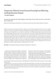
Fig. 2. Initial brain MRI. Axial T1-weighted image shows hypo-intense thickened dura mater (A). Axial T2-weighted image shows isointense dura mater with hyperintense border (B).
Hearing level (dB)
Hearing level (dB)
A 54-year-old woman presented with a progressive visual disturbance and hearing impairment, especially in the left ear, over the previous month. She also complained of recent intensification of a throbbing headache in her left fronto-temporal area that had been present for 6 months. An audiologic test revealed bilateral, down-sloping, moderate, sensorineural hearing loss with a mean threshold of 50 dB in the pure tone audiogram (Fig. 1A). Word recognition scores were 100% in the right ear and 24% in the left. Ophthalmic examination showed both peripapillary edema and atrophy, which was consistent with optic neuropathy. Other neurologic examinations yielded normal findings. Laboratory tests showed an elevated erythrocyte sedimentation rate (ESR) to 47 mm/hour (normal range, 0 to 20 mm/hour), a C-reactive protein (CRP) level up to 4.66 mg/dL (normal range