Review: Current Perspective From Vulnerable Plaque to Vulnerable Patient A Call for New Definitions and Risk Assessment Strategies: Part II Morteza Naghavi, MD; Peter Libby, MD; Erling Falk, MD, PhD; S. Ward Casscells, MD; Silvio Litovsky, MD; John Rumberger, MD; Juan Jose Badimon, PhD; Christodoulos Stefanadis, MD; Pedro Moreno, MD; Gerard Pasterkamp, MD, PhD; Zahi Fayad, PhD; Peter H. Stone, MD; Sergio Waxman, MD; Paolo Raggi, MD; Mohammad Madjid, MD; Alireza Zarrabi, MD; Allen Burke, MD; Chun Yuan, PhD; Peter J. Fitzgerald, MD, PhD; David S. Siscovick, MD; Chris L. de Korte, PhD; Masanori Aikawa, MD, PhD; K.E. Juhani Airaksinen, MD; Gerd Assmann, MD; Christoph R. Becker, MD; James H. Chesebro, MD; Andrew Farb, MD; Zorina S. Galis, PhD; Chris Jackson, PhD; Ik-Kyung Jang, MD, PhD; Wolfgang Koenig, MD, PhD; Robert A. Lodder, PhD; Keith March, MD, PhD; Jasenka Demirovic, MD, PhD; Mohamad Navab, PhD; Silvia G. Priori, MD, PhD; Mark D. Rekhter, PhD; Raymond Bahr, MD; Scott M. Grundy, MD, PhD; Roxana Mehran, MD; Antonio Colombo, MD; Eric Boerwinkle, PhD; Christie Ballantyne, MD; William Insull, Jr, MD; Robert S. Schwartz, MD; Robert Vogel, MD; Patrick W. Serruys, MD, PhD; Goran K. Hansson, MD, PhD; David P. Faxon, MD; Sanjay Kaul, MD; Helmut Drexler, MD; Philip Greenland, MD; James E. Muller, MD; Renu Virmani, MD; Paul M Ridker, MD; Douglas P. Zipes, MD; Prediman K. Shah, MD; James T. Willerson, MD Abstract—Atherosclerotic cardiovascular disease results in ⬎19 million deaths annually, and coronary heart disease accounts for the majority of this toll. Despite major advances in treatment of coronary heart disease patients, a large number of victims of the disease who are apparently healthy die suddenly without prior symptoms. Available screening and diagnostic methods are insufficient to identify the victims before the event occurs. The recognition of the role of the vulnerable plaque has opened new avenues of opportunity in the field of cardiovascular medicine. This consensus
From The Center for Vulnerable Plaque Research, University of Texas—Houston, The Texas Heart Institute, and President Bush Center for Cardiovascular Health, Memorial Hermann Hospital, Houston (M. Naghavi, S.W.C., S.L., M.M., A.Z., J.T.W.); The Leducq Center for Cardiovascular Research, Department of Medicine, Brigham and Women’s Hospital, Harvard Medical School, Boston, Mass (P.L., M.A.); Department of Cardiology and Institute of Experimental Clinical Research, Aarhus University, Aarhus, Denmark (E.F.); Experimental Cardiology Laboratory, Vascular Biology of the University Medical Center in Utrecht, the Netherlands (G.P.); Ohio State University (J.R.); the Zena and Michael A. Wiener Cardiovascular Institute, Mount Sinai Medical Center, New York, NY (Z.F.); Cardiac Catheterization Laboratory at the VA Medical Center, University of Kentucky, Lexington (P.M.); Cardiovascular Division, Department of Medicine, Brigham and Women’s Hospital, Harvard Medical School, Boston, Mass (P.H.S.); Division of Cardiology, New England Medical Center, Boston, Mass (S.W.); Department of Medicine, Section of Cardiology, Tulane University School of Medicine, New Orleans, La (P.R.); Department of Cardiovascular Pathology, Armed Forces Institute of Pathology, Washington, DC (A.B., A.F., R.V.); Department of Radiology, University of Washington, Seattle (C.Y.); Stanford University Medical Center Stanford, Calif (P.J.F.); Cardiovascular Health Research Unit, University of Washington, Seattle (D.S.S.); Department of Cardiology, Athens Medical School, Athens, Greece (C.S.); Catheterization Laboratory, Thorax Center, Erasmus University, Rotterdam, the Netherlands (C.L.d.K.); Division of Cardiology, Department of Medicine, University of Turku, Finland (K.E.J.A.); Institute of Arteriosclerosis Research and the Institute of Clinical Chemistry and Laboratory Medicine, Central Laboratory, Hospital of the University of Münster, Munich, Germany (G.A.); Department of Clinical Radiology, University of Münster, Munich, Germany (C.R.B.); Mayo Clinic Medical School, Jacksonville, Fla (J.H.C.); Department of Medicine, Division of Cardiology, Emory University School of Medicine, Atlanta, Ga (Z.S.G.); Bristol Heart Institute, Bristol University, Bristol, United Kingdom (C.J.); Cardiology Division, Massachusetts General Hospital and Harvard Medical School, Boston, Mass (I.-K.J.); Department of Internal Medicine II, Cardiology, University of Ulm, Ulm, Germany (W.K.); University of Kentucky, Lexington, Ky (R.A.L.); R.L. Roudebush VA Medical Center, Indianapolis, Ind (K.M.); School of Public Health, University of Texas—Houston, Houston, Texas (J.D.); Division of Cardiology, University of California Los Angeles, Los Angeles, Calif (M. Navab); Fondazione Salvatore Maugeri, University of Pavia, Pavia, Italy (S.G.P.); Department of Cardiovascular Therapeutics, Pfizer Global Research and Development, Ann Arbor Laboratories, Ann Arbor, Mich (M.D.R.); Paul Dudley White Coronary Care System at St. Agnes HealthCare, Baltimore, Md (R.B.); Center for Human Nutrition, University of Texas Health Science Center, Dallas (S.M.G.); Lenox Hill Hospital, New York, NY (R.M.); Catheterization Laboratories, Ospedale San Raffaele and Emo Centro Cuore Columbus, Milan, Italy (A.C.); Human Genetics Center, Institute of Molecular Medicine, Houston, Tex (E.B.); Department of Medicine, Baylor College of Medicine, Houston, Tex (C.B., W.I.); Minneapolis Heart Institute and Foundation, Minneapolis, Minn (R.S.S.); Division of Cardiology, University of Maryland School of Medicine, Baltimore, Md (R.V.); Karolinska Institute, Center for Molecular Medicine, Karolinska Hospital, Stockholm, Sweden (G.K.H.); Section of Cardiology, University of Chicago, Ill (D.P.F.); Vascular Physiology and Thrombosis Research Laboratory of the Atherosclerosis Research Center, Cedars-Sinai Medical Center, Los Angeles, California (S.K.); Cardiology Department, Hannover University, Hannover, Germany (H.D.); Department of Medicine, Feinberg School of Medicine, Northwestern University, Chicago, Ill (P.G.); UCLA School of Medicine and Cedars-Sinai Medical Center, Los Angeles, Calif (P.K.S.); Massachusetts General Hospital, Harvard Medical School and CIMIT (Center for Integration of Medicine and Innovative Technology), Boston, Mass (J.E.M.); Cardiovascular Division, Division of Preventive Medicine, Brigham and Women’s Hospital, Boston, Mass (P.M.R.); and Indiana University School of Medicine, Krannert Institute of Cardiology, Indianapolis (D.P.Z.). Many of the authors of this work, in addition to their research activities, have served as consultants to and/or employees of pharmaceutical, medical equipment, and other related companies. This article is Part II of a 2-part article. Part I appeared in the October 7, 2003 issue of Circulation (Circulation. 2003;108:1664 –1672). Guest editor for this article was Eugene Braunwald, MD, Brigham and Women’s Hospital, Boston, Mass. Correspondence to Morteza Naghavi, MD, Association for Eradication of Heart Attack, P.O. Box 20345, Houston, TX 77225-0345. E-mail
[email protected] © 2003 American Heart Association, Inc. Circulation is available at http://www.circulationaha.org
DOI: 10.1161/01.CIR.0000087481.55887.C9
1772
Naghavi et al
Vulnerable Patient: Part II
1773
document concludes the following. (1) Rupture-prone plaques are not the only vulnerable plaques. All types of atherosclerotic plaques with high likelihood of thrombotic complications and rapid progression should be considered as vulnerable plaques. We propose a classification for clinical as well as pathological evaluation of vulnerable plaques. (2) Vulnerable plaques are not the only culprit factors for the development of acute coronary syndromes, myocardial infarction, and sudden cardiac death. Vulnerable blood (prone to thrombosis) and vulnerable myocardium (prone to fatal arrhythmia) play an important role in the outcome. Therefore, the term “vulnerable patient” may be more appropriate and is proposed now for the identification of subjects with high likelihood of developing cardiac events in the near future. (3) A quantitative method for cumulative risk assessment of vulnerable patients needs to be developed that may include variables based on plaque, blood, and myocardial vulnerability. In Part I of this consensus document, we cover the new definition of vulnerable plaque and its relationship with vulnerable patients. Part II of this consensus document will focus on vulnerable blood and vulnerable myocardium and provide an outline of overall risk assessment of vulnerable patients. Parts I and II are meant to provide a general consensus and overviews the new field of vulnerable patient. Recently developed assays (eg, C-reactive protein), imaging techniques (eg, CT and MRI), noninvasive electrophysiological tests (for vulnerable myocardium), and emerging catheters (to localize and characterize vulnerable plaque) in combination with future genomic and proteomic techniques will guide us in the search for vulnerable patients. It will also lead to the development and deployment of new therapies and ultimately to reduce the incidence of acute coronary syndromes and sudden cardiac death. We encourage healthcare policy makers to promote translational research for screening and treatment of vulnerable patients. (Circulation. 2003;108:1772-1778.) Key Words: coronary disease 䡲 plaque 䡲 myocardial infarction 䡲 atherosclerosis 䡲 death, sudden
I
n Part I of this consensus document, we have introduced the concept of vulnerable patient as defined by plaque, blood, and myocardial vulnerability. Vulnerable plaque was extensively discussed in Part I. Here we discuss the definition of vulnerable blood and vulnerable myocardium and present an outline for overall risk assessment of vulnerable patients.
Vulnerable (Thrombogenic) Blood Serum Markers of Atherosclerosis and Inflammation Serum markers may predict a patient’s risk of acute cardiovascular complications (Table 1). C-reactive protein (CRP) is an independent risk factor and a powerful predictor of future coronary events in the asymptomatic population1–3 and in patients with stable and unstable disease. Although CRP is a nonspecific marker of systemic inflammation, it activates endothelium and accumulates in the plaque, suggesting an important role in plaque inflammation.4,5 Circulating interleukin-6 levels, which are elevated in patients with acute coronary syndromes, also predict the risk of future coronary events in such patients.6 Recently, investigators have shown that high plasma concentrations of soluble CD40 ligand may indicate an increased vascular risk in apparently healthy women.7 Likewise, Hwang et al8 showed in a large population-based sample of individuals that circulating levels of soluble intracellular adhesion molecule were predictive of future acute coronary events. Markers of systemic inflammation, such as soluble adhesion molecules, circulating bacterial endotoxin, soluble human heat-shock protein 60, and antibodies to mycobacterial heat-shock protein 65, may predict an increased risk of atherosclerosis.9 Pregnancy-associated plasma protein A (PAPP-A) is present in unstable plaques, and its circulating levels are elevated in patients with acute coronary syndromes.10 Increased serum levels of PAPP-A may reflect instability of atherosclerotic plaques.11
With major advances in high-throughput genomics and proteomics research, future studies will undoubtedly identify new risk and protective factors and biomarkers that can be used for screening purposes. A recent study suggested an association between several genetic polymorphisms and clinical outcomes, some of which can be possibly related to plaque, blood, and myocardial vulnerability.12 The tools and knowledge base made possible by the Human Genome Project allow the field to move beyond one or a few single-nucleotide polymorphisms in a priori candidate genes. Genome-wide linkage analyses have been TABLE 1. Serological Markers of Vulnerability (Reflecting Metabolic and Immune Disorders) ● Abnormal lipoprotein profile (eg, high LDL, low HDL, abnormal LDL and HDL size density, lipoprotein 关a兴, etc) ● Nonspecific markers of inflammation (eg, hsCRP, CD40L, ICAM-1, VCAM-1, P-selectin, leukocytosis, and other serological markers related to the immune system; these markers may not be specific for atherosclerosis or plaque inflammation) ● Serum markers of metabolic syndrome (eg, diabetes or hypertriglyceridemia) ● Specific markers of immune activation (eg, anti-LDL antibody, anti-HSP antibody) ● Markers of lipid peroxidation (eg, ox-LDL and ox-HDL) ● Homocysteine ● PAPP-A ● Circulating apoptosis marker(s) (eg, Fas/Fas ligand, not specific to plaque) ● ADMA/DDAH ● Circulating nonesterified fatty acids (eg, NEFA) hsCRP indicates high-sensitivity CRP; CD40L, CD40 ligand; ICAM, intracellular adhesion molecule; VCAM, vascular cell adhesion molecule; MMP, matrix metalloproteinases; TIMP, tissue inhibitors of MMPs; LDL, low-density lipoprotein; HDL, high-density lipoprotein; HSP, heat shock protein; ADMA, asymmetric dimethylarginine; ADMA, dimethylarginine dimethylaminohydrolase; and NEFA, nonesterified fatty acids.
1774
Circulation
October 14, 2003
TABLE 2. Blood Markers of Vulnerability (Reflecting Hypercoagulability) ● Markers of blood hypercoagulability (eg, fibrinogen, D-dimer, and factor V Leiden) ● Increased platelet activation and aggregation (eg, gene polymorphisms of platelet glycoproteins IIb/IIIa, Ia/IIa, and Ib/IX) ● Increased coagulation factors (eg, clotting of factors V, VII, and VIII; von Willebrand factor; and factor XIII) ● Decreased anticoagulation factors (eg, proteins S and C, thrombomodulin, and antithrombin III) ● Decreased endogenous fibrinolysis activity (eg, reduced t-PA, increased PAI-1, certain PAI-1 polymorphisms) ● Prothrombin mutation (eg, G20210A) ● Other thrombogenic factors (eg, anticardiolipin antibodies, thrombocytosis, sickle cell disease, polycythemia, diabetes mellitus, hypercholesterolemia, hyperhomocysteinemia) ● Increased viscosity ● Transient hypercoagulability (eg, smoking, dehydration, infection, adrenergic surge, cocaine, estrogens, postprandial, etc) t-PA indicates tissue plasminogen activator; PAI, type 1 plasminogen activator inhibitor.
carried out for coronary artery calcification,13 and genomewide association studies for myocardial infarction are already a reality.14 Further studies are needed to address the relationship between single-nucleotide polymorphisms in components of each of the plaque, blood, and myocardial vulnerabilities and future outcomes (acute coronary syndromes and sudden cardiac death). However, ongoing proteomic research on serum samples of vulnerable patients collected from prospective studies before the onset of symptoms is most promising.
Coagulation/Anticoagulation System The importance of the coagulation system in the outcome of plaque complications was recently reemphasized by Karnicki et al,15 who in a porcine model demonstrated that the role assigned to lesion-bound tissue factor was not physically realistic and that blood borne factors must have a major role in thrombus propagation. A hematologic disorder is rarely the sole cause of coronary thrombosis and myocardial infarction. Inflammation promotes thrombosis and vice versa.16 Extensive atherosclerosis may be associated with increased blood thrombogenicity, but the magnitude of thrombogenicity varies from patient to patient, and unstable plaques are much more thrombogenic than stable ones (Table 2). Some platelet polymorphisms, such as glycoprotein IIIa P1(A2),17 Ib ␣ gene-5T/C Kozak,18 high factor V and factor VII clotting,19 have been reported as independent risk factors for myocardial infarction. Reiner et al20 recently reviewed the associations of known and potential genetic susceptibility markers for intermediate hemostatic phenotypes with arterial thrombotic disease. Other conditions that lead to a hypercoagulable state are diabetes mellitus, hypercholesterolemia, and cigarette smoking. High levels of circulating tissue factor may be the
mechanism of action responsible for the increased thrombotic complications associated with the presence of these cardiovascular risk factors.21 Acute coronary syndromes are associated with proinflammatory and prothrombotic conditions that involve a prolonged increase in the levels of fibrinogen, CRP, and plasminogen activator inhibitor.22,23 A number of blood abnormalities, including antithrombin III deficiency, protein C or S deficiency, and resistance to activated protein C (also known as factor V Leiden), have been implicated as causes of venous thrombosis. The risk of arterial thrombosis is only modestly increased in these conditions, but these abnormalities are thought to interact with traditional risk factors for arterial thrombosis. Venous and arterial thromboses are prominent features of the antiphospholipid syndrome.24,25 The main antibodies of this syndrome are the anticardiolipin antibody, the lupus anticoagulant, and the IgG antibodies against prothrombin and 2-glycoprotein.24,25 In the nephrotic syndrome, proteinuria results in abnormal concentration and activity of coagulation factors. Moreover, the associated hypoalbuminemia, thrombocytosis, and hypercholesterolemia may induce arterial and venous thrombosis.26 The importance of the coagulation/fibrinolytic system is highlighted by several autopsy studies that have shown a high prevalence of old plaque disruptions without infarctions. Therefore, an active fibrinolytic system may be able to prevent luminal thrombosis in some cases of plaque disruption.27,28 A transient shift in the coagulation and anticoagulation balance is likely to be an important factor in plaque-blood interaction, resulting in an acute event. “Triggers” such as exercise and smoking, which are associated with catecholamine release, may increase the risk of plaque thrombosis.29 Similarly, metabolic factors, such as postprandial metabolic changes, are associated with increased blood coagulability.30 Likewise, estrogen replacement therapy can lead to a hypercoagulable state.31 Finally, plasma viscosity, as well as fibrinogen and white blood cell counts, is positively associated with CHD events as shown by Koenig et al.32 Furthermore, Junker et al33 showed a positive relationship between plasma viscosity and the severity of coronary heart disease (CHD).
Vulnerable Myocardium Ischemic Vulnerable Myocardium Without Prior Atherosclerosis-Derived Myocardial Damage Abrupt occlusion of a coronary artery is a common cause of sudden death. It often leads to acute myocardial infarction or exacerbation of chest pain.34,35 Extensive studies in experimental animals and increasing clinical evidence indicate that autonomic nervous activity has a significant role in modifying the clinical outcome with coronary occlusion.30,36,37 Susceptibility of the myocardium to acute ischemia was reviewed by Airaksinen,38 who emphasized the key role of autonomic tone in the outcome after plaque rupture. Sympathetic hyperactivity favors the genesis of life-threatening ventricular tachyarrhythmias, whereas vagal activation exerts an antifibrillatory effect. Strong afferent stimuli from the
Naghavi et al TABLE 3. Conditions and Markers Associated With Myocardial Vulnerability With atherosclerosis-derived myocardial ischemia as shown by: ECG abnormalities:
Vulnerable Patient: Part II
TABLE 4. Available Techniques for Electrophysiological Risk Stratification of Vulnerable Myocardium Diagnostic criteria: Arrhythmia
During rest
QT dispersion
During stress test
QT dynamics
Silent ischemia (eg, ST changes on Holter monitoring)
T-wave alternans
Perfusion and viability disorder: PET scan SPECT Wall motion abnormalities
Ventricular late potentials Heart rate variability Diagnostic techniques: Noninvasive
Echocardiography
Resting ECG
MR imaging
Stress ECG
X-ray ventriculogram
Ambulatory ECG
MSCT
Signal-averaged ECG
Without atherosclerosis-derived myocardial ischemia: Sympathetic hyperactivity
1775
Surface high-resolution ECG Invasive
Impaired autonomic reactivity
Programmed ventricular stimulation
Left ventricular hypertrophy
Real-time 3D magnetic-navigated activation map
Cardiomyopathy (dilated, hypertrophic, or restrictive) Valvular disease (aortic stenosis and mitral valve prolapse) Electrophysiological disorders: Long-QT syndrome, Brugada syndrome, Wolff-Parkinson-White syndrome, sinus and atrioventricular conduction disturbances, catecholaminergic polymorphic ventricular tachycardia, T-wave alternans, drug-induced torsades de pointes Commotio cordis Anomalous origination of a coronary artery Myocarditis Myocardial bridging MSCT indicates multislice computed tomography; PET, positron emission tomography; and SPECT, single-photon emission computed tomography.
ischemic myocardium may impair the arterial baroreflex and lead to hemodynamic instability.39 There seems to be a wide interindividual variation in the type and severity of autonomic reactions during the early phase of abrupt coronary occlusion, a critical period for out-of-hospital cardiac arrest. The pre-existing severity of a coronary stenosis, adaptation or preconditioning to myocardial ischemia, habitual physical exercise, -blockade, and gender seem to affect autonomic reactions and the risk of fatal ventricular arrhythmias.38,40,41 Recent studies have documented a hereditary component for autonomic function, and genetic factors may also modify the clinical presentation of acute coronary occlusion.42,43 Table 3 depicts conditions and markers associated with myocardial vulnerability.
Ischemic Vulnerable Myocardium With Prior Atherosclerosis-Derived Myocardial Damage (Chronic Myocardial Damage) Any type of atherosclerosis-related myocardial injury, such as ischemia, an old or new myocardial infarction, inflammation, and/or fibrosis, potentially increases the patient’s vulnerability to arrhythmia and sudden death. In
the past few decades, a number of diagnostic methods have been developed for imaging cardiac ischemia and for assessing the risk of developing a life-threatening cardiac arrhythmia. In patients with a history of ischemic heart disease, ischemic cardiomyopathy is the ultimate form of myocardial damage. With the advent of new, effective treatments for hypertension and more efficient management of acute myocardial infarction, deaths resulting from stroke and acute myocardial infarction have steadily decreased.44 More patients are now surviving acute events, but some develop heart failure or ischemic cardiomyopathy later with the potential for fatal arrhythmias. It is also important to remember that in a significant number of patients, sudden cardiac death is the first manifestation of underlying heart disease, and it is still responsible for ⬎450 000 deaths annually in the United States.
Nonischemic Vulnerable Myocardium A smaller subset of patients experience fatal arrhythmia as a result of diseases other than coronary atherosclerosis. The various forms of cardiomyopathy (dilated, hypertrophic, restrictive, and right ventricular) account for most noncoronary cardiac deaths. Other underlying pathological processes include valvular heart disease, such as aortic stenosis and primary electrical disturbances (long-QT syndromes, Brugada syndrome, Wolff-Parkinson-White syndrome, sinus and atrioventricular conduction disturbances, catecholaminergic polymorphic ventricular tachycardia, and congenital and drug-induced long QT syndromes with torsades de pointes), and, infrequently, commotio cordis from chest trauma. Less common pathological conditions include anomalous origin of a coronary artery, myocarditis, and myocardial bridging (Table 3). Circulating nonesterified fatty acids are another risk factor
1776
Circulation
October 14, 2003
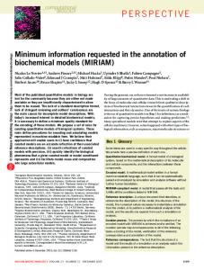
The “VP Pyramid.” This pyramid illustrates a speculative roadmap in search of vulnerable patients (numbers represent population in the United States). The major need is to develop noninvasive, relatively inexpensive, readily available, and accurate screening/diagnostic tools allowing multistep screening of an apparently healthy population and those with known atherosclerosis but whose risks for acute events are uncertain. Modified with permission from the AEHA.
for sudden death in middle-aged men, as is elevated serum concentration of CRP; serum measurements may help screening for vulnerable myocardium.45 Recently, the Task Force on Sudden Cardiac Death, organized by the European Society of Cardiology, issued a report that includes detailed diagnostic and therapeutic recommendations for a large number of cardiomyopathic conditions capable of causing sudden cardiac death.46 Table 4 provides electrophysiological diagnostic criteria and techniques for detection of myocardial vulnerability.
Risk Assessment for Vulnerable Patients Traditional Risk Assessment Strategies Despite extensive studies and development of several risk prediction models, traditional CHD risk factors fail to predict development of CHD in a large group of cases (25%47 to 50%3,48,49). Risk prediction models developed on the basis of data from long-term population-based follow-up studies may not be able to predict short-term risks for individual persons. The recent report by Ridker et al,3 who noted a greater impact
of an inflammatory marker such as serum CRP than LDL levels, is of interest. Several risk factor assessment models (eg, Framingham,50 Sheffield,51,52 New Zealand,53,54 Canadian,55 British,56 European,57 Dundee,58 Munster [PROCAM],59 and MONICA60) have been developed. However, all of them are based on the traditional risk factors known to contribute to the chronic development of atherosclerosis. Addition of emerging risk factors, particularly those indicative of the activity of the disease (ie, plaque inflammation), may allow individualized risk assessments to be made. The traditional risk assessment has been shown to predict long-term outcome in large populations. However, they fall short in predicting near-future events particularly in individual clinical practice. For example, a high Framingham Risk Score, although capable of forecasting an adverse cardiovascular event in 10 years, clearly falls short in accurately predicting events in individual patients and cannot provide a clear clinical route for cardiologists to identify and treat, to prevent near future victims of acute coronary syndromes and sudden death. The same is true for
Naghavi et al coronary evaluations using electrocardiography, myocardial perfusion tests, and coronary angiography. A positive test for coronary stenosis or reversible perfusion defect (ischemia), although considered as a major risk factor, must be coupled in the future with emerging methods of risk assessment for detection of vulnerable patients to predict more accurately the near-future outcome and prognosis. Those who have no indication of coronary stenosis or myocardial ischemia and who may even lack traditional risk factors may benefit from the techniques now under development that evaluate plaque biology and inflammation.
New Risk Assessment Strategies We propose a Cumulative Vulnerability Index based on the following: ● ● ●
Vulnerable plaque/artery Vulnerable blood (prone to thrombosis) Vulnerable myocardium (prone to life-threatening arrhythmia)
This proposal is by no means intended to disregard the predictive value of traditional risk assessment strategies that have been proven in predicting long-term outcome but instead to strengthen their value in providing higher accuracy, especially for near-term outcomes. Atherosclerosis is a diffuse and multisystem, chronic inflammatory disorder involving vascular, metabolic, and immune systems with various local and systemic manifestations. Therefore, it is essential to assess total vulnerability burden and not just search for a single, unstable coronary plaque. A composite risk score (eg, a vulnerability index), that comprises the total burden of atherosclerosis and vulnerable plaque in the coronaries (and aorta and carotid, femoral, etc, arteries), and that includes blood and myocardial vulnerability factors, should be a more accurate method of risk stratification. Such a vulnerability index would indicate the likelihood that a patient with certain factors would have a clinical event in the coming year. Use of the state-of-the-art bioinformatics tools such as neural networks may provide substantial improvement for risk calculations.61 The information used for developing such risk stratification in the future is likely to come from a combination of smaller prospective studies (eg, from new imaging techniques) and retrospective cohort studies (eg, for serum factors) in which the risks for near future cardiovascular events can be quantitatively calculated. A few such studies have been conducted or are underway.2,62
In Search of the Vulnerable Patient The ideal method for screening vulnerable patients should be (1) inexpensive, (2) relatively noninvasive, (3) widely reproducible, (4) readily applicable to an asymptomatic population, and (5) capable of adding predicted value to measurements of established risk factors. Such a method should provide a cost-effective, stepwise approach designed to further stratify risk and provide reliable diagnosis and pathways for monitoring therapy. Obviously, these goals are hard to achieve with today’s tools. However, it is well within our
Vulnerable Patient: Part II
1777
reach, if academia and industry in the field of cardiovascular medicine undertake a coordinated effort to embark on developing new screening and diagnostic techniques to identify vulnerable patients (Figure).
Acknowledgments We are indebted to Valentin Fuster, MD, and Salim Yusuf, MD, for their insightful reviews and thoughtful comments. We also greatly appreciate the administrative support provided by Jennifer Harris, Philip Ralston, and Nadhir Kosa, PhD.
References 1. Ridker PM, Cushman M, Stampfer MJ, et al. Inflammation, aspirin, and the risk of cardiovascular disease in apparently healthy men. N Engl J Med. 1997;336:973–979. 2. Ridker PM, Hennekens CH, Buring JE, et al. C-reactive protein and other markers of inflammation in the prediction of cardiovascular disease in women. N Engl J Med. 2000;342:836 – 843. 3. Ridker PM, Rifai N, Rose L, et al. Comparison of C-reactive protein and low-density lipoprotein cholesterol levels in the prediction of first cardiovascular events. N Engl J Med. 2002;347:1557–1565. 4. Pasceri VWJ, Yeh ET. Direct proinflammatory effect of C-reactive protein on human endothelial cells. Circulation. 2000;102:2165–2168. 5. Verma SLS, Badiwala MV, Weisel RD, et al. Endothelin antagonism and interleukin-6 inhibition attenuate the proatherogenic effects of C-reactive protein. Circulation. 2002;105:1890 –1896. 6. Koukkunen H, Penttila K, Kemppainen A, et al. C-reactive protein, fibrinogen, interleukin-6 and tumour necrosis factor-␣ in the prognostic classification of unstable angina pectoris. Ann Med. 2001;33:37– 47. 7. Schonbeck U, Varo N, Libby P, et al. Soluble CD40L and cardiovascular risk in women. Circulation. 2001;104:2266 –2268. 8. Hwang SJ, Ballantyne CM, Sharrett AR, et al. Circulating adhesion molecules VCAM-1, ICAM-1, and E-selectin in carotid atherosclerosis and incident coronary heart disease cases: the Atherosclerosis Risk In Communities (ARIC) study. Circulation. 1997;96:4219 – 4225. 9. Kiechl S, Egger G, Mayr M, et al. Chronic infections and the risk of carotid atherosclerosis : prospective results from a large population study. Circulation. 2001;103:1064 –1070. 10. Bayes-Genis A, Conover CA, Overgaard MT, et al. Pregnancy-associated plasma protein A as a marker of acute coronary syndromes. N Engl J Med. 2001;345:1022–1029. 11. Beaudeux JL, Burc L, Imbert-Bismut F, et al. Serum plasma pregnancyassociated protein A: a potential marker of echogenic carotid atherosclerotic plaques in asymptomatic hyperlipidemic subjects at high cardiovascular risk. Arterioscler Thromb Vasc Biol. 2003;23:e7– e10. 12. Yamada Y, Izawa H, Ichihara S, et al. Prediction of the risk of myocardial infarction from polymorphisms in candidate genes. N Engl J Med. 2002; 347:1916 –1923. 13. Lange LA, Lange EM, Bielak LF, et al. Autosomal genome-wide scan for coronary artery calcification loci in sibships at high risk for hypertension. Arterioscler Thromb Vasc Biol. 2002;22:418 – 423. 14. Ozaki K, Ohnishi Y, Iida A, et al. Functional SNPs in the lymphotoxin-␣ gene that are associated with susceptibility to myocardial infarction. Nat Genet. 2002;32:650 – 654. 15. Karnicki K, Owen WG, Miller RS, et al. Factors contributing to individual propensity for arterial thrombosis. Arterioscler Thromb Vasc Biol. 2002;22:1495–1499. 16. Libby P, Simon DI. Inflammation and thrombosis: the clot thickens. Circulation. 2001;103:1718 –1720. 17. Barakat K, Kennon S, Hitman GA, et al. Interaction between smoking and the glycoprotein IIIa P1(A2) polymorphism in non-ST-elevation acute coronary syndromes. J Am Coll Cardiol. 2001;38:1639 –1643. 18. Douglas H, Michaelides K, Gorog DA, et al. Platelet membrane glycoprotein Ib␣ gene –5T/C Kozak sequence polymorphism as an independent risk factor for the occurrence of coronary thrombosis. Heart. 2002;87:70 –74. 19. Redondo M, Watzke HH, Stucki B, et al. Coagulation factors II, V, VII, and X, prothrombin gene 20210G3 A transition, and factor V Leiden in coronary artery disease: high factor V clotting activity is an independent risk factor for myocardial infarction. Arterioscler Thromb Vasc Biol. 1999;19:1020 –1025. 20. Reiner AP, Siscovick DS, Rosendaal FR. Hemostatic risk factors and arterial thrombotic disease. Thromb Haemost. 2001;85:584 –595.
1778
Circulation
October 14, 2003
21. Sambola A, Osende J, Hathcock J, et al. Role of risk factors in the modulation of tissue factor activity and blood thrombogenicity. Circulation. 2003;107:973–977. 22. Passoni F, Morelli B, Seveso G, et al. Comparative short-term prognostic value of hemostatic and inflammatory markers in patients with non-ST elevation acute coronary syndromes. Ital Heart J. 2002;3:28 –33. 23. Hoffmeister HM, Heller W, Seipel L. Activation markers of coagulation and fibrinolysis: alterations and predictive value in acute coronary syndromes. Thromb Haemost. 1999;82:76 –79. 24. Vaarala O, Puurunen M, Manttari M, et al. Antibodies to prothrombin imply a risk of myocardial infarction in middle-aged men. Thromb Haemost. 1996;75:456 – 459. 25. Jouhikainen T, Pohjola-Sintonen S, Stephansson E. Lupus anticoagulant and cardiac manifestations in systemic lupus erythematosus. Lupus. 1994; 3:167–172. 26. Osula S, Bell GM, Hornung RS. Acute myocardial infarction in young adults: causes and management. Postgrad Med J. 2002;78:27–30. 27. Burke AP, Kolodgie FD, Farb A, et al. Healed plaque ruptures and sudden coronary death: evidence that subclinical rupture has a role in plaque progression. Circulation. 2001;103:934 –940. 28. Mann J, Davies MJ. Mechanisms of progression in native coronary artery disease: role of healed plaque disruption. Heart. 1999;82:265–268. 29. Servoss SJ, Januzzi JL, Muller JE. Triggers of acute coronary syndromes. Prog Cardiovasc Dis. 2002;44:369 –380. 30. Silveira A. Postprandial triglycerides and blood coagulation. Exp Clin Endocrinol Diabetes. 2001;109:S527–S532. 31. McNagny SE, Wenger NK. Postmenopausal hormone-replacement therapy. N Engl J Med. 2002;346:63– 65. 32. Koenig W, Sund M, Filipiak B, et al. Plasma viscosity and the risk of coronary heart disease: results from the MONICA-Augsburg Cohort Study, 1984 to 1992. Arterioscler Thromb Vasc Biol. 1998;18:768 –772. 33. Junker R, Heinrich J, Ulbrich H, et al. Relationship between plasma viscosity and the severity of coronary heart disease. Arterioscler Thromb Vasc Biol. 1998;18:870 – 875. 34. Myerburg RJ, Kessler KM, Castellanos A. Sudden cardiac death: structure, function, and time-dependence of risk. Circulation. 1992; 85(suppl I):I-2–I-10. 35. Kannel WB, Doyle JT, McNamara PM, et al. Precursors of sudden coronary death: factors related to the incidence of sudden death. Circulation. 1975;51:606 – 613. 36. Schwartz PJ, Vanoli E, Zaza A, et al. The effect of antiarrhythmic drugs on life-threatening arrhythmias induced by the interaction between acute myocardial ischemia and sympathetic hyperactivity. Am Heart J. 1985; 109:937–948. 37. Vanoli E, De Ferrari GM, Stramba-Badiale M, et al. Vagal stimulation and prevention of sudden death in conscious dogs with a healed myocardial infarction. Circ Res. 1991;68:1471–1481. 38. Airaksinen KE. Autonomic mechanisms and sudden death after abrupt coronary occlusion. Ann Med. 1999;31:240 –245. 39. Airaksinen KE, Tahvanainen KU, Eckberg DL, et al. Arterial baroreflex impairment in patients during acute coronary occlusion. J Am Coll Cardiol. 1998;32:1641–1647. 40. Billman GE, Schwartz PJ, Stone HL. The effects of daily exercise on susceptibility to sudden cardiac death. Circulation. 1984;69:1182–1189. 41. Burke AP, Farb A, Malcom GT, et al. Plaque rupture and sudden death related to exertion in men with coronary artery disease. JAMA. 1999;281: 921–926. 42. Jouven X, Desnos M, Guerot C, et al. Predicting sudden death in the population: the Paris Prospective Study I. Circulation. 1999;99: 1978 –1983.
43. Singh JP, Larson MG, O’Donnell CJ, et al. Heritability of heart rate variability: the Framingham Heart Study. Circulation. 1999;99: 2251–2254. 44. Claessens C, Claessens P, Claessens M, et al. Changes in mortality of acute myocardial infarction as a function of a changing treatment during the last two decades. Jpn Heart J. 2000;41:683– 695. 45. Jouven X, Charles MA, Desnos M, et al. Circulating nonesterified fatty acid level as a predictive risk factor for sudden death in the population. Circulation. 2001;104:756 –761. 46. Priori SG, Aliot E, Blomstrom-Lundqvist C, et al. Task Force on Sudden Cardiac Death of the European Society of Cardiology. Eur Heart J. 2001;22:1374 –1450. 47. Magnus P, Beaglehole R. The real contribution of the major risk factors to the coronary epidemics: time to end the “only-50%” myth. Arch Intern Med. 2001;161:2657–2660. 48. Lefkowitz RJ, Willerson JT. Prospects for cardiovascular research. JAMA. 2001;285:581–587. 49. Nieto FJ. Cardiovascular disease and risk factor epidemiology: a look back at the epidemic of the 20th century. Am J Public Health. 1999;89: 292–294. 50. Anderson KM, Odell PM, Wilson PW, et al. Cardiovascular disease risk profiles. Am Heart J. 1991;121:293–298. 51. Ramsay LE, Haq IU, Jackson PR, et al. Targeting lipid-lowering drug therapy for primary prevention of coronary disease: an updated Sheffield table. Lancet. 1996;348:387–388. 52. Wallis EJ, Ramsay LE, Ul Haq I, et al. Coronary and cardiovascular risk estimation for primary prevention: validation of a new Sheffield table in the 1995 Scottish health survey population. BMJ. 2000;320:671– 676. 53. 1996 National Heart Foundation clinical guidelines for the assessment and management of dyslipidaemia. Dyslipidaemia Advisory Group on behalf of the Scientific Committee of the National Heart Foundation of New Zealand. N Z Med J. 1996;109:224 –231. 54. Jackson R. Updated New Zealand cardiovascular disease risk-benefit prediction guide. BMJ. 2000;320:709 –710. 55. McCormack JP, Levine M, Rangno RE. Primary prevention of heart disease and stroke: a simplified approach to estimating risk of events and making drug treatment decisions. CMAJ. 1997;157:422– 428. 56. Joint British recommendations on prevention of coronary heart disease in clinical practice: summary. British Cardiac Society, British Hyperlipidaemia Association, British Hypertension Society, British Diabetic Association. BMJ. 2000;320:705–708. 57. Wood D, De Backer G, Faergeman O, et al. Prevention of coronary heart disease in clinical practice: recommendations of the Second Joint Task Force of European and other Societies on Coronary Prevention. Atherosclerosis. 1998;140:199 –270. 58. Tunstall-Pedoe H. The Dundee coronary risk-disk for management of change in risk factors. BMJ. 1991;303:744 –747. 59. Assmann G, Cullen P, Schulte H. Simple scoring scheme for calculating the risk of acute coronary events based on the 10-year follow-up of the prospective cardiovascular Munster (PROCAM) study. Circulation. 2002;105:310 –315. 60. Manhem K, Dotevall A, Wilhelmsen L, et al. Social gradients in cardiovascular risk factors and symptoms of Swedish men and women: the Goteborg MONICA Study 1995. J Cardiovasc Risk. 2000;7:359 –368. 61. Voss R, Cullen P, Schulte H, et al. Prediction of risk of coronary events in middle-aged men in the Prospective Cardiovascular Münster Study (PROCAM) using neural networks. Int J Epidemiol. 2002;31:1253–1264. 62. Arad YSL, Goodman K, Newstein D, et al. Prediction of coronary events with electron beam computed tomography. J Am Coll Cardiol. 2000;36: 1253–1260.