2899 The Journal of Experimental Biology 213, 2899-2911 © 2010. Published by The Company of Biologists Ltd doi:10.1242/jeb.043307
Review Oxidative stress in cold-induced hyperthyroid state P. Venditti*, L. Di Stefano and S. Di Meo Department of the Biological Sciences, Section of Physiology, University ‘Federico II’ of Naples, 80134, Naples, Italy *Author for correspondence (
[email protected])
Accepted 4 May 2010
SUMMARY Exposure of homeothermic animals to low environmental temperature is associated with oxidative stress in several body tissues. Because cold exposure induces a condition of functional hyperthyroidism, the observation that tissue oxidative stress also happens in experimental hyperthyroidism, induced by 3,5,3⬘-triiodothyronine (T3) treatment, suggests that this hormone is responsible for the oxidative damage found in tissues from cold-exposed animals. Examination of T3-responsive tissues, such as brown adipose tissue (BAT) and liver, shows that changes in factors favoring oxidative modifications are similar in experimental and functional hyperthyroidism. However, differences are also apparent, likely due to the action of physiological regulators, such as noradrenaline and thyroxine, whose levels are different in cold-exposed and T3-treated animals. To date, there is evidence that biochemical changes underlying the thermogenic response to cold as well as those leading to oxidative stress require a synergism between T3- and noradrenaline-generated signals. Conversely, available results suggest that thyroxine (T4) supplies a direct contribution to cold-induced BAT oxidative damage, but contributes to the liver response only as a T3 precursor. Key words: cold exposure, thyroid hormone, oxidative stress, ROS production, antioxidant capacity, mitochondria, BAT, liver.
INTRODUCTION
It is now widely accepted that aerobic metabolism implies the formation of radicals and other reactive oxygen species (ROS) (Chance et al., 1979; Davies, 1995). When organic molecules are oxidized, to obtain the energy essential for life, oxygen is reduced to form water by concerted four-electron transfer. However, oxygen can also undergo univalent reduction by one-electron transfer which allows the formation of the superoxide radical anion (O•– 2 ). This is converted by superoxide dismutase (SOD) into hydrogen peroxide (H2O2), which can be turned into the highly reactive hydroxyl radical (•OH). Radicals and other ROS can oxidize various cellular substances, including DNA, proteins and lipids (Halliwell and Gutteridge, 1998). The oxidative effects of ROS are minimized by a fairly complex system of antioxidant defenses (Yu, 1994; Davies, 2000). Although ROS can be employed in living systems for useful purposes, such as cellular signal transduction (Poli et al., 2004), ROS generation that exceeds the cell antioxidant capacity results in oxidative stress (Sies, 1997), a deleterious process which appears to be related to many animal disorders. Among these, overproduction of ROS appears to promote the development of cancer (Halliwell, 2007), and cardiovascular (Fearon and Faux, 2009) and neurodegenerative (Tsang and Chung, 2009) diseases. In addition, it is well established that thyroid hormone-induced oxidative stress in target tissues is responsible for some complications of experimental hyperthyroidism, including thyrotoxic myopathy (Asayama and Kato, 1990), liver injury (Videla, 2000) and alterations of heart electrical activity (Venditti and Di Meo, 2006). Moreover, a large volume of circumstantial evidence indicates that, in comparison with euthyroid tissues, hyperthyroid tissues have a higher content of Fe2+ complexes and polyunsaturated fatty acids (PUFAs), that increase their susceptibility to oxidants and the extent of the oxidative damage
they suffer following oxidative challenge both in vitro and in vivo (Venditti and Di Meo, 2006). It has long been known that a condition of functional hyperthyroidism can be induced in homeothermic animals by physiological modification of thyroid activity. Exposure of rats to low environmental temperatures is associated with increases in thyroxine (T4) metabolism (Galton and Nisula, 1969) and serum levels of 3,5,3⬘-triiodothyronine (T3) (Nejad et al., 1972), which was thought to be the main factor responsible for the heat produced by non-shivering thermogenesis (Jansky, 1963). More recent and fragmentary information is available about the effects of such physiological modification on tissue oxidative damage. The present review, after examining experimental evidence indicating that cold exposure induces oxidative stress in animal tissues, focuses on a possible thyroid hormone role in the coldinduced alteration of the pro-oxidant–antioxidant balance of brown adipose and hepatic tissues. Cold-induced oxidative stress
Oxidatively damaged lipids (Gutteridge, 1995), proteins (Pacifici and Davis, 1990) and DNA (Shigenaga et al., 1990) have been suggested as indexes of oxidative stress in biological systems. However, information concerning the effect of cold on DNA oxidation is lacking, that concerning protein oxidation is scant, and only lipid peroxidation has been frequently used as an index of tissue oxidative stress in cold-exposed animals. This choice is due to the special susceptibility of PUFAs to ROS attack and the simplicity with which by-products of lipid peroxidation, such as malondialdehyde (MDA), thiobarbituric acid-reactive substances (TBARS) and hydroperoxides (HPs) are measured. Although there are many results indicating that cold exposure induces oxidative stress in several tissues, there are some conflicting reports.
THE JOURNAL OF EXPERIMENTAL BIOLOGY
2900 P. Venditti, L. Di Stefano and S. Di Meo Sometimes, the discrepancies seem to depend on the species and tissue examined and on treatment duration, short-term cold exposure not being generally able to induce tissue oxidative damage. Early investigation showed that short-term (24h) exposure of rats to low temperature increased the MDA level in the brain and decreased it in the liver, whereas after 3days lipid peroxidation returned to the control level (Lomakina, 1980). It was also found that 4h at 4°C did not influence the endogenous levels of TBARS or fluorescent lipofuscine-like products in mouse liver (Mileva et al., 2002). Thus, a study investigating the effect of acute cold exposure (1, 10 and 100h duration) on protein oxidation in a small mammal model, the short-tailed field vole Microtus agrestis, showed that the levels of protein-bound carbonyls in the small intestine and liver increased only after 10h cold exposure (Selman et al., 2002). This pattern was not found in brown adipose tissue (BAT), plasma, kidney and skeletal muscle (gastrocnemious), whose levels of protein carbonyls were not modified even after 100h cold exposure. A lack of changes in spontaneous lipid peroxidation was also found in BAT of rats chronically exposed to cold (Mory et al., 1983). Conversely, examination of the content of lipid peroxidation products in rat plasma, adrenal glands, brown fat, heart, thymus and liver in various periods of cold acclimation (2, 5, 24h and 5, 10, 15 and 49days) showed that an elevation in such content happened after about 24h cold exposure even though it did not coincide for the various tissues (Kolosova et al., 1995). The levels of MDA, measured via the thiobarbituric acid reaction, were found to be significantly increased in brain, heart, kidney, liver and small intestine of rats exposed for 3weeks at 7–8°C (Kaushik and Kaur, 2003). Increased lipid peroxidation in the rat lungs was found to be associated with prolonged (8days) exposure to low environmental temperatures (Tnimov et al., 1984). Recent work showed a significant increase in the levels of protein-bound carbonyls and HPs in rat liver, heart and skeletal (gastrocnemious) muscle, after 2days of cold exposure (Venditti et al., 2004a). Further significant increases of such levels were found in the tissues after 10days of cold exposure (Venditti et al., 2004a). The increase in liver protein and lipid oxidation in rats exposed for 10days to cold was confirmed by subsequent work (Venditti et al., 2006a). The aforementioned results indicate that long-term cold exposure certainly induces oxidative stress in liver and small intestine of shorttailed field vole. Cold-induced oxidative stress also occurs in rat tissues such as heart, kidney, small intestine, thymus, adrenal glands and lungs. Moreover, some reports indicate that oxidative stress occurs in other rat tissues such as liver, brain, BAT and skeletal muscle, even though it is not clear why some researchers were not able to show this. A possible explanation for the disagreement between these results is the different accuracy of the methods used for lipid peroxidation determination. Thus, the method for evaluation of TBARS is inaccurate and supplies results which differ according to the assay conditions used (Halliwell and Gutteridge, 1998). However, on the whole the available information supports the idea that exposure to low environmental temperatures induces oxidative stress in several animal tissues. Mechanisms underlying tissue oxidative stress
In aerobic cells O2 is mainly consumed through its four-electron reduction to water by cytochrome c oxidase. This occurs without the release of any intermediate in the O2 reduction. However, despite the efficiency of the mitochondrial electron transport system, the nature of the alternating one-electron oxidation–reduction reactions it catalyses predisposes electron carriers to side reactions, in which
an electron is transferred to O2 directly, instead of to the next electron carrier in the chain, generating O•– 2 . Numerous oxidases in the cytosol, endoplasmic reticulum and outer mitochondrial membrane also contribute to O2 consumption and lead to O•– 2 and H2O2 generation (Halliwell and Gutteridge, 1998). Furthermore, in most (if not all) mammalian cells, another free radical, containing nitrogen and named nitric oxide (NO• ), is synthesized in a NO synthase (NOS)-catalyzed reaction (Knowles and Moncada, 1994). At physiological concentration NO• is relatively unreactive, but it may be converted to a number of more reactive derivatives, collectively known as reactive nitrogen species (RNS). Thus, reacting with superoxide, NO• produces peroxynitrite (ONOO–), a powerful oxidant, able to damage many biological molecules and decompose to release small amounts of •OH radicals (Radi et al., 2002). Substances that neutralize the potential ill effects of free radicals are generally grouped in the so-called antioxidant defense system (Yu, 1994; Davies, 2000). Such a system includes both low molecular weight free radical scavengers and complex enzyme arrays involved in scavenging free radicals, terminating chain reactions and removing or repairing damaged cell constituents. Free radical scavengers include endogenous factors such as reduced glutathione (GSH) and other tissue thiols, hemoproteins, coenzyme Q, bilirubin and urates, and a variety of factors deriving from nutritional sources, such as the antioxidant vitamins E and C. Antioxidant enzymatic defenses include a cascade of enzymes such as SOD, catalyzing the conversion of superoxide radical to H2O2, catalase (CAT), converting the peroxide to water, and glutathione peroxidase (GPX), reducing organic peroxides at the expense of glutathione, whose reduced form is reinstated by glutathione reductase (GR). Oxidative stress results from a disturbance of the normal cell balance between ROS and RNS production and the capacity to neutralize their action. Thus, to understand why oxidative stress develops, it would be helpful to both examine the changes in tissue capacity to generate and neutralize free radicals and establish the factors promoting such changes. Because available information on the effects of cold on tissue antioxidant systems is extensive, information on ROS production is scarce and that on RNS production is lacking, it is not possible to perform an exhaustive examination of the reasons why oxidative stress occurs in some tissues but not in others. Thus, an adequate analysis can be performed on BAT and liver, but not for other tissues responsive to thyroid hormone treatment and cold exposure, such as skeletal muscle. Moreover, only recently has a link been proposed between the enhancement in serum levels of T3 and oxidative stress induced by exposure to low environmental temperatures (Venditti et al., 2004a). This is surprising enough especially in the light of what is known about the role of thyroid hormone in heat production and its side effects in homeothermic animals. Heat production is commonly classified as either obligatory or adaptive, the former resulting from energetic transformations inherent to life occurring in many tissues, and the latter being the process by which additional heat, needed to preserve body temperature in a cold environment, is obtained. BAT is the primary site for adaptive thermogenesis (Foster and Frydman, 1979), but other tissues, including liver (Goglia et al., 1983) and skeletal muscle (Guernsey and Stevens, 1977), are involved. It is well established that the thyroid hormone plays an essential role in adaptive thermogenesis, even though the underlying mechanisms are still incompletely understood (Silva, 2001). It is also well
THE JOURNAL OF EXPERIMENTAL BIOLOGY
Functional hyperthyroidism and oxidative stress established that the hypermetabolic state typical of animals made experimentally hyperthyroid by hormonal treatment is associated with increased oxidative damage of target tissues (Venditti and Di Meo, 2006). In addition, biochemical changes, leading to an enhancement in the metabolic activity of hyperthyroid tissues such as the increased content in mitochondrial respiratory chain components, favor the development of oxidative damage (Venditti and Di Meo, 2006). On this basis, it is conceivable that thyroid hormones can play a major role in cold-induced oxidative stress in hepatic tissue, which is responsive to their action (Oppenheimer, 1999). Conversely, it is not clear whether this also happens in BAT, whose response to thyroid hormones is strongly dependent on type 2 5⬘-iodothyronine deiodinase (D2) (Carvalho et al., 1991). However, to establish a link between BAT and liver oxidative stress and cold-induced hyperthyroid state it is necessary to verify that in the two tissues oxidative stress also develops in experimental hyperthyroidism through biochemical mechanisms similar to those found in functional hyperthyroidism.
2901
proteins such as histones which make it less susceptible to ROS attack (Richter et al., 1988). Finally, 8-oxo-dG is rapidly repaired by a specific 8-oxoguanine DNA glycosylase/lyase (Lu et al., 1997) and increased oxidative stress induces enhanced repair of DNA oxidative damage (Yamaguchi et al., 1996). Oxidative stress mechanisms in functional and experimental hyperthyroidism
It is apparent that there are some discrepancies in the results dealing with thyroid hormone-induced oxidative damage, which seem to depend on factors such as the iodothyronine used and treatment duration. However, on the whole such results suggest that experimental hyperthyroidism, like functional hyperthyroidism, induces oxidative stress in liver and BAT. It remains to be established whether a common mechanism, indicating a role for thyroid hormone in oxidative stress induced by cold in the two tissues, exists. Brown adipose tissue
Oxidative damage in experimental hyperthyroidism
Information on thyroid hormone effects on oxidative damage of interscapular BAT are scant since only a recent study has shown that 5days of T4 administration (300mgkg–1 body mass) to euthyroid rats, but not of T3, increased lipid peroxidation in rat BAT (Petrovic et al., 2003). More extensive work has been performed on thyroid hormoneinduced oxidative stress in rat liver. T3-induced hyperthyroidism was found to be associated with altered lipid peroxidation indexes including: elevated levels of TBARS (Fernández et al., 1985; Venditti et al., 1997; Tapia et al., 1999; Huh et al., 1998; Venditti et al., 1999a; Das and Chainy, 2001) and HPs (Venditti et al., 1997; Venditti et al., 1999a) in homogenates, and augmented spontaneous chemiluminescence in homogenates (Fernández et al., 1985) and in the in situ tissue (Fernández et al., 1988). Conversely, no change in TBARS was found in liver homogenate from rats made hyperthyroid by the addition of T4 to their drinking water over a 4week period (Asayama et al., 1987). Interestingly, following the above treatment the significant increase in oxidative metabolism observed in rat liver following T4 (Venditti et al., 2006a) and T3 (Venditti et al., 2006a; Fernández et al., 1985) treatment was not found. Decreased liver content of Ne-(malondialdehyde)lysine (MDAL), an adduct generated by the reaction between MDA and protein lysine residues, was found in T3-treated rats (Venditti et al., 2006a). However, this result does not indicate decreased oxidative stress in hyperthyroid liver because protein degradation seems to be the most relevant variable in determining MDAL levels (Guerrero et al., 1999). Hyperthyroidism also promotes protein oxidation in rat liver as shown by the enhanced tissue content of protein-bound carbonyls (Tapia et al., 1999). While protein damage was already shown after 3days of T3 treatment, oxidative damage to DNA, estimated as 8oxo-7,8-dihydro-2⬘-deoxyguanosine (8-oxo-dG), was not found to be increased in rat liver (Pamplona et al., 1999) after 10days of T3 treatment. Conversely, it was subsequently reported that longer term (25days) T4 treatment was associated with increased 8-oxodG hepatic levels (Andican et al., 2004). Some explanations can be provided for the lack of an increase in 8-oxo-dG after the shorter term treatment. Firstly, it is likely that most of the H2O2, generated by various cellular sources, is intercepted by cytosolic antioxidants before it reaches the nucleus in which the vast majority of genomic DNA is located. Secondly, nuclear DNA is extensively covered by
ROS production Cold exposure
Early studies (Sekhar et al., 1987) showed that the rate of H2O2 generation by BAT mitochondria was tripled in 30day acclimated rats. Mitochondria should be the principal source of O2 radicals in activated BAT, whereas smaller amounts would arise from peroxisomes (Cannon et al., 1982) and microsomes (Ramasarma, 1982; Sekhar et al., 1990). Increased activity of the H2O2-producing enzyme monoamino oxidase (MAO) was also found in coldexposed rats (Petrovic et al., 2003). The high increase in H2O2 production by the BAT mitochondrial respiratory chain induced by cold is surprising enough considering that cold-induced thermogenesis in BAT is associated with upregulation of uncoupling protein-1 (UCP1) (Petrovic et al., 2003) and uncoupling protein-3 (UCP3) (Larkin et al., 1997), two members of a subfamily of mitochondrial carriers, which are expressed in BAT and seem to be able to control ROS production. This is demonstrated by the observation that UCP1 inhibition actives H2O2 generation in brown fat mitochondria (NègreSalvayre et al., 1997) and ROS production in UCP3 knockout mice is higher than that in wild-type mice (Vidal-Puig et al., 2000). A possible explanation for this discrepancy is that in cold BAT the UCPs reduce the rate of ROS production without bringing back the rate to the control levels. This could happen because of a strong cold-induced increase in the levels of the mitochondrial autoxidizable carriers, but to date experimental support for such an idea is not available. On the other hand, it has been observed that 24h cold exposure causes a significant increase in mitochondrial protein content in rat BAT (Petrovic et al., 2003). This indicates that the rate of ROS flow from the mitochondrial to the cytoplasmic compartment is higher than that found by measuring the specific H2O2 generation rate (nmolmin–1mg–1 mitochondrial protein). Hormonal treatment
Less information is available on ROS production in experimental hyperthyroidism. The MAO activity was found to decrease following T3 treatment of euthyroid rats and remain unchanged following T4 treatment (Petrovic et al., 2003). Unfortunately, ROS production by the mitochondrial respiratory chain in BAT from hyperthyroid animals has not been determined. However, an increase in UCP1 following T3, but not T4, treatment has been reported (Petrovic et al., 2003; Branco et al., 1999). This result suggests that mitochondrial ROS production may not be increased by T3 treatment, thus
THE JOURNAL OF EXPERIMENTAL BIOLOGY
2902 P. Venditti, L. Di Stefano and S. Di Meo providing an explanation for the finding that T3, unlike T4, does not induce oxidative stress in brown adipose tissue. Antioxidant system
Examining the changes induced by cold exposure or hormonal treatment in components of the antioxidant defense system, it must be emphasized that although in conditions such as low dietary introduction of micronutrients, the lowering of levels of enzymes or other substances with antioxidant activity can cause oxidative stress, in other conditions antioxidant depletion is not the cause but rather the consequence of the oxidative stress. Cold exposure
The available data show a remarkable variability in the effects of cold on the activities of some antioxidant enzymes reported by different researchers (Table1). Such variability seems sometimes to depend on acclimation temperature and duration. Thus, 6h cold exposure resulted in a decrease in copper, zincdependent superoxide dismutase (Cu,ZnSOD) activity and a considerable increase in manganese-dependent superoxide dismutase (MnSOD) activity in comparison with controls acclimated to 30°C (Petrovic et al., 1989), whereas it resulted in a decrease in both enzymes in comparison with controls acclimated to 22°C (Davidovic et al., 1999). Over a period of 21days, Cu,ZnSOD and MnSOD specific activities (activities per mg of protein) were higher than those in animals acclimated to 22°C (Davidovic et al., 1999). Conversely, over the same period total SOD activity (activity per mg of tissue) was higher than that in controls adapted to 30°C, whereas specific activity was unchanged in comparison with controls because of the increase in tissue protein content (Petrovic et al., 1987). These data were subsequently confirmed by Barja de Quiroga and colleagues (Barja de Quiroga et al., 1991), who found that, in rats acclimated to 6°C for 21days, SOD activity per mg of tissue reached around 400% of the values of controls acclimated at 28°C, and protein concentration was around three times higher in cold-acclimated rats than in controls. Similar results were obtained for CAT activity, whereas total activities of selenium (Se)-dependent and Se-independent GPX and GR only doubled and their specific activities showed moderate decreases (Barja de Quiroga et al., 1991). Specific CAT activity after 21days of exposure to cold was also unchanged in comparison with controls kept at 22°C (Davidovic et al., 1999). Because the BAT protein content increased, it is conceivable that total enzyme activity increased after cold exposure. In a study dealing with the effects of long-term cold exposure, the CAT specific activity was unchanged after 35days in the cold, but it increased remarkably after more prolonged exposure periods (75 and 105days). Conversely, GPX activity increased after 35days and GR activity increased only after 105days (Spasic et al., 1993). Because the authors reported the protein concentration of the BAT, it is possible to calculate the total enzyme activities, which were always higher in comparison with respective controls. Thus, the above data only indicate that the activities of the antioxidant enzymes undergo fluctuations during cold acclimation. Total vitamin E content was unchanged during all periods of cold exposure, whereas GSH level, determined after 105days of cold exposure, was higher in comparison with controls (Spasic et al., 1993). Increased GSH content was also found after 45days in the cold (Buzadzic et al., 1999). An increase in GSH levels seems to happen even after shorter periods of cold exposure. Indeed, Mory and colleagues (Mory et al., 1983) and Barja de Quiroga and
colleagues (Barja de Quiroga et al., 1991) found that these levels were already higher than controls at day 15 and 25, respectively, of cold exposure. Despite the apparent increase in the effectiveness of the antioxidant defense system, cold BAT exhibited a greater susceptibility to in vitro peroxidation, assayed by tissue incubation in the presence of ascorbate and FeSO4 (Barja de Quiroga et al., 1991). Actually, other cold-induced changes in the biochemical characteristics of the tissues could modify their susceptibility to oxidative challenge. The extent of the peroxidative processes depends on the degree of unsaturation of PUFAs in membrane phospholipids (North et al., 1994). Thus, the high susceptibility of the cold BAT to oxidative stress induced in vitro was attributed to the increased PUFA content of the tissue after cold acclimation (Ogawa et al., 1987; Mory et al., 1988). On the whole, the pattern observed in BAT is difficult to explain even in the light of the uncertainty about the extent of in vivo lipid peroxidation in cold-exposed rats. The increases found in the activities of some antioxidant enzymes could be expected as a result of the strong increase in ROS production. Indeed, it is known that cells may activate de novo synthesis of antioxidant enzymes to cope with the oxidative stress they encounter (Sies, 1993). However, other parameters, such as GSH level, whose decrease is often considered to be indicative of oxidative stress (Yu, 1994), are increased after different periods of cold exposure. The increase in GSH level in BAT has a clear adaptive character because it can counteract the oxidative challenge BAT encounters after cold exposure. However, the mechanisms through which GSH levels increase, despite the high ROS production by various cellular sources, remain to be understood, even though the increase found after 21days in the cold is consistent with the concomitant increases in GPX and GR activities (Barja de Quiroga et al., 1991). Recent reports suggest that the increase in GSH levels could be due to enhanced production of NO• in BAT. In fact, it has been found that cold exposure, which is known to stimulate noradrenaline release from sympathetic nerve terminals in BAT, leads to a significant increase in endothelial NOS mRNA in this tissue (Kikuchi-Utsumi et al., 2002). It has also been found that NO• induces in vivo BAT glutathione synthesis through the activation of glutamate–cysteine ligase (GCL) (Petrovic et al., 2008). Hormonal treatment
The effects of experimental hyperthyroidism on the BAT antioxidant system have been scarcely investigated. Available data concern only some antioxidant enzymes, which also exhibit different responsiveness to T3 and T4. Indeed, MnSOD activity increased after T3, but not T4, treatment of euthyroid rats, Cu,ZnSOD activity was decreased by T3 and not modified by T4, and CAT activity was decreased by both T3 and T4 treatment (Petrovic et al., 2003). The effects of T3 and T4 treatment on UCP1 content, and MAO and MnSOD activities are consistent with the observation that only T4 increases lipid peroxidation, even though it remains to be understood why the two treatments induce different effects. A possible explanation can be provided by the observation that high plasma levels of T4 inhibit D2 (Silva and Larsen, 1983). This suggests that T3 is lacking in BAT from T4-treated rats, and, because occupancy of T3 nuclear receptors is dependent on T3 concentration, this should explain why T4 treatment does not affect the UCP1 content as well as MnSOD and MAO activities. Comparing the results concerning antioxidant enzyme levels with those obtained following cold exposure (Table1) it is apparent that
THE JOURNAL OF EXPERIMENTAL BIOLOGY
Functional hyperthyroidism and oxidative stress
2903
Table 1. Effect of cold exposure and hormonal treatment on tissue antioxidants Liver Cold exposure
BAT Hormonal treatment
Cold exposure
Hormonal treatment
MnSOD
F T4 4 weeks f T4 15 days } T4 15 days
F 6 h (4–30°C) f 6 h (6–22°C)* F 21 days (6–22°C)*
F T3 5 days (22°C) }T4 5 days (22°C)
Cu,ZnSOD
f T4 4 weeks f T4 15 days } T4 15 days f T3 3 days
f 6 h (4–30°C) f 6 h (6–22°C)* F 21 days (6–22°C)*
f T3 5 days (22°C) } T4 5 days (22°C)
f 21 days (7–25°C)†
f T3 7 days
F (4–30°C) F 21 days (5–28°C)
CAT
f 21 days (7–25°C)† } 35, 75, 105 days (4–22°C)‡
f T4 4 weeks f T3 7 days f T4 15 days } T4 15 days f T3 1 day
F 21 days (6–22°C)* F 21 days (6–28°C) F 35, 75, 105 days (4–22°C)‡
GPX
F 2 days (4–24°C) F 10 days (4–24°C) } 10 days (4–24°C) f 21 days (7–25°C)† } 35, 75, 105 days (4–22°C)‡
F T3 6 days } T3 10 days } T3 3 days f T4 4 weeks f T4 15 days } T4 15 days f T3 1 day
F 21 days (6–28°C) F 35, 75, 105 days (4–22°C)‡
} 2 days (4–24°C) F 10 days (4–24°C) f 21 days (7–25°C)† } 35, 105 days (4–22°C)‡ F 75 days (4–22°C)
F T3 6 days } T3 10 days } T3 3 days f T3 2 days
F 21 days (6–28°C) F 35, 75, 105 days (4–22°C)‡
} 2 days (4–24°C) } 10 days (4–24°C) F 10 days (4–24°C) } 35, 75, 105 days (4–22°C)‡
F T3 10 days } T3 10 days
} 35, 75, 105 days (4–22°C)‡
CoQ9
F 2 days (4–24°C) F 10 days (4–24°C)
F T4 3 weeks } T4 3 weeks F T3 10 days } T4 10 days
GSH
f 10 days (4–24°C) f 21 days (7–25°C)† f 45 days (5–22°C) } 105 days (4–22°C)‡
f T3 3–10 days f T4 10 days f T3+T4 3 days f T4 15 days } T4 15 days f T3 3 days
} 2 days (4–24°C) f 10 days (4–24°C)
f T3 10 days
Total SOD
GR
Vitamin E
CA
f T3 5 days (22°C) f T4 5 days (22°C)
F 15 days (5–25°C) F 21 days (6–28°C) F 45 days (5–22°C) F 105 days (4–22°C)‡
BAT, brown adipose tissue; SOD, superoxide dismutase; CAT, catalase; GPX, glutathione peroxidase; GR, glutathione reductase; CoQ9, coenzyme Q9; GSH, reduced glutathione; CA, total antioxidant capacity. The temperatures at which cold-exposed animals and controls were housed are shown in parentheses. Comparison with control values was performed using enzyme activities and scavenger concentrations expressed for tissue weight. *The comparison for data reported in this work was performed assuming that in BAT the protein concentration is increased by cold exposure (Barja de Quiroga et al., 1991; Spasic et al., 1993). † The comparison for data reported in this work was performed assuming that in the liver protein concentration is not significantly modified by cold exposure. ‡ The values of the activities in this work were calculated by multiplying specific activities for protein concentration in BAT. F increased; f decreased; } unchanged.
there is concordance only between the effects of prolonged cold exposure and T3 treatment on MnSOD activity. If one considers that both treatments increase UCP content in BAT, whereas only cold exposure leads to increased mitochondrial protein content, it is apparent that T3 is only in part responsible for the BAT response to low environmental temperatures. T4 treatment, like cold exposure, increases lipid peroxidation, but produces effects on antioxidant
enzymes different from those elicited by cold. It is possible that this discrepancy is due to different conversion degree of T4 to T3 via D2, which is inhibited by the substrate in T4-treated rats, and is increased up to about 50 times by noradrenaline in cold-exposed animals (Silva and Larsen, 1983). In conclusion, it is possible to think that, the biochemical changes affecting BAT susceptibility to oxidative stress, like those
THE JOURNAL OF EXPERIMENTAL BIOLOGY
2904 P. Venditti, L. Di Stefano and S. Di Meo inducing thermogenic effects (Bianco et al., 2005), require a synergism between T3- and noradrenaline-generated signals, which does not happen in thyroid hormone-treated animals in which sympathetic nervous activity is normal or diminished (Beley et al., 1973; Prange et al., 1970). Liver ROS production Cold exposure
Despite the scarcity of results concerning cold-linked ROS production by the mitochondrial respiratory chain and other hepatic sources there is convincing evidence for increased radical production in cold liver. Actually, the rates of mitochondrial H2O2 release during basal (State 4) respiration with Complex II-linked substrate (succinate) were found to gradually increase after 2 and 10days of cold exposure, whereas with Complex I-linked substrates (pyruvate plus malate) the rates were found to be significantly increased only after 10days. During ADP-stimulated (State 3) respiration with both Complex I- and Complex II-linked substrates, the rates were not modified by cold exposure (Venditti et al., 2004b; Venditti et al., 2006a; Venditti et al., 2006b). Studies of three liver mitochondrial fractions, resolved by differential centrifugation, showed that, in the cold liver, changes in H2O2 release rates were generally higher in mitochondrial subpopulations than in the whole mitochondrial population (Venditti et al., 2004c). This apparent discrepancy can be explained, at least in part, by the cold-induced decrease in the percentage content of the heaviest mitochondrial fraction (Venditti et al., 2004c), which is characterized by the highest rate of H2O2 release and the highest susceptibility to both oxidative challenge and Ca2+-induced swelling (Venditti et al., 2002). Despite this, mitochondrial ROS release seems to strongly contribute to liver oxidative stress because the cold-induced increase in tissue mitochondrial proteins (Venditti et al., 2004b; Venditti et al., 2004c; Venditti et al., 2006a) results in an increased flux of ROS from the mitochondrial to the cytosolic compartment. It has also been reported that the increased ROS release by microsomes contributes to liver oxidative damage (Venditti et al., 2006a). It is worth noting that in control rats the overall microsomal production (6.30nmolmin–1g–1 liver) was about 87.5% and 36.8% of the mitochondrial one during State 4 respiration sustained by succinate (7.2nmolmin–1g–1 liver) and pyruvate/malate (17.1nmolmin–1g–1 liver), respectively. The above values, obtained in highly artificial conditions, do not allow us to deduce that the endoplasmic reticulum contributes less effectively than mitochondria to cold-linked tissue oxidative damage in vivo. Conversely, it is possible to think that this contribution increases in cold-exposed rats, in which the overall microsomal H2O2 production was about 55% of the mitochondrial one during pyruvate/malate-sustained State 4 respiration. Hormonal treatment
There are only two papers dealing with microsomal ROS production in hyperthyroidism. The first reported an increased rate of NADPHsupported generation of superoxide radical by microsomal fractions from rat liver after 2 (30%) and 7days (67%) of T3 treatment of euthyroid rats (Fernández et al., 1985). This report was in agreement with a concomitant elevation in microsomal NADPH oxidase activity which had been found to be associated with O•– 2 production (Fong et al., 1973). The T3-induced increase in the rate of NADPHdependent H2O2 production by liver microsomes was confirmed by more recent work (Venditti et al., 2006a).
The only work dealing with the rates of H2O2 production by monoamine oxidase reported no significant change of such rates after treatment with T3 or T4 (Venditti et al., 2006a). In contrast, several papers showed that hyperthyroidism induced an increase in mitochondrial ROS generation. Swaroop and Ramasarma (Swaroop and Ramasarma, 1985) found a stimulation of H2O2 generation by euthyroid rat treatment with a single dose or 3 daily doses of T4. Subsequently, it was found that liver submitochondrial particles from T3-treated rats exhibited an enhanced rate of succinate or NADH-supported O•– 2 production, and succinate-supported H2O2 production in the absence and in the presence of antimycin-A (AA), a mitochondrial respiration inhibitor (Fernández and Videla, 1993). More recent studies showed that, after 10days of T3 administration to hypothyroid (Venditti et al., 2003a) and euthyroid rats (Venditti et al., 2006a; Venditti et al., 2006b), the rate of succinate and pyruvate/malate supported H2O2 release by intact mitochondria increased in both basal and ADP-stimulated respiration. Interestingly, it was also found that thyroid hormone, which increases the number of liver peroxisomes (Just et al., 1982), increased the rate of urate-supported AA-insensitive H2O2 generation by preparations of submitochondrial particles (Fernández and Videla, 1993), suggesting a peroxisome role in hyperthyroid state-linked enhancement in liver ROS production. Sites of ROS production Cold exposure
Hydrogen peroxide produced within intact mitochondria is partially removed by H2O2-metabolizing enzymes located in the matrix and by hemoproteins located in the inner membrane. Therefore, determination of the rate of hydrogen peroxide release does not allow us to deduce anything about its rate of production, even though it is obvious that the respiratory chain must produce more H2O2 than that released by intact organelles. A method to measure the capacity of mitochondria to remove H2O2 was developed, but its failure to express this capacity as the rate of H2O2 removal did not permit calculation of the hydrogen peroxide production rate (Venditti et al., 2001). However, the observation that the H2O2 removal capacity of liver mitochondria increased in cold-exposed rats (Venditti et al., 2004b; Venditti et al., 2004c; Venditii et al., 2006b) suggested that the cold accelerated Complex I- and Complex II-linked H2O2 production during both basal and ADP-stimulated respiration in hepatic tissue. Within the mitochondrial matrix, H2O2 rises by dismutation of O•– 2 radical catalyzed by MnSOD (Fridovich, 1995). O2•– generation seems to occur at a Fe-S center (N2) of Complex I (Turrens and Boveris, 1980) and at a segment between NADH dehydrogenase and ubiquinone/cytochrome b of Complex III (Loschen et al., 1971), and Complex I seems to be the major site of ROS generation by rat liver mitochondria in the euthyroid state (Venditti et al., 2004b). However, it is conceivable that the contribution of ROS generators in liver mitochondria can be modified by cold. Information on this matter was obtained by analyzing the effects of respiratory inhibitors and substrates on mitochondrial H2O2 production in cold-exposed rats. The results of this analysis failed to show cold-induced modification of the contribution of autoxidizable carriers to ROS production because treatment of liver mitochondria with respiratory inhibitors caused similar changes in H2O2 release in control and cold-exposed rats (Venditti et al., 2004b; Venditti et al., 2006b). Hormonal treatment
The observation that the H2O2 removal capacity of rat liver mitochondria increased in the T3-induced hyperthyroid state
THE JOURNAL OF EXPERIMENTAL BIOLOGY
Functional hyperthyroidism and oxidative stress (Venditti et al., 2003a) demonstrated that even triiodothyronine treatment accelerates H2O2 production during basal and ADPstimulated respiration in hepatic tissue. The analysis of H2O2 production in the presence of Complex I- and Complex II-linked substrates and inhibitors of the respiratory chain suggested that pyruvate/malate is the most effective substrate and Complex I the major site of ROS generation in liver mitochondria irrespective of thyroid state (Venditti et al., 2003a). Biochemical changes determining increased ROS production
The available results indicate that both cold exposure and T3 treatment increase H2O2 generation from rat liver mitochondria, but it is not clear why this happens. Although the idea is widely shared that in healthy tissues the majority of ROS are produced by mitochondria, few studies have been performed concerning the physiological factors which regulate the rate of mitochondrial free radical production. It has frequently been assumed that this rate increases in proportion to the rate of O2 consumption according to an apparent relationship between oxidative damage and metabolic state in animals subjected to thyroid hormone treatment (Asayama and Kato, 1990), cold exposure (Barja de Quiroga, 1992), exercise (Barja de Quiroga, 1992) and caloric restriction (Sacher, 1977). In effect, in tissues from hyperthyroid rats there is an increase in both mitochondrial O2 consumption and H2O2 production (Venditti et al., 2003a; Venditti et al., 2003b; Venditti et al., 2003c). However, H2O2 generation by liver mitochondria from cold-exposed rats appears to be accelerated without any change in O2 consumption rate (Venditti et al., 2004b), although cold exposure enhances tissue respiration (Venditti et al., 2004a). Moreover, despite some conflicting reports, differences in both whole-body metabolic rate (McCarter et al., 1985) and mitochondrial O2 consumption (Lambert et al., 2004) between calorie-restricted and ad libitum-fed rats have not been reported, while the rate of mitochondrial H2O2 release seems to be reduced by caloric restriction (Gredilla et al., 2001; Drew et al., 2002). On the other hand, the dependence of ROS production on O2 consumption should require that the percentage of total electron flow escaping from the respiratory chain to reduce O2 to oxygen radical (the mitochondrial free radical leak) is not substantially modified during the transition from basal to ADP-stimulated respiration. Conversely, studies on several tissues, including BAT, spleen, thymus (Nègre-Salvayre et al., 1997), liver (Sekhar et al., 1990; Venditti et al., 2003a), heart (Herrero and Barja, 1997; Venditti et al., 2003c) and skeletal muscle (Venditti et al., 1999b; Venditti et al., 2003b) have shown decreased ROS production during ADP-stimulated respiration, indicating that free radical leak in the various tissues strongly decreases in State 3. On the other hand, this agrees with the observation that the rate of mitochondrial ROS generation is related to the degree of reduction of the autoxidizable electron carriers (Boveris and Chance, 1973; Loschen et al., 1971), which decreases strongly during the State 4 to State 3 transition together with the increased electron flow through the respiratory chain (Tzagoloff, 1982). Cold exposure
The above reasoning suggests that the enhancement in ROS production found in mitochondria from cold liver could be due to an increase in the content and degree of reduction of the autoxidizable electron carriers. Information on the effects of cold on the mitochondrial content of respiratory chain components is poor and that on their degree of reduction is lacking. However, the effects of inhibitors of the
2905
respiratory chain, which render the concentration of the autoxidizable electron carriers the only factor affecting ROS production, indicated that an increase in the concentration of both carriers happens after 10days of cold exposure (Venditti et al., 2004b; Venditti et al., 2006b). Hormonal treatment
It is worth noting that even measurements of H2O2 generation rate, in the presence of inhibitors of the respiratory chain, suggest that the transition between the euthyroid and hyperthyroid state is associated with an increase in the mitochondrial concentration of the reduced form of the autoxidizable electron carriers during both basal and ADP-stimulated respiration (Venditti et al., 2004b; Venditti et al., 2006b). These results are consistent with previous reports showing that long-term effects of thyroid hormone on mitochondrial respiration are due to increases in the content (Horrum et al., 1985; Horrum et al., 1986; Nishiki et al., 1987; Venditti et al., 2003d) and the degree of reduction (Bronk, 1966; Horrum et al., 1985) of electron carriers. Thus, the results strongly support the idea that the increase in mitochondrial ROS generation, underlying cellular oxidative damage, is a side effect of thyroid hormone-induced biochemical changes by which the liver increases its metabolic capacity. Finally, the observation that the mitochondrial content of autoxidizable carriers increases in liver from cold-exposed and T3treated rats suggests that T3 brings about the biochemical changes underlying the increased mitochondrial generation of ROS found in functional and experimental hyperthyroidism. Antioxidant system Cold exposure
After cold exposure, levels of antioxidant system components are extremely variable, showing a strong dependence on treatment duration with a general trend to restore control values for long-term exposure (Table1). After 21days at 7–8°C, total SOD activity underwent a decrease (Kaushik and Kaur, 2003) likely due to enzyme inactivation by oxygen radicals according to observations of SOD inactivation by •OH and O•– radicals (Hodgson and Fridovich, 1975; Pigeolet et 2 al., 1990). Even CAT activity decreased after 21days at 7–8°C (Kaushik and Kaur, 2003), but remained unchanged in rats exposed to the cold for 35, 75 and 105days (Spasic et al., 1993). GPX activity was increased after 2days (Venditti et al., 2004a), slightly increased (Venditti et al., 2004a) and unchanged (Venditti et al., 2006a) after 10days, and decreased after 21days (Kaushik and Kaur, 2003) in the cold. In experiments using more prolonged cold exposure, GPX activity was unchanged after 35, 75 and 105days (Spasic et al., 1993). GR activity was unchanged after 2days (Venditti et al., 2004a), increased after 10days (Venditti et al., 2004a; Venditti et al., 2006a), decreased after 21days (Kaushik and Kaur, 2003), and unchanged after 35, increased after 75 and again unchanged after 105days (Spasic et al., 1993) in the cold. Vitamin E content was unchanged after 2days (Venditti et al., 2004a), unchanged (Venditti et al., 2004a) or slightly increased after 10days (Venditti et al., 2006a) and unchanged after 35, 75 and 105days at 4°C (Spasic et al., 1993). A progressive increase in rat liver ubiquinone concentration over a period of 40days at low environmental temperatures (0–5°C) has been reported (Aithal et al., 1968). Coenzyme Q9 (CoQ9) levels were increased after 2days (Venditti et al., 2004a) and 10days (Venditti et al., 2004a; Venditti et al., 2006a) in the cold, whereas
THE JOURNAL OF EXPERIMENTAL BIOLOGY
2906 P. Venditti, L. Di Stefano and S. Di Meo CoQ10 levels were unchanged after 2days (Venditti et al., 2004a) and unchanged (Venditti et al., 2006a) or increased (Venditti et al., 2004a) after 10days in the cold. Liver GSH content was decreased after 10days at 4°C (Venditti et al., 2006a), 21days at 7–8°C (Kaushik and Kaur, 2003) and 45days at 5°C (Buzadzic et al., 1999), but was unchanged after 105days at 4°C (Spasic et al., 1993). The liver depletion of GSH found up to 45days of cold exposure is of great interest because the tripeptide is involved in many metabolic processes, including the prevention of oxidation of thiol groups of the proteins, that are essential for the stability and function of such macromolecules. Under normal steady-state conditions, the liver GSH concentration is maintained by continuous synthesis and use. The former mainly depends on g-glutamylcysteine synthetase (g-GCS), the enzyme that catalyses the first and rate-limiting step in GSH synthesis, whereas the latter is carried out by processes such as oxidation to glutathione disulfide (GSSG), conjugation reactions, enzymatic breakdown initiated by g-glutamyl-transferase (g-GT), and transport of GSH across canalicular and sinusoidal membranes (Kaplowitz et al., 1985). Thus, there are several possible explanations for the decrease in liver GSH content found following cold exposure. It is conceivable that such a change can be due to reduced synthesis or increased degradation of GSH during stress conditions. Reports on the effects of short- and long-term cold exposure on g-GCS and gGT activities are lacking. However, it has been reported that hepatic GSH level is decreased, whereas g-GCS activity is increased and gGT activity is not modified by water-immersion restraint, suggesting that GSH depletion is not due to depressed synthesis or increased breakdown following water-immersion restraint stress (Alptekin et al., 1996). Conversely, it is likely that liver GSH content is reduced because of increased utilization in both conjugation reactions favored by increased activity of GST and redox reactions by which lipid peroxidation products are eliminated. Another possible explanation is that the decrease in liver GSH content is the result of a cold-induced stimulation of GSH efflux from the liver. It is well known that during cold exposure plasma levels of adrenaline significantly increase (Avakian et al., 1984). It has also been observed that, in isolated rat liver preparation, GSH efflux across the sinusoidal plasma membrane is rapidly stimulated by increasing adrenaline levels in the perfusion fluid (Sies and Graf, 1985). Such results suggest that a hormonally controlled mechanism of GSH efflux from the liver could be used by the rat to deliver GSH to tissues that need it the most. Thus, an intriguing possibility is that BAT is able to conserve GSH and limit oxidative damage during cold stress at the expense of the hepatic GSH. On the whole, the cold-induced changes in liver antioxidants do not seem to follow a coherent pattern. On the other hand, even with more consistent data, measurements of selected antioxidants provide limited information on the total antioxidant status of a tissue. Indeed, this approach does not permit a complete appraisal of the synergistic cooperation of the various components of the antioxidant system. Therefore, the question of how antioxidant system effectiveness is modified in cold liver requires a different solution. An appropriate approach to resolve the above question would be to challenge tissue with specific free radicals in vitro to determine overall antioxidant capacity. Studies performing such experiments showed that total antioxidant capacity was not modified after 2days and was decreased after 10days in the cold (Venditti et al., 2004a). This result indicates that changes in the individual components of the antioxidant defense system strongly reduce the global efficacy of such a system in hepatic tissue from cold-exposed rats. This reduction could strengthen the effect of increased free radical activity, resulting in an enhancement
in lipid and protein oxidative damage. Because cold exposure also seems to increase both the unsaturation degree of PUFAs and the Fe2+ complex content (Venditti et al., 2004a; Venditti et al., 2006a), which favors oxidative processes, it is conceivable that cold liver exhibits an enhancement in sensitivity to oxidants and the extent of the oxidative damage it suffers following further oxidative challenge. Indeed, using a technique of enhanced luminescence, it was shown that susceptibility to oxidants is higher in liver homogenates from cold-exposed rats than in those from control rats (Venditti et al., 2004a; Venditti et al., 2006a). Hormonal treatment
Even the changes in liver antioxidants induced by thyroid hormone treatment do not offer a coherent pattern. MnSOD activity increased after T4 treatment (Asayama et al., 1987), whereas Cu,ZnSOD and total SOD activities decreased after T4 (Asayama et al., 1987) and T3 treatment (Fernández et al., 1988), respectively. CAT activity decreased after both T4 (Asayama et al., 1987) and T3 treatment (Fernández et al., 1988). Conversely, GPX activity decreased after T4 treatment (Asayama et al., 1987), but increased (Morini et al., 1991) and remained unchanged (Fernández et al., 1991; Venditti et al., 1997) after T3 treatment. The changes induced by T3 treatment in liver GR activities shown in the various laboratories (Fernández et al., 1991; Morini et al., 1991; Venditti et al., 1997) were consistent with those found for GPX activities. Discrepant results were also obtained by studying the effects of hormonal treatment on vitamin E and CoQ9 levels. Indeed, vitamin E levels were unmodified (Venditti et al., 1999a) and increased (Venditti et al., 2006a) following T3 treatment. CoQ9 levels were increased in mild and unchanged in severe hyperthyroidism induced by T4 treatment (Ikeda et al., 1984), and were increased in mild hyperthyroidism induced by T3 but not by T4 treatment (Venditti et al., 2006a). GSH levels were found to decrease in liver from rats made hyperthyroid by T3 (Fernández et al., 1988; Fernández et al., 1991; Morini et al., 1991; Venditti et al., 1999a; Younes et al., 1980), T4 (Venditti et al., 2006a), and T3+T4 (1:3) (Huh et al., 1998) treatment. Total antioxidant capacity exhibited a significant decrease in liver (Venditti et al., 1997; Venditti et al., 1999a) from T3-treated rats. It is worth noting that the effects of T4 treatment on some components of the antioxidant system are age dependent. Thus, whereas Cu,ZnSOD, MnSOD, CAT and GPX activities, and GSH content decreased when 75day old rats were subjected to 15days of treatment, no significant change was found when 15day old rats were treated (Saicic et al., 2006). Moreover, a study dealing with the time course of the T3 effect on components of the rat liver antioxidant system showed that Cu,ZnSOD, CAT and GR activities decreased after 72, 24 and 48h, respectively. Conversely, GPX activity decreased after 1day, returned to control values after 48h and again decreased after 72h. GSH content decreased after 72h (Chattopadhyay et al., 2007). Comparison of the effects on liver of cold and hormonal treatment
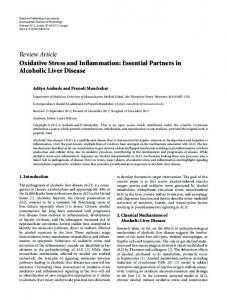
In the light of the several similar effects of cold exposure and T3 treatment on hepatic tissue, it is reasonable to think that in both conditions triiodothyronine brings about the biochemical changes leading to tissue oxidative damage. On the other hand, there are some substantial differences in the mechanisms underlying the increases in liver O2 consumption and ROS production in response to hormone administration or stimulation of thyroid activity (Fig.1). In fact, in cold-exposed rats the increase in liver respiration is due mainly to mitochondrial proliferation (Venditti et al., 2006a),
THE JOURNAL OF EXPERIMENTAL BIOLOGY
Functional hyperthyroidism and oxidative stress
2907
Fig.1. Pathway of oxidative damage in brown adipose tissue (BAT) and liver cells elicited by 3,5,3⬘-triiodothyronine (T3) treatment or cold exposure. Following thyroid hormone treatment, T3 binds to thyroid hormone receptors (TRs), which stimulate the expression of nuclear and mitochondrial (not shown) genes. Among T3-regulated genes a pivotal role is played by peroxisomal proliferator-activated receptor-g coactivator (PGC-1), which enters the nucleus and activates the expression of components of the respiratory chain. This leads, together with increased O2 consumption, to accelerated reactive oxygen species (ROS) production, not counteracted by cell antioxidant capacity (CA), and to cell oxidative damage. Following cold exposure, noradrenaline (NE), released by the sympathetic nervous system further increases PGC-1 expression leading to activation of the cAMP response element binding protein (CREB), a potent inducer of PGC-1. It is possible that this is the signal for mitochondrial proliferation, which contributes to increased ROS production. In BAT cells, the increase in uncoupling protein (UCP) level balances enhanced ROS production after T3 treatment, but not after cold exposure. T4, thyroxine. F increased; f decreased; } unchanged. + and – represent activating and inhibiting effects, respectively.
in agreement with the observation that the light mitochondrial fraction, characterized by a low content of respiratory carriers and low oxidative capacity, increases in chilled liver (Venditti et al., 2004c). Conversely, in the T3-treated rats the increase in liver respiration is due exclusively to the enhancement in mitochondrial oxidative capacity (Venditti et al., 2004c), likely linked to the higher mitochondrial content of respiratory chain components. The light mitochondrial fraction, which is also characterized by a low content of autoxidizable electron carriers, exhibits a low capacity to produce ROS (Venditti et al., 2004c). Therefore, it is understandable that the enhancement in liver ROS production, like O2 consumption, depends mainly on mitochondrial proliferation in cold-exposed animals and exclusively on the mitochondrial capacity to produce ROS in T3-treated rats. Another important difference has been shown by a study in which the effects of 10day cold exposure and T3 treatment were compared (Venditti et al., 2006a). In this study the highest levels of hydroperoxides and protein-bound carbonyls were found following T3 treatment and cold exposure, respectively. Amino acids able to generate carbonyl groups in tissue proteins can be subjected either to oxidative damage by ROS or to attack by reactive carbonyl compounds, formed by carbohydrate and fatty acid oxidation. Thus, indirect damage, resulting from carbonyl–amine reaction, can interfere with that due to direct oxidative reactions to a degree that is dependent on the extent of tissue peroxidative and glycoxidative processes. Therefore, the lower levels of protein carbonyls in T3treated rats were consistent with higher levels of hydroperoxides, Ne-(carboxymethyl)lysine and Ne-(carboxyethyl)lysine they exhibited (Venditti et al., 2006a). However, this did not clarify why differences in the extent of the lipid oxidative damage occurred in animals whose serum levels of T3 were not significantly different. The differences in the liver response to physiologically and experimentally induced serum T3 level increases suggested that other factors, whose concentrations were differentially affected by
T3 treatment and cold exposure, contributed to oxidative stress associated with experimental and functional hyperthyroidism. Serum T4 levels undergo a transitory increase, returning to values not significantly different from controls after 5days of cold exposure (Goglia et al., 1983), whereas, following T3 treatment, they fall to negligible values (Venditti et al., 2006a). T4 seems to have an intrinsic biological activity since it permits survival in the cold of hypothyroid rats without being converted to T3 (Du Breuil and Galton, 1978; Cageao et al., 1992), and is necessary for the survival of rats treated with replacement doses of T3 (Cageao et al., 1992). These results suggest T4 involvement in tissue oxidative stress associated with cold exposure. The available data show that some parameters affecting liver susceptibility to oxidative stress are differentially affected by T3 or T4 treatment. However, it is possible that this is due to other differences in the experimental protocols, such as the strain, age and sex of the animals, and the dose and duration of hormonal treatment. Therefore, the role of iodothyronines in the liver response to cold can be investigated by comparing the response with those elicited by T3 or T4 treatment in animals subjected to the same experimental conditions. On the other hand, because T4 is able to bind to nuclear receptors for T3, even though its affinity is smaller, conclusions about the role of thyroxine can be drawn only if T4 treatment gives rise to changes opposite to or greater than those produced by treatment with an equal dose of T3. Unfortunately, this is difficult to do when measuring parameters which depend on numerous factors whose relative contribution is not well defined. Thus, in recent studies, comparing the effects of 10days of T3 or T4 treatment of euthyroid rats on liver oxidative metabolism and oxidative damage (Venditti et al., 2006a; Venditti et al., 2006b), the only results suggesting differential effects of T3 and T4 concerned parameters more directly dependent on gene activity, such as protein levels. Indeed, it was found that liver GPX activity, which was not affected by cold exposure, was increased
THE JOURNAL OF EXPERIMENTAL BIOLOGY
2908 P. Venditti, L. Di Stefano and S. Di Meo by T3 and decreased by T4 treatment (Venditti et al., 2006a), whereas mitochondrial protein content was remarkably increased by cold exposure, slightly increased by T4 treatment, but not affected by T3 treatment (Venditti et al., 2006a). Furthermore, SDSPAGE analysis of protein-bound carbonyls in liver mitochondria showed oxidative damage in a 133kDa protein fraction, whose expression seemed to be related to serum T4 (Venditti et al., 2006b). Although the T4-induced changes could supply an explanation of the different effects elicited by cold exposure and T3 treatment on the above parameters, there was not enough evidence to indicate a role of T4 in the liver response to cold. However, they constituted an interesting starting point for subsequent experimental work, performed on hypothyroid rats treated with T4, whose conversion to T3 was prevented by deiodinase inhibitors. The results of this study did not support the idea that thyroxine was a factor responsible for cold-linked mitochondrial proliferation. Indeed, T4 treatment did not increase mitochondrial proteins that, conversely, were increased by cold exposure of T3-treated rats in which T4 plasma levels were low (Venditti et al., 2009). Plasma levels of catecholamines increase remarkably during cold exposure (Storm et al., 1981; Peralta et al., 2003), whereas plasma levels of noradrenaline are low or normal in hyperthyroidism and those of adrenaline are not affected by thyroid state (Stoffer et al., 1973). Thus, catecholamines constitute other factors exhibiting different plasma concentrations in functional and experimental hyperthyroidism. BAT activation by cold exposure (Foster, 1986) or noradrenaline infusion (Foster and Frydman, 1979) accounts for about 40% of increased metabolic rate. This indicates that cold-induced, noradrenaline-mediated non-shivering thermogenesis occurs in other tissues, as demonstrated by a fall in liver and muscle mitochondria respiration elicited by injection of adrenergic-receptor blockers to cold-acclimated rats (Zaninovich et al., 2003). On the other hand, in cold-exposed rats increased autooxidation of catecholamines could be a source of ROS generation, which should be lacking in hyperthyroid rats. However, these results do not demonstrate that catecholamines contribute to the cold-induced increase in liver respiration and oxidative damage through mitochondrial proliferation. To date, the available data indicate that mitochondrial proliferation requires the concomitant action of more factors and that T3 contributes to such a phenomenon in conditions, such as cold exposure, in which an additional factor is present. Indeed, it has been observed that, whereas T3 treatment of hypothyroid rats does not induce mitochondrial proliferation, 2day cold exposure of T3-treated rats induces a significant increase in mitochondrial protein content and cytochrome oxidase activity of hepatic tissue which is associated with a decrease in mitochondrial cytochrome c oxidase (CO) activity (Venditti et al., 2009). On the other hand, indirect evidence is available supporting the idea that the additional factor promoting mitochondrial proliferation could be noradrenaline. It is known that the expression of the respiratory apparatus is controlled by nuclear regulatory proteins including nuclear respiratory factors 1 and 2 (NRF-1 and NRF-2), and the peroxisomal proliferator-activated receptor-g coactivator (PGC-1). NRF-1 and NRF-2 are transcriptional factors which have been linked to the transcriptional control of many genes involved in mitochondrial function and biogenesis (Scarpulla, 2002). PGC-1 is a transcriptional coactivator which appears to play a role as an intermediary between environmental stimuli and transcriptional responses (Scarpulla, 2002). PGC-1 enhances the regulation of the UCP1 gene dependent on peroxisome proliferator-activated receptor-a (PPARa) (Barberà et al., 2001). Moreover, PGC-1
powerfully induces mRNA for NRF-1 and NRF-2 and binds to and coactivates NRF-1, and increases its transcriptional activity on target genes, including the promoter for mitochondrial transcription factor A (mtTFA), a direct regulator of mitochondrial replication and transcription (Wu et al., 1999). It has been shown that T3 triggers processes, such as mitochondrial biogenesis, adaptive thermogenesis and hepatic gluconeogenesis (Yen, 2001), which resemble those regulated by PGC-1, which, in turn, interacts with several nuclear hormone receptors including thyroid hormone receptor- (Puigserver et al., 1998). Significant increases in PGC-1 protein levels after T3 treatment (5days) were found in several tissues, including liver (Irrcher et al., 2003). Although a recent report indicates that thyroid hormone-mediated gene expression patterns are not completely dependent on PGC-1 activation (Wulf et al., 2007), the above data support the idea that a thyroid hormone-mediated activation of PGC-1 might favour tissue adaptation to endocrine signal. On the other hand, additional factors might contribute to PGC-1 activation in response to other signals, such as low environmental temperature. Indeed, PGC-1 mRNA is induced in the mouse brown fat by cold and -adrenergic agonist treatment which mimics the cold-induced sympathetic innervation of brown fat (Puigserver et al., 1998), supporting the view that PGC-1 is a transcriptional coactivator of adaptive thermogenesis. To illustrate how PGC-1 could link the external environment to mitochondrial biogenesis and gene expression, a model has been proposed in which noradrenaline, released following cold exposure, leads to the induction of PGC-1 expression by increasing cellular cAMP (Wu et al., 1999). In fact, it has been reported (Scarpulla, 2006) that cAMP is able to activate a cAMP response element binding protein (CREB), a potent inducer of PGC-1a, through phosphorylation by protein kinase A. The determination of expression levels of liver PGC-1, NRF-1 and NRF-2 proteins in hypothyroid rats, subjected for 10days to hormonal treatment (T4 or T3) or T3 treatment and 2day cold exposure, has shown that changes in PGC-1 and NRF-2 levels induced by treatments are largely matched by parallel changes in CO activity. Conversely, no relationship has been found between NRF-1 levels and CO activities, because of the unexpected fall in NRF-1 levels following cold exposure of T3-treated rats. This result disagrees with the previous observation that the biogenesis of muscle mitochondria stimulated by PGC-1 requires the function of NRF-1 (Wu et al., 1999). On the other hand, the finding that the cold-induced increase in CO activity in zebra fish muscle is associated with increased NRF-1 mRNA levels but unmodified PGC-1 mRNA levels (McClelland et al., 2006) suggests that the regulatory proteins and the mechanisms involved in mitochondrial biogenesis can be species and tissue dependent. In summary, these data supply strong indication that in rat liver PGC-1 and NRFs are responsible for the increases in mitochondrial respiratory chain components induced by thyroid hormone in experimental and functional hyperthyroidism. Further investigation needs to be carried out to clarify their possible role, as well as catecholamine involvement in cold-induced mitochondrial proliferation. LIST OF ABBREVIATIONS AA BAT CAT CoQ9 CO CREB D2 GCL
antimycin-A brown adipose tissue catalase coenzyme Q9 cytochrome c oxidase cAMP response element binding protein type 2 5⬘-iodothyronine deiodinase glutamate–cysteine ligase
THE JOURNAL OF EXPERIMENTAL BIOLOGY
Functional hyperthyroidism and oxidative stress GPX GR GSH GSSG HPs MAO MDA MDAL mtTFA NO• NOS NRF O•– 2 8-oxo-dG PGC-1 PPARa PUFA RNS ROS SOD T3 T4 TBARS UCP g-GCS g-GT
glutathione peroxidase glutathione reductase reduced glutathione glutathione disulfide hydroperoxidases monoamine oxidase malondialdehyde Ne-(malonyldialdehyde)lysine mitochondrial transcription factor A nitric oxide nitric oxide synthase nuclear respiratory factor superoxide radical anion 8-oxo-7,8-dihydro-2⬘-deoxyguanosine peroxisomal proliferator-activated receptor-g coactivator peroxisome proliferator-activated receptor-a polyunsaturated fatty acid reactive nitrogen species reactive oxygen species superoxide dismutase 3,5,3⬘-triiodothyronine thyroxine thiobarbituric acid-reactive substances uncoupling protein g-glutamylcysteine synthetase g-glutamyl-transferase
REFERENCES Aithal, H. N., Joshi, V. C. and Ramasarma, T. (1968). Effect of cold exposure on the metabolism of ubiquinone in the rat. Biochim. Biophys. Acta 162, 66-72. Alptekin, N., Seçkin, S., Dogru-Abbasoglu, S., Yelkenci, F., Koçak-Toker, N., Toker, G. and Uysal, M. (1996). Lipid peroxides, glutathione, g-glutamylcysteine synthetase, and g-glutamyltranspeptidase activities in several tissues of rats following water-immersion stress. Pharmacol. Res. 34, 167-169. Andican, G., Gelis¸gen, R., Civelek, S., Seven, A., Seymen, O., Altug, T., Yigit, G. and Burçak, G. (2004). Oxidative damage to nuclear DNA in hyperthyroid rat liver: inability of vitamin C to prevent the damage. J. Toxicol. Environ. Health A 67, 413420. Asayama, K. and Kato, K. (1990). Oxidative muscular injury and its relevance to hyperthyroidism. Free Radic. Biol. Med. 8, 293-303. Asayama, K., Dobashi, K., Hayashibe, H., Megata, Y. and Kato, K. (1987). Lipid peroxidation and free radical scavengers in thyroid dysfunction in the rat: a possible mechanism of injury to heart and skeletal muscle in hyperthyroidism. Endocrinology 121, 2112-2118. Avakian, E. V., Horvath, S. M. and Colburn, R. W. (1984). Influence of age and cold stress on plasma catecholamine levels in rats. J. Auton. Nerv. Syst. 10, 127-133. Barberà, M. J., Schluter, A., Pedraza, N., Iglesias, R., Villaroya, F. and Giralt, M. (2001). Peroxisome proliferator-activated receptor alpha activated transcription of the brown fat uncoupling protein-1 gene. A link between of the thermogenic and lipid oxidation pathways in the brown fat cell. J. Biol. Chem. 276, 1486-1493. Barja de Quiroga, G. (1992). Brown fat thermogenesis and exercise: two examples of physiological oxidative stress? Free Radic. Biol. Med. 13, 325-340. Barja de Quiroga, G., Lopez-Tores, M., Perez-Campo, R., Abelenda, M., Paz Nava, M. and Puerta, M. L. (1991). Effect of cold acclimation on GSH, antioxidant enzymes and lipid peroxidation in brown adipose tissue. Biochem. J. 277, 289-292. Beley, A., Rochette, L. and Bralet, J. (1973). Effect of treatment by thyroxine and propylthiouracil on the rate of formation of norepinephrine in eight peripheral organs of the rat. Arch. Int. Physiol. Biochim. 81, 287-298. Bianco, A. C., Maia, A. L., da Silva, W. S. and Christoffolete, M. A. (2005). Adaptive activation of thyroid hormone and energy expenditure. Biosci. Rep. 25, 191-208. Boveris, A. and Chance, B. (1973). The mitochondrial generation of hydrogen peroxide. Biochem. J. 134, 707-716. Boveris, A., Oschino, N. and Chance, B. (1972).The cellular production of hydrogen peroxide. Biochem. J. 128, 617-630. Branco, M., Ribeiro, M., Negrão, N. and Bianco, A. C. (1999). 3,5,3⬘-triiodothyronine actively stimulates UCP in brown fat under minimal sympathetic activity. Am. J. Physiol. 276, E179-E187. Bronk, J. R. (1966). Thyroid hormone: effects on electron transport. Science 153, 638639. Buzadzic, B., Korac, B. and Petrovic, V. M. (1999). The effect of adaptation to cold and re-adaptation to room temperature on the level of glutathione in rat tissues. J. Therm. Biol. 24, 373-377. Cageao, L. F., Mignone, I. R., Ricci, C. R., Brignone, C. C., Brignone, J. A. and Zaninovich, A. A. (1992). Effects of thyroid hormones on mitochondrial oxygen consumption in brown adipose tissue and heart from cold-exposed hypothyroid rats. Acta Endocrinol. 127, 72-75. Cannon, B., Alexon, S. and Nedergaard, J. (1982). Peroxisomal -oxidation in brown fat. Ann. NY Acad. Sci. 386, 40-57. Carvalho, S. D., Kimura, E. T., Bianco, A. C. and Silva, J. E. (1991). Central role of brown adipose tissue thyroxine 5⬘-deiodinase on thyroid hormone-dependent thermogenic response to cold. Endocrinology, 128, 2149-2159.
2909
Chance, B., Sies, H. and Boveris, A. (1979). Hydroperoxide metabolism in mammalian organs. Physiol. Rev. 5, 527-605. Chattopadhyay, S., Sahoo, D. K., Subudhi, U. and Chainy, G. B. (2007). Differential expression profiles of antioxidant enzymes and glutathione redox status in hyperthyroid rats: a temporal analysis. Comp. Biochem. Physiol. C Pharmacol. Toxicol. Endocrinol. 146, 383-391. Das, K. and Chainy, G. B. N. (2001). Modulation of rat liver mitochondrial antioxidant defence system by thyroid hormone. Biochim. Biophys. Acta 1537, 1-13. Davidovic, V., Djokic, I., Petrovic, N., Durasevic, S. and Cvijic, G. (1999). Activity of antioxidant enzymes in rat skeletal muscle and brown fat: effect of cold and propanolol. J. Therm. Biol. 24, 385-389. Davies, K. J. A. (1995). Oxidative stress: the paradox of aerobic life. Biochem. Soc. Symp. 61, 1-31. Davies, K. J. A. (2000). Oxidative stress, antioxidant defenses, and damage removal, repair, and replacement systems. IUBMB Life 50, 279-289. Drew, B., Phaneuf, S., Dirks, A., Selman, C., Gredilla, R., Lezza, A., Barja, G. and Leeuwenburgh, C. (2002). Effects of aging and caloric restriction on mitochondrial energy production in gastrocnemious muscle and heart. Am. J. Physiol. 284, R474R480. Du Breuil, A. and Galton, V. A. (1978). Thyroxine: studies concerning its intrinsic physiological activity. Acta Endocrinol. 88, 87-93. Fearon, I. M. and Faux, S. P. (2009). Oxidative stress and cardiovascular disease: novel tools give (free) radical insight. J. Mol. Cell. Cardiol. 47, 372-381. Fernández, V. and Videla, L. A. (1993). Influence of hyperthyroidism on superoxide radical and hydrogen peroxide production by rat liver submitochondrial particles. Free Radic. Res. Commun. 18, 329-335. Fernández, V., Barrientos, X., Kipreos, K., Valenzuela, A. and Videla, L. A. (1985). Superoxide radical generation, NADPH oxidase activity, and cytochrome P-450 content of rat liver microsomal fractions in a experimental hyperthyroid state: relation to lipid peroxidation. Endocrinology 117, 496-501. Fernández, V., Llesuy, S., Solari, L., Kipreos, K., Videla, L. A. and Boveris, A. (1988). Chemiluminescent and respiratory responses related to thyroid hormoneinduced liver oxidative stress. Free Radic. Res. Commun. 5, 77-84. Fernández, V., Simizu, K., Barros, S. B. M., Azzalis, L. A., Pimentel, R., Junqueira, V. B. C. and Videla, L. A. (1991). Effects of hyperthyroidism on rat liver glutathione metabolism: Related enzymes activities, efflux, and turnover. Endocrinology 129, 8591. Fong, K.-L., McCay, P. B., Poyer, J. L., Keele, B. B. and Misra, H. (1973). Evidence that peroxidation of lysosomal membranes is initiated by hydroxyl free radicals produced during flavin enzyme activity. J. Biol. Chem. 248, 7792-7797. Foster, D. O. (1986). Quantitative role of brown adipose tissue in thermogenesis. In Brown Adipose Tissue (ed. P. Trayurn and D. G. Nicholls), pp. 201-204. London: Edward Arnold. Foster, D. O. and Frydman, M. L. (1979). Tissue distribution of cold-induced thermogenesis in conscious warm- or cold-acclimated rats revaluated from changes in tissue blood flow: the dominant role of brown adipose tissue in the replacement of shivering by nonshivering thermogenesis. Can. J. Physiol. Pharmacol. 57, 257-270. Fridovich, I. (1995). Superoxide radical and superoxide dismutases. Annu. Rev. Biochem. 64, 97-112. Galton, V. A. and Nisula, B. C. (1969). Thyroxine metabolism and thyroid function in the cold-adapted rat. Endocrinology 85, 79-86. Goglia, F., Liverini, G., De Leo, T. and Barletta, A. (1983). Thyroid state and mitochondrial population during cold exposure. Pflügers Arch. 396, 49-53. Gredilla, R., Barja, G. and López-Torres, M. (2001). Effect of short-term caloric restriction on H2O2 production and oxidative DNA damage in rat liver mitochondria and location of the free radical sources. J. Bioenerg. Biomembr. 33, 279-287. Guernsey, D. L. and Stevens, E. D. (1977). The cell membrane sodium pump as a mechanism for increasing thermogenesis during cold acclimation in rats. Science 186, 908-910. Guerrero, A., Pamplona, R., Portero-Otin, M., Barja, G. and López-Torres, M. (1999). Effect of thyroid status on lipid composition and peroxidation in the mouse liver. Free Radic. Biol. Med. 26, 73-80. Gutteridge, J. M. C. (1995). Lipid peroxidation and antioxidants as biomarkers of tissue damage. Clin. Chem. 41, 1819-1828. Halliwell, B. (2007). Oxidative stress and cancer: have we moved forward? Biochem. J. 401, 1-11. Halliwell, B. and Gutteridge, J. M. C. (1998). Free Radicals in Biology and Medicine. Oxford: Oxford University Press. Herrero, A. and Barja, G. (1997). ADP-regulation of mitochondrial free radical production is different with complex I- or complex II-linked substrates: implications for the exercise paradox and brain hypermetabolism. J. Bioenerg. Biomembr. 29, 241249. Hodgson, E. K. and Fridovich, I. (1975). The interaction of bovine erythrocyte superoxide dismutase with hydrogen peroxide: inactivaction of the enzyme. Biochemistry 14, 5294-5298. Horrum, M. A., Tobin, R. B. and Ecklund, E. (1985). Thyroxine-induced changes in rat liver mitochondrial cytochromes. Mol. Cell. Endocrinol. 41, 163-169. Horrum, M. A., Tobin, R. B. and Ecklund, E. (1986). Thyroxine-induced changes in rat liver mitochondrial ubiquinone. Biochem. Biophys. Res. Commun. 138, 381-386. Huh, K., Kwon, T. H., Kim, J. S. and Park, J. M. (1998). Role of the hepatic xanthine oxidase in thyroid dysfunction: effect of thyroid hormones in oxidative stress in rat liver. Arch. Pharm. Res. 21, 236-249. Ikeda, S., Hamada, N., Moril, H., Inaba, M. and Yamakawa, J. (1984). Serum and tissue coenzyme Q9 in rats with thyroid dysfunction. Horm. Metab. Res. 16, 585-588. Irrcher, I., Adhihetty, P. J., Sheehan, T., Joseph, A. M. and Hood, D. A. (2003). PPARgamma coactivator-1alpha expression during thyroid hormone- and contractile activity-induced mitochondrial adaptations. Am. J. Physiol. 284, 1669-1677. Jansky, L. (1963). Body organ thermogenesis of the rat during exposure to cold and at maximal metabolic rate. Fed. Proc. 5, 1297-1302.
THE JOURNAL OF EXPERIMENTAL BIOLOGY
2910 P. Venditti, L. Di Stefano and S. Di Meo Just, W. W., Hartl, F. U. and Schimassk, H. (1982). Rat liver peroxisomes. I. New peroxisome population induced by thyroid hormones in the liver of male rats. Eur. J. Cell Biol. 26, 249-254. Kaplowitz, N., Aw, T. Y. and Ookhtens, M. (1985). The regulation of hepatic glutathione. Annu. Rev. Pharmacol. Toxicol. 25, 715-744. Kaushik, S. and Kaur, J. (2003). Chronic cold exposure affects the antioxidants defense system in various rat tissues. Clin. Chim. Acta 333, 69-77. Kikuchi-Utsumi, K., Gao, B., Ohinata, H., Hashimoto, M., Yamamoto, N. and Kuroshima, A. (2002). Enhanced gene expr ession of endothelial nitric oxide synthase in brown adipose tissue during cold exposure. Am. J. Physiol. 282, R623R626. Knowles, R. G. and Moncada, S. (1994). Nitric oxide synthases in mammals. Biochem. J. 298, 249-258. Kolosova, N. G., Kolpakov, A. R. and Panin, L. E. (1995). Tocopherol level and lipid peroxidation in Wistar rat tissues during adaptation to cold. Vopr. Med. Khim. 41, 1619. Lambert, A. J., Wang, B., Yardley, Y., Edwards, J. and Merry, B. J. (2004). The effect of aging and caloric restriction on mitochondrial protein density and oxygen consumption. Exp. Gerontol. 39, 289-295. Larkin, S., Mull, E., Miao, W., Pittner, R., Albrandt, K., Moore, C., Young, A., Denaro, M. and Beaumont, K. (1997). Regulation of the third member of the uncoupling protein family, UCP3, by cold and thyroid hormone. Biochem. Biophys. Res. Commun. 240, 222-227. Lomakina, L. V. (1980). Effects of low temperature on lipid peroxidation and intensity of proteolysis in rat brain and liver. Ukr. Biochim. Zh. 52, 305-308. Loschen, G., Flohé, L. and Chance, B. (1971). Respiratory chain linked H2O2 production in pigeon heart mitochondria. FEBS Lett. 18, 261-264. Lu, R., Nash, H. M. and Verdine, G. L. A. (1997). A mammalian DNA repair enzyme that excises oxidatively damaged guanines maps to a locus frequently lost in lung cancer. Curr. Biol. 7, 397-407. McCarter, R. J., Masoro, E. J. and Yu, B. P. (1985). Does food restriction retard aging by reducing the metabolic rate? Am. J. Physiol. 248, E488-E490. McClelland, G. B., Craig, P. M., Dhekney, K. and Dipardo, S. (2006). Temperatureand exercise-induced gene expression and metabolic enzyme changes in skeletal muscle of adult zebrafish (Danio rerio). J. Physiol. 577, 739-751. Mileva, M., Bakalova, R., Tancheva, L. and Galabov, A. S. (2002). Effect of immobilation, cold and cold-restraint stress on liver monooxygenase activity and lipid peroxidation of influenza virus-infected mice. Arch. Toxicol. 76, 96-103. Morini, P., Casalino, E., Sblano, C. and Landriscina, C. (1991). The response of rat liver lipid peroxidation, antioxidant enzyme activities and glutathione concentration to the thyroid hormone. Int. J. Biochem. 23, 1025-1030. Mory, G., Bal, D. and Ricquier, D. (1983). Parallel increase of ascorbic acid and glutathione contents in brown adipose tissue during chronic cold exposure. Experientia 39, 734-735. Mory, G., Gawer, M. and Kader, J.-C. (1988). Effect of noradrenaline chronic administration on brown fat phospholipids. Biosci. Rep. 8, 465-469. Nègre-Salvayre, A., Hirtz, C., Carrera, G., Cazenave, R., Troly, M., Salvayre, R., Pénicaud, L. and Casteilla, L. (1997). A role for uncoupling protein-2 as a regulator of mitochondrial hydrogen peroxide generation. FASEB J. 11, 809-815. Nejad, I. F., Bollinger, J. A., Mitnich, M. and Reichlin, S. (1972). Importance of T3 (triiodothyronine) secretion in altered states of thyroid function in the rat: cold exposure, subtotal thyroidectomy, and hypophysectomy. Trans. Assoc. Am. Physicians 85, 295-308. Nishiki, K., Erecinska, M., Wilson, D. F. and Cooper, S. (1978). Evaluation of oxidative phosphorylation in hearts from euthyroid, hypothyroid and hyperthyroid rats. Am. J. Physiol. 235, C212-C219. North, J. A., Spector, A. A. and Buettner, G. R. (1994). Cell fatty acid composition affects free radical formation during lipid peroxidation. Am. J. Physiol. 267, C177C188. Ogawa, K., Ohno, T. and Kuroshima, A. (1987). Muscle and brown adipose tissue fatty acid profiles in cold-exposed rats. Jpn. J. Physiol. 37, 783-796. Oppenheimer, J. H. (1999). Evolving concepts of thyroid hormone action. Biochimie 81, 539-543. Pacifici, R. E. and Davis, K. J. A. (1990). Protein degradation as an index of oxidative stress. Methods Enzymol. 186, 485-502. Pamplona, R., Portero-Otín, M., Ruiz, C., Bellmunt, M. J., Requena, J. R., Thorpe, S. R., Baynes, J. W., Romero, M., López-Torres, M. and Barja, G. (1999). Thyroid status modulates glycoxidative and lipoxidative modifications of tissue proteins. Free Radic. Biol. Med. 27, 901-910. Peralta, J. G., Finocchietto, P. V., Converso, D., Schöpfer, F., Carreras, M. C. and Poderoso, J. J. (2003). Modulation of mitochondrial nitric oxide synthase and energy expenditure in rats during cold acclimation. Am. J. Physiol. 284, H2375H2383. Petrovic, N., Cvijic, G. and Davidovic, V. (2003). Thyroxine and tri-iodothyronine differently affect uncoupling protein-1 content and antioxidant enzyme activities in rat interscapular brown adipose tissue. J. Endocrinol. 176, 31-38. Petrovic, V. M., Spasic, M. B., Saicic, Z. S., Milic, B. and Radojicic, R. (1987). Superoxide dismutase activity in the cytosol of brown adipose tissue and liver during an acute and chronic exposure of rats to cold. Bull. Acad. Serb. Sci. 27, 99-108. Petrovic, V. M., Saicic, Z. S., Radojicic, R., Buzadzic, B. and Spasic, M. B. (1989). Difference in the inducibility between copper zinc containing superoxide dismutase and manganese containing superoxide dismutase in brown adipose tissue of the rat exposed or adapted to cold; correlation with tissue hyperplasia. Iugoslav. Physiol. Pharmacol. Acta 25, 33-38. Petrovic, V., Korac, A., Buzadzic, B., Vasilijevic, A., Jankovic, A., Micunovic, K. and Korac, B. (2008). Nitric oxide regulates mitochondrial re-modelling in interscapular brown adipose tissue: ultrastructural and morphometric-stereologic studies. J. Microsc. 232, 542-548. Pigeolet, E., Corbisier, P., Houbion, A., Lambert, D., Michiels, C., Raes, M., Zachary, M. D. and Remacle, J. (1990). Glutathione peroxidase, superoxide
dismutase, and catalase inactivation by peroxides and oxygen-derived free radicals. Mech. Ageing Dev. 51, 283-296. Poli, G., Leonarduzzi, G., Biasi, F. and Chiarpotto, E. (2004). Oxidative stress and cell signalling. Curr. Med. Chem. 11, 1163-1182. Prange, A. J., Jr, Meek, J. L. and Lipton, M. A. (1970). Catecholamines: diminished rate of synthesis in rat brain and heart after thyroxine pretreatment. Life Sci. 9, 901907. Puigserver, P., Wu, Z., Park, C. W., Graves, R., Wright, M. and Spiegelman, B. M. (1998). A cold-inducible coactivator of nuclear receptors linked to adaptive thermogenesis. Cell 92, 829-839. Radi, R., Cassina, A., Hodara, R., Quijana, C. and Castro, L. (2002). Peroxynitrite reactions and formation in mitochondria. Free Radic. Biol. Med. 33, 1451-1464. Ramasarma, T. (1982). Generation of H2O2 in biomembranes. Biochim. Biophys. Acta 649, 69-93. Richter, C., Park, J. W. and Ames, B. N. (1988)Normal oxidative damage to mitochondrial and nuclear DNA is extensive. Proc. Natl. Acad. Sci. USA 85, 64656467. Sacher, G. A. (1977). Life table modification and life prolongation. In Handbook of the Biology of Aging (ed. C. E. Finch and L. Hajflick), pp. 582-638. New York: Van Nostrand Reinhold. Saicic, Z. S., Mijalkovic, D. N., Nikolic, A. L., Blagojevic, D. P. and Spasic, M. B. (2006). Effect of thyroxine on antioxidant defense system in the liver of different aged rats. Physiol. Res. 55, 561-568. Scarpulla, R. C. (2002). Nuclear activators and coactivators in mammalian mitochondrial biogenesis. Biochim. Biophys. Acta 1576, 1-14. Scarpulla, R. C. (2006). Nuclear control of respiratory gene expression in mammalian cells. J. Cell. Biochem. 97, 673-683. Sekhar, B. S., Kurup, K. R. and Ramasarma, T. (1987). Generation of hydrogen peroxide by brown adipose tissue. J. Bioenerg. Biomembr. 19, 397-407. Sekhar, B. S., Kurup, K. R. and Ramasarma, T. (1990). Microsomal redox systems in brown adipose tissue: High lipid peroxidation, low cholesterol biosynthesis and no detectable cytochrome P-450. Mol. Cell. Biochem. 92, 147-157. Selman, C., Grune, T., Stolzing, A., Jakstadt, M., McLaren, J. S. and Speakman, J. R. (2002). The consequences of acute cold exposure on protein oxidation and proteasome activity in short-tailed field vole, Microtus agrestis. Free Radic. Biol. Med. 33, 259-265. Shigenaga, M. K., Park, J.-W., Kundy, K. C., Gimeno, C. J. and Ames, B. N. (1990). In vivo oxidative DNA damage: measurement of 8-hydroxy-2’deoxyguanosine in DNA and urine by high-performance liquid chromatography with electrochemical detection. Methods Enzymol. 186, 521-530. Sies, H. (1993). Strategies of antioxidant defense. Eur. J. Biochem. 215, 213-219. Sies, H. (1997). Oxidative stress: oxidants and antioxidants. Exp. Physiol. 82, 291-295. Sies, H. and Graf, P. (1985). Hepatic thiol and glutathione efflux under the influence of vasopressin, phenylephrine and adrenaline. Biochem. J. 226, 545-549. Silva, J. E. (2001). The multiple contributions of thyroid hormone to heat production. J. Endocrinol. Invest. 108, 35-37. Silva, J. E. and Larsen, P. R. (1983). Adrenergic activation of triiodothyronine production in brown adipose tissue. Nature 305, 712-713. Silva, J. E. and Larsen, P. R. (1986). Interrelationships among thyroxine, growth hormone, and the sympathetic nervous system in the regulation of 5⬘-iodothyronine deiodinase in rat brown adipose tissue. J. Clin. Invest. 77, 1214-1223. Spasic, M. B., Saicic, Z. S., Buzadzic, B., Korac, B., Blagojevic, D. and Petrovic, V. M. (1993). Effect of long-term exposure to cold on the antioxidant defense system in the rat. Free Radic. Biol. Med. 15, 291-299. Stoffer, S. S., Jiang, N. S., Gorman, C. A. and Picker, G. M. (1973). Plasma catecholamines in hypothyroidism and hyperthyroidism. J. Clin. Endocrinol. Metab. 36, 587-589. Storm, H., van Hardelved, C. and Kassenaar, A. A. H. (1981). Thyroid hormonecatecholamines interrelationship during exposure to cold. Acta Endocrinol. 97, 91-97. Swaroop, A. and Ramasarma, T. (1985). Heat exposure and hypothyroid conditions decrease hydrogen peroxide generation in liver mitochondria. Biochem. J. 226, 403408. Tapia, G., Cornejo, P., Fernández, V. and Videla, L. A. (1999). Protein oxidation in thyroid hormone-induced liver oxidative stress: relation to lipid peroxidation. Toxicol. Lett. 106, 209-214. Tnimov, M. Kh., Semeniuk, A. V., Voronina, N. P., Shishkina, L. N. and Maianskii, D. N. (1984). Effect of low temperature on lipid peroxidation in the lungs and on pulmonary macrophage activity in rats. Biull. Eksp. Biol. Med. 98, 591-593. Tsang, A. H. and Chung, K. K. (2009). Oxidative and nitrosative stress in Parkinson’s disease. Biochim. Biophys. Acta 1792, 643-650. Turrens, J. F. and Boveris, A. (1980). Generation of superoxide anion by the NADH dehydrogenase of bovine heart mitochondria. Biochem. J. 191, 421-427. Tzagoloff, A. (1982). Mitochondria. Plenum Press, New York. Venditti, P. and Di Meo, S. (2006). Thyroid hormone-induced oxidative stress. Cell. Mol. Life Sci. 63, 414-434. Venditti, P., Balestrieri, M., Di Meo, S. and De Leo, T. (1997). Effect of thyroid state on lipid peroxidation, antioxidant defences, and susceptibility to oxidative stress in rat tissues. J. Endocrinol. 155, 151-157. Venditti, P., Daniele, M. C., Masullo, P. and Di Meo, S. (1999a). Antioxidantsensitive triiodothyronine effects on characteristics of rat liver mitochondrial population. Cell. Physiol. Biochem. 9, 38-52. Venditti, P., Masullo, P. and Di Meo, S. (1999b). Effect of training on H2O2 release by mitochondria from rat skeletal muscle. Arch. Biochem. Biophys. 372, 315-320. Venditti, P., Masullo, P. and Di Meo, S. (2001). Hemoproteins affects H2O2 removal from rat tissues. Int. J. Biochem. Cell Biol. 33, 293-301. Venditti, P., Costagliola, I. R. and Di Meo, S. (2002). H2O2 production and response to stress conditions by mitochondrial fractions from rat liver. J. Bioenerg. Biomembr. 34, 115-125. Venditti, P., De Rosa, R. and Di Meo, S. (2003a). Effect of thyroid state on H2O2 production by rat liver mitochondria. Mol. Cell. Endocrinol. 205, 185-192.
THE JOURNAL OF EXPERIMENTAL BIOLOGY
Functional hyperthyroidism and oxidative stress Venditti, P., Puca, A. and Di Meo, S. (2003b). Effect of thyroid state on rate and sites of H2O2 production in rat skeletal muscle mitochondria. Arch. Biochem. Biophys. 411, 121-128. Venditti, P., Puca, A. and Di Meo, S. (2003c). Effects of thyroid state on H2O2 production by rat heart mitochondria: sites of production with Complex I and Complex II-linked substrates. Horm. Metab. Res. 35, 55-61. Venditti, P., De Rosa, R. and Di Meo, S. (2003d). Effect of thyroid state on susceptibility to oxidants and swelling of mitochondria from rat tissues. Free Radic. Biol. Med. 35, 485-494. Venditti, P., De Rosa, R., Portero-Otin, M., Pamplona, R. and Di Meo, S. (2004a). Cold-induced- hyperthyroidism produces oxidative damage in rat tissues and increases susceptibility to oxidants. Int. J. Biochem. Cell Biol. 36, 1319-1331. Venditti, P., De Rosa, R. and Di Meo, S. (2004b). Effect of cold-induced hyperthyroidism on H2O2 production and susceptibily to stress conditions of rat liver mitochondria. Free Radic. Biol. Med. 36, 348-358. Venditti, P., De Rosa, R., Caldarone, G. and Di Meo, S. (2004c). Functional and biochemical characteristics of mitochondrial fractions from rat liver in cold-induced oxidative stress. Cell. Mol. Life Sci. 61, 3104-3116. Venditti, P., Pamplona, R., Ayala, V., De Rosa, R., Caldarone, G. and Di Meo, S. (2006a). Differential effects of experimental and cold-induced hyperthyroidism on factors inducing rat liver oxidative damage. J. Exp. Biol. 209, 817-825. Venditti, P., Pamplona, R., Portero-Otin, M., De Rosa, R. and Di Meo, S. (2006b). Effect of experimental and cold exposure induced hyperthyroidism on H2O2 production and susceptibility to oxidative stress of rat liver mitochondria. Arch. Biochem. Biophys. 447, 11-22. Venditti, P., Bari, A., Di Stefano, L., Cardone, A., Della Ragione, F., D’Esposito, M. and Di Meo, S. (2009). Involvement of PGC-1, NRF-1, and NRF-2 in metabolic
2911
response by rat liver to hormonal and environmental signals. Mol. Cell. Endocrinol. 305, 22-29. Vidal-Puig, A. J., Grujic, D., Zhang, C. Y., Hagen, T., Boss, O., Ido, Y., Szczepanik, A., Wade, J., Mootha, V., Cortright, R. et al. (2000). Energy metabolism in uncoupling protein 3 gene knockout mice. J. Biol. Chem. 275, 1625816266. Videla, L. A. (2000). Energy metabolism, thyroid calorigenesis, and oxidative stress: Functional and cytotoxic consequences. Redox Rep. 5, 265-275. Wu, Z., Puigserver, P., Andersson, U., Zhang, C., Adelmant, G., Mootha, V., Troy, A., Cinti, S., Lowell, B., Scarpulla, R. C. and Spiegelman, B. M. (1999). Mechanisms controlling mitochondrial biogenesis and function through the thermogenic coactivator PGC-1. Cell 98, 115-124. Wulf, A., Harneit, A. and Weitzel, J. M. (2007). T3-mediated gene expression is independent of PGC-1alpha. Mol. Cell. Endocrinol. 270, 57-63. Yamaguchi, R., Hirano, T., Asami, S., Sugita, A. and Kasai, H. (1996). Increase in 8-hydroxyguanine repair activity in the rat kidney after the administration of a renal carcinigen, ferric nitroloacetate. Environ. Health Perspec. 104. 651-653. Yen, P. M. (2001). Physiological and molecular basis of thyroid hormone action. Physiol. Rev. 81, 1097-1142. Younes, M., Schlichting, R. and Siegers, C. P. (1980). Glutathione S-transferase activities in rat liver: effect of some factors influencing the metabolism of xenobiotics. Pharmacol. Res. Commun. 12, 115-129. Yu, B. P. (1994). Cellular defenses against damage from reactive oxygen species. Physiol. Rev. 74, 139-162. Zaninovich, A. A., Rebagliati, I., Raíces, M., Ricci, C. and Hagmüller, K. (2003). Mitochondrial respiration in muscle and liver from cold-acclimated hypothyroid rats. J. Appl. Physiol. 95, 1584-1590.
THE JOURNAL OF EXPERIMENTAL BIOLOGY