IOP PUBLISHING
SCIENCE AND TECHNOLOGY OF ADVANCED MATERIALS
Sci. Technol. Adv. Mater. 14 (2013) 025003 (8pp)
doi:10.1088/1468-6996/14/2/025003
Rigid microenvironments promote cardiac differentiation of mouse and human embryonic stem cells Armin Arshi1 , Yasuhiro Nakashima1 , Haruko Nakano1,2 , Sarayoot Eaimkhong3,4 , Denis Evseenko5,6,7 , Jason Reed4 , Adam Z Stieg4,8 , James K Gimzewski3,4,6,8 and Atsushi Nakano1,2,6,7,9 1
Department of Molecular, Cell and Developmental Biology, University of California, Los Angeles, CA 90095, USA 2 Eli and Edythe Broad Center of Regenerative Medicine and Stem Cell Research, University of California, Los Angeles, CA 90095, USA 3 Department of Chemistry and Biochemistry, University of California, Los Angeles, CA 90095, USA 4 California NanoSystems Institute, University of California, Los Angeles, CA 90095, USA 5 Department of Orthopaedic Surgery, University of California, Los Angeles, CA 90095, USA 6 Jonsson Comprehensive Cancer Center, University of California, Los Angeles, CA 90095, USA 7 Molecular Biology Institute, University of California, Los Angeles, CA 90095, USA 8 WPI Center for Materials Nanoarchitectonics (MANA), National Institute for Materials Science (NIMS), Tsukuba, Ibaraki 305-0044, Japan E-mail:
[email protected]
Received 24 February 2013 Accepted for publication 21 March 2013 Published 11 April 2013 Online at stacks.iop.org/STAM/14/025003 Abstract While adult heart muscle is the least regenerative of tissues, embryonic cardiomyocytes are proliferative, with embryonic stem (ES) cells providing an endless reservoir. In addition to secreted factors and cell–cell interactions, the extracellular microenvironment has been shown to play an important role in stem cell lineage specification, and understanding how scaffold elasticity influences cardiac differentiation is crucial to cardiac tissue engineering. Though previous studies have analyzed the role of matrix elasticity on the function of differentiated cardiomyocytes, whether it affects the induction of cardiomyocytes from pluripotent stem cells is poorly understood. Here, we examine the role of matrix rigidity on cardiac differentiation using mouse and human ES cells. Culture on polydimethylsiloxane (PDMS) substrates of varied monomer-to-crosslinker ratios revealed that rigid extracellular matrices promote a higher yield of de novo cardiomyocytes from undifferentiated ES cells. Using a genetically modified ES system that allows us to purify differentiated cardiomyocytes by drug selection, we demonstrate that rigid environments induce higher cardiac troponin T expression, beating rate of foci, and expression ratio of adult α- to fetal β- myosin heavy chain in a purified cardiac population. M-mode and mechanical interferometry image analyses demonstrate that these ES-derived cardiomyocytes display functional maturity and synchronization of beating when co-cultured with neonatal cardiomyocytes harvested from a developing embryo. Together, these data identify matrix stiffness as an independent factor that instructs not only the maturation of already differentiated cardiomyocytes but also the
9
Present address: Biomedical Sciences Research Building, Room # 490D, 615 Charles E Young Drive South, Los Angeles, CA 90095, USA. Content from this work may be used under the terms of the Creative Commons Attribution-NonCommercial-ShareAlike 3.0 licence. Any further distribution of this work must maintain attribution to the author(s) and the title of the work, journal citation and DOI.
1468-6996/13/025003+08$33.00
1
© 2013 National Institute for Materials Science
Printed in the UK
Sci. Technol. Adv. Mater. 14 (2013) 025003
A Arshi et al
induction and proliferation of cardiomyocytes from undifferentiated progenitors. Manipulation of the stiffness will help direct the production of functional cardiomyocytes en masse from stem cells for regenerative medicine purposes. Keywords: cardiac differentiation pluripotent embryonic stem cell matrix elasticity,
drug-selected cardiomyocyte, synchronization, mechanical interferometry S Online supplementary data available from stacks.iop.org/STAM/14/025003/mmedia
monitored for emergence of the cardiac lineage, and scored for cardiomyocyte activity using several quantitative methods. In all cases, the growth and lineage specification of mouse and human pluripotent stem cells showed a relative dependence on substrate elasticity, whereby the highest yield of cardiomyocytes occurred under the rigid microenvironmental conditions of the standard tissue culture plate. This effect was also observed in the culture of a more select population of ES-derived cardiomyocytes, which was purified using a genetic drug selection system [13]. Together, such findings give insight into the role of the gradually stiffening physical microenvironment in cardiac development and the potential uses of ES and iPS cells for tissue engineering purposes.
1. Introduction Stem cell lineage specification is regulated by three types of inductive signal: soluble factors secreted by cells that diffuse through extracellular fluid, cell–cell interactions that vary within a complex multicellular organism, and extracellular matrix (ECM). Soluble growth factors and cell membrane-bound factors such as PDGF, TGF-β, VEGF, BMP and Notch signaling have been implicated as having multivariable effects on cardiac development [1–3]. Insoluble ECM proteins are also known to affect the microenvironmental cues during cardiovascular development [4, 5]. Such developmental cues have been intensely exploited in traditional tissue culture methods to simulate a biological process in vitro. However, recent work has also suggested the relevance of a fourth factor—interactions between the stem cell and its physical microenvironment. In the living organism, adult stem cells, as part of regenerative or developmental processes are believed to egress away from their native environment and embed themselves within a range of tissue microenvironments to facilitate maturation [6]. For example, na¨ıve mesenchymal stem cells (MSCs) show extreme sensitivity to tissue-level elasticity when cultured under various microenvironments [7]. The cell lineages differentiating from MSCs is known to reflect the extracellular microenvironment in terms of elasticity; soft matrices mimicking brain tissue and rigid matrices resembling bone, for example, are neurogenic and osteogenic, respectively. While several studies have discerned how the physical microenvironment influences the phenotype and behavior of mature cardiomyocytes [8–11], little is known about its role in determining the cardiac lineage from an undifferentiated state. Here, we investigated the role of matrix elasticity on cardiac lineage specification from undifferentiated pluripotent stem cells. These forces are quantified by the elastic modulus (E). While some reported the typical range expected for high-yield myogenic lineage specification to be between 1 and 20 kPa [12], others have reported that neonatal cardiomyocytes demonstrate the greatest contractile function on very rigid matrices [9, 10]. To recapitulate the varied matrix elasticities of these extracellular environments, we coated standard tissue-culture plates with polydimethylsiloxane (PDMS), an elastic and chemically inert siloalkane polymer, of synthetically tunable stiffness. Embryoid bodies (EBs) representative of in vivo growth conditions were plated onto these synthetic substrates,
2. Materials and methods 2.1. Elastic substrate synthesis PDMS substrates of 0.5 cm thickness were created in standard six-well tissue culture plates according to the manufacturer’s specifications (Dow Corning). Briefly, various ratios of PDMS base-to-crosslinking agent (either 10:1 or 50:1) were mixed to alter the substrates’ elastic modulus, degassed for 30 min to prevent bubble formation, and cured at 65 ◦ C for 3 h. The PDMS was then soaked in molecular biology grade 95% ethanol overnight to extract unwanted siloxane monomers. Both PDMS and standard tissue culture substrates were treated with oxygen plasma for 30 s for sterilization and uniform surface modification. Elastic moduli (E) were determined before and after 2D culture in liquid media using plate-to-plate rheometry. 2.2. Mouse ES cell culture and differentiation Undifferentiated wild type mouse E14Tg2a (ATCC) cells and N kx2-5neoR/+ mouse ES [13] cells were maintained on mouse embryonic fibroblast feeder cells in high-glucose Dulbecco’s modified Eagle medium (DMEM, Gibco) supplemented with 15% fetal calf serum (FCS), 1×non-essential amino acids (Invitrogen), 25 mmol L−1 HEPES, 2 mmol L−1 l-glutamine, 0.1 mg ml−1 penicillin/streptomycin, 0.1 mmol L−1 – 1 2-mercaptoethanol and 1000 U ml leukemia inhibitory factor (LIF, ESGRO-TM, Chemicon International, Inc.). ES cells were differentiated in hanging drops (600 cells per 20 µl−1 ) without LIF under non-adherent conditions. E14Tg2a wild type (day 2) and N kx2-5neoR/+ (day 5) embryoid bodies (EBs) were plated onto PDMS and standard 2
Sci. Technol. Adv. Mater. 14 (2013) 025003
A Arshi et al
tissue culture dishes coated with 10 µg ml–1 fibronectin (Sigma). Media were changed every 2 days during adherent culture. N kx2-5neoR/+ EBs were purified by adding G418 neomycin antibiotic (600 µg ml–1 ) for 6 days.
chain (β-MHC) was performed on cDNA reverse transcribed from mRNA collected from EB cultures at day 10 for wild type cells or day 16 for Nkx2-5neoR/+ cells. qRT-PCR was performed using the following primer designs: Mouse cTnT: forward: 50 GGAAATAGATGAAACTGTC; reverse: 50 GCTCCCACTATCCAAAC, Mouse α-MHC: forward: 50 GAGATTTCTCCAACCCAG; reverse: 50 TCTGACTTTCGGAGGTACT, Mouse β-MHC: forward: 50 CTACAGGCCTGGGCTTACC; reverse: 50 TCTCCTTCTCAGACTTCCGC, Human cTnT: forward: 50 CAGAGCGGAAAAGTGGGAAGA; reverse: 50 TCGTTGATCCTGTTTCGGAGA.
2.3. Human ES cell culture and differentiation Human H1, H9 (WiCell) and UCLA1-6 (UCLA stem cell core) [14] ES cells were grown on mouse embryonic fibroblast feeder in DMEM (Gibco) media supplemented with 20% knockout (KO) serum replacement (Gibco) and 8 ng ml–1 bFGF (Sigma). EBs were differentiated according to previous reports with minor modification [15]. Briefly, human ES cells were dissociated with TrypLE Select (Invitrogen) for 5 min and 20 000 human ES cells were differentiated in StemPro 34 (Gibco) supplemented with 10 µM Y27632 (Rho kinase inhibitor IV, EMD Chemicals), 3 ng ml−1 Activin A (Peprotech), 5 ng ml−1 bFGF, 10 ng ml−1 BMP4 (Peprotech) and 5 µM IWR-1 (EMD Chemicals), for the first 4 days. EBs were then cultured in StemPro 34 supplemented with 10 ng ml−1 bFGF, 5 ng ml–1 vascular endothelial growth factor (VEGF, Peprotech) and 5 µM IWR-1 for 5 additional days before plating onto elastic substrates. Usage of all human ES cell lines is approved by the UCLA Embryonic Stem Cell Research Oversight (ESCRO) Committee and the Institutional Review Boards (IRB, approval #2009-006-04).
All expression values were quantitatively normalized by expression of the GAPDH housekeeping gene. Relative expression values were calculated using the delta–delta Ct method [17]. 2.6. Transplantation of ES-derived cardiac foci After culture and drug selection until day 16, EBs were washed with trypsin for 2 min to facilitate dissociation from the substrate surface and transferred into a small conical tube. Light centrifugation was performed for 5 min until individual EBs were reseeded onto a new layer of neonatal cardiomyocytes that were in culture for 5 days in ES media without LIF and neomycin.
2.4. Preparation of neonatal cardiomyocytes
2.7. Analysis of cardiac synchronization
Neonatal hearts were dissected, washed in HBSS, and bathed in 0.5 mg ml−1 trypsin-HBSS overnight at 4 ◦ C for pre-digestion as previously described [16]. The heart tissues were then washed in warm medium and digested with 0.5 mg ml−1 type II collagenase (Worthington Corp.) in HBSS for 10 min at 37 ◦ C for four times. Dissociated cells then underwent two rounds of differential plating onto plastic tissue culture dishes for 1 h at 37 ◦ C and 5% CO2 to remove adherent non-cardiomyocytes. Non-adherent cardiomyocytes were then plated onto tissue culture dishes coated with 10 µg ml–1 fibronectin at 75% confluency prior to co-culture with ES-derived cardiomyocytes. All experiments were performed following the guidelines and protocol approved by the Animal Research Committee for care and use of laboratory animals (Animal Research Committee approval #2008–143-11B).
Synchronization between ES-derived and neonatal cardiomyocytes post-transplantation was examined both by M-mode image analysis and mechanical interferometry imaging (MII) [18]. M-mode image analysis was performed using customized software [19] on videos of beating cardiomyocytes in culture obtained using a Sony HDR-SR11 camcorder. MII was performed by culturing cells on silicon substrates and subsequent observation under a Michelson interferometer with an adjustable mirror in the reference arm to permit measurements in the media-filled observation chamber. Areas of interest were selected and analyzed for vertical motion through changes in interference pattern using a customized image-processing algorithm using MATLAB.
3. Results 3.1. PDMS substrates model physical microenvironments of varied matrix elasticity
2.5. Quantifying cardiac proliferation and function Single cells were isolated from EB culture via trypsinization, fixed with 0.5% PFA, and analyzed for expression of cardiac troponin T (cTnT). The relative abundance of cells was quantified with by FACS analysis using BD∗ LSR FACS Diva for PE-conjugated rabbit anti-mouse cTnT primary antibody. Extracellular staining was performed by blocking the cells with a 0.1% saponine 3% FCS/PBS buffer, labeled with a 1:500 dilution of rabbit anti-cTnT antibody, and conjugated with 1:1000 dilution of anti-rabbit PE antibody. mRNA transcription profiles for cardiac troponin T (cTnT), alpha myosin heavy chain (α-MHC), and beta myosin heavy
While several elastomeric substrates have been used for the analyses of the role of matrix elasticity of the behavior of the cells, we selected PDMS for its modifiable surface properties, ease of synthesis, biocompatibility with mammalian cell culture, and established use [20, 21]. PDMS substrates were synthesized on six-well tissue culture plates according to manufacturer’s instructions. Scaffolds of varying stiffness were created using different ratios of the PDMS base-to-curing agent, rinsed with ethanol overnight to extract siloxane monomers, and O2 plasma 3
Sci. Technol. Adv. Mater. 14 (2013) 025003
A Arshi et al
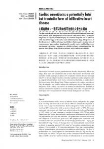
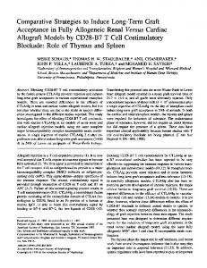
Figure 1. Experimental design for ES stiffness culture. (A) Elastic modulus of PDMS substrates of varying base-to-curing-agent ratio were rheometrically characterized before and after culture in liquid media relative to 3 GPa standard TC plate. ES cells are formed into EBs via a hanging drop method, marking the onset of differentiation and (B) landed onto substrates. (C) EBs show similar adhesion profile to PDMS and tissue culture plates. All culture conditions show greater than 85% adhesion of individual EBs with no statistically significant difference between groups ( p > 0.05).
treated for uniform surface chemistry prior to culture. We related the PDMS base-to-curing agent ratio to the elastic modulus (E) both before and after cell culture in liquid media using rheometry, and a nonlinear relationship was established in accord with previously reported data [22]. Only substrates corresponding to an elastic modulus of ∼1000 (10:1 PDMS base-to-crosslinking agent) and ∼10 kPa (50:1 PDMS base-to-crosslinking agent) were used for experiments (figure 1(A)). Standard polystyrene tissue culture (TC) plates were used to recapitulate a high stiffness environment, with an elastic modulus on the order of 3 GPa [9]. Mouse EBs were then landed onto 2D culture environments for differentiation after several days (figure 1(B)). Although the PDMS substrates are hydrophobic, fibronectin coating appeared to mitigate this effect, as each condition showed a similar adhesion profile (figure 1(C)). Moreover, EBs showed comparable morphologies and differentiation profiles when cultured on either PDMS or standard TC plates.
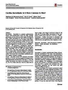
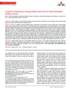
at which point cells were dissociated and collected for further analysis. EBs adherent to PDMS substrates started to spontaneously beat and express cTnT with clear striation patterns at day 8 (figure 2(B)). To quantitatively determine the relative number of cardiomyocytes, fluorescence-activated cell sorting (FACS) analysis was performed on cultured cells fluorescently stained for intracellular expression of cTnT (figure 2(C)). EBs cultured on substrates softer than the traditional tissue culture plates yielded fewer cTnT+ cells than those grown on standard, highly-stiff culture plates (figure 2(D)). Consistently, qRT-PCR showed the highest cTnT expression relative to the GAPDH housekeeping gene on a rigid substrate (figure 2(E)). A previous study reported that fully-differentiated murine cardiomyocytes show enhanced functional maturation in beating rate and contractile force on an extremely rigid substrate [9]. Our data further demonstrate that a rigid environment enhances the induction of cardiomyocytes from undifferentiated ES cells. We next examined whether matrix stiffness also regulates the cardiac differentiation of human ES cells. As human ES cell lines display tremendous variability in their lineage differentiation capabilities, we first tested eight human ES cell lines for their cardiac differentiation efficiency. H1, H9 and UCLA1-6 human ES cell lines were differentiated in EBs using Wnt inhibitor [15], and cardiac differentiation was examined at day 15 by FACS analysis for the expression of cardiac specific myosin using MF20 antibody. Among human ES cell lines tested, H9 and UCLA4 consistently showed efficient cardiac induction (figures 2(F), and (G)), and hence the effect of the matrix rigidity was examined using these two cell lines. Cardiomyocytes were induced from H9 and UCLA4 EBs using Wnt inhibitor [15] for 5 days and plated on matrices of varied rigidity for 10 days. qRT-PCR showed that
3.2. Rigid substrates promote induction of cardiomyocytes from an undifferentiated state Using this culture setup, we sought to quantify the effect of the stiffness of the extracellular matrix on cardiac lineage specification of undifferentiated ES cells. With our hands, mouse ES cells start to differentiate into cardiac, vascular and hematopoietic lineage as early as day 4 after formation of EBs in hanging drop culture [23, 24]. At 2 days of differentiation, when mouse ES cells are not yet committed to cardiac lineage, EBs were plated onto fibronectin-coated 2D substrates of varying stiffness: the rigid TC plate, 10:1 PDMS, and 50:1 PDMS (figure 2(A)). They were maintained in a differentiation culture medium and observed until day 10, 4
Sci. Technol. Adv. Mater. 14 (2013) 025003
A Arshi et al
Figure 2. Undifferentiated ES cells show greatest cardiac differentiation on rigid substrates. (A) Timeline of culture and analysis of ES-derived cardiomyocytes from stiffness culture. (B) Fluorescent staining of cTnT (green) on adherent EBs shows organized striation patterns and is a suitable marker for analysis. (C) Representative FACS analysis profiles and (D) cumulative data of cTnT+ cells demonstrate a correlation between high rigidity and cardiac proliferation from a pluripotent state (n = 5). (E) qRT-PCR analysis of cultured cells for cTnT mRNA expression relative to GAPDH shows that the rigidity of the 2D substrate determines cardiac induction in both murine (n = 4) and human (n = 3) ES cell lines. FACS quantification of MF20+ cells in day 15 EB generated from indicated human ES cell lines (F). Representative FACS profiles show induction of MF20+ cardiomyocytes at day 15 of EB formation (G). All values are mean ±SD, where ∗ indicates p-value < 0.05 and ∗∗ indicates p-values < 0.01.
stiffer substrates also induce stronger cardiac differentiation in human ES cell models (figure 2(E)). Thus, human ES cells respond to matrix elasticity similarly to mouse ES in vitro differentiation models.
effect of matrix stiffness on the differentiation and function of a purified cardiac population, we next employed a drug selection method using N kx2-5neoR/+ knockin ES cells, in which the neomycin resistance gene is targeted into the cardiac specific N kx2-5 allele [13]. This drug selection system yields purified beating cardiac foci with enrichment of multiple downstream targets of N kx2-5, including ANF, Mlc2v and HOP. Such high yields are particularly important for the efficient production of functional cardiac tissue en masse for regenerative medicine purposes.
3.3. Rigid matrices promote differentiation of functional cardiomyocytes at later stages One limitation of the ES in vitro differentiation system is that it generates various cell types simultaneously. To ascertain the 5
Sci. Technol. Adv. Mater. 14 (2013) 025003
A Arshi et al
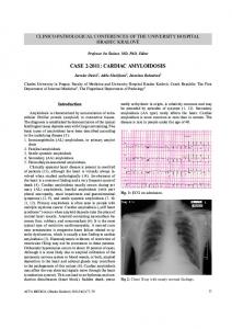
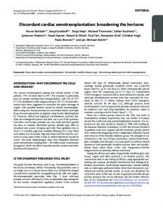
Figure 3. Nkx2-5 drug-selected cardiomyocytes show greatest cardiac maturation on rigid substrates. (A) N kx2-5neoR/+ ES cells were formed into EBs, landed onto PDMS and/or TC plate substrates, and subjected to drug selection. After 6 days under neomycin selection, ES-derived cardiac foci showed greater maturity on rigid matrices via (B) cTnT mRNA expression relative to GAPDH (n = 3) and (C) beating rate (n = 3). Cardiac foci also demonstrated a higher expression of adult α-MHC relative to fetal β-MHC, verifying their mature cardiac phenotype (D). Expression analyses of MLC2v (ventricular myocytes) and SLN (atrial myocytes) indicate that rigid and soft environments induce ventricular and atrial myocytes, respectively (n = 3) (E). MLC2v and SLN expression levels are normalized to those of 50:1 and TC plate, respectively. All values are mean ± SD, where ∗ indicates p-value < 0.05 and ∗∗ indicates p-value < 0.01.
expression analyses of chamber-specific markers revealed that MLC2v+ ventricular myocytes are strongly induced in rigid environments whereas SLN+ atrial myocytes are dominant in softer environments (figure 3(E)), indicating that matrix elasticity induces the cardiac differentiation in an atrial-ventricular lineage specific manner. Together, these results suggest that increased microenvironmental stiffness not only promotes differentiation of cardiomyocytes but also supports their functional maturation at later stages.
N kx2-5neoR/+ EBs were formed via a hanging drop method, landed onto substrates at day 5, and cultured with neomycin at day 10 (figure 3(A)) for 6 days, resulting in the detachment of all non-cardiac lineages and the selective enrichment of beating cardiomyocyte populations (supplementary video 1, available from stacks.iop.org/STAM/ 14/025003/mmedia). qRT-PCR from drug-selected cardiomyocytes indicated that cTnT expression was higher on the rigid substrates (figure 3(B)). We also observed the highest beating rate in cells cultured on the extremely rigid TC plate substrate, with rates of up to 100 bpm (figure 3(C)). Although suggestive, the beating rate of ES-derived cardiomyocytes alone is not an accurate indicator of cardiac maturation. To further examine whether stiffer environments improve the functional maturity of ES-derived cardiomyocytes, we examined the relative expression of two differentially expressed isoforms of the myosin heavy chain (MHC) contractile protein, α-MHC and β-MHC. In mice, the α-MHC gene is predominantly expressed in the adult myocardium and is 4 kilobases upstream of β-MHC, which is expressed only during late fetal life [25]. The relative expression of these two genes is tightly controlled and indicative of the maturity of the cardiomyocytes. Misregulation of the ratio of α and β-MHCs, known as fetal gene reactivation, has been correlated with a wide variety of pathological conditions of the heart including hypertrophy and infarction [26, 27]. qRT-PCR analysis of these two genes showed that cardiomyocytes derived from drug selection and culture on more rigid substrates have the highest α/β-MHC expression ratio (figure 3(D)). Interestingly,
3.4. ES-derived cardiac foci display functional synchrony with native cardiomyocytes To test the functionality of these cultured cardiomyocytes, we transplanted the ES-derived cardiac foci at day 16 of drug selection onto a layer of neonatal cardiomyocytes obtained from the hearts of newborn mice and imaged using light microscopy (figure 4(A)). The onset of synchronized beating between these two cell types suggests that the cells may be transplantable and useful for tissue regeneration purposes and medical therapeutics. Here, we observed that drug-selected cardiomyocytes transplanted from TC plate, 10:1 PDMS and 50:1 PDMS stiffness conditions electrically coupled with neonatal cardiomyocytes for synchronized beating between the EB and neonatal cardiomyocyte (CM), as observed visually and confirmed via M-mode image analysis of contractile frequency pattern in each region (figures 4(B) and (C)). Mechanical interferometry imaging (MII), a recently developed real-space technique for the quantitative detection 6
Sci. Technol. Adv. Mater. 14 (2013) 025003
A Arshi et al
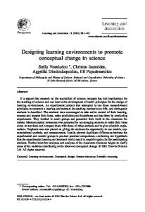
Figure 4. Transplanted cardiomyocyte foci display synchrony with the neonatal cardiomyocyte feeder layer. Cardiac foci derived from drug-selection (EB) and stiffness culture were transplanted onto a monolayer of neonatal cardiomyocytes (CM) at day 16, as shown in figure 3(A). (A) Representative light microscopic image of EB cultured on CM. The M-mode image was obtained at the red line. (B), (C) After 2 days, M-mode image analysis of videos of co-cultured samples confirms synchronization of beating frequency between EB (B) and neonatal cardiomyocyte (C) layers. Beating is largely regular with minor arrhythmia. (D)–(F) MII analyses at the beating regions of EBs transplanted onto CM (D). EB and CM initially beat asynchronously (E), but become fully synchronized after 48 h in co-culture (F).
of nanoscale mechanical displacements, was used to confirm the electrical coupling between ES-derived and neonatal cardiomyocytes [18]. In this setup, cells were cultured on silicon substrates in a custom-built fluid compensation cell maintained at 37 ◦ C and 5% CO2 for real-time imaging of the vertical movement of cardiac foci while in culture. Under MII, clustered cells of EBs normally appear dark and become lighter when vertical heights change, while individual neonatal cardiomyocytes change from light to dark upon contraction (figure 4(D), supplementary video 2, available from stacks.iop.org/STAM/14/025003/mmedia). MII analyses of cultured cells revealed that ES-derived and neonatal cardiomyocytes initially beat asynchronously (figure 4(E)), and become fully synchronized after 48 h in co-culture (figure 4(F), supplementary video 2).
4. Discussion Collectively, these data suggest that rigid extracellular matrices support the differentiation of pluripotent ES cells into functional cardiomyocytes in two ways: cardiac lineage induction and functional maturation. This paper is, to our knowledge, the first report of the role of matrix elasticity in de novo cardiogenesis from undifferentiated ES cells, and the first demonstration of mechanical interferometry imaging as a tool for microscale characterization of cardiac differentiation and maturation. Previous studies have demonstrated the influence of the physical microenvironment on the function of already differentiated cardiomyocytes isolated from embryos or ES cells [9, 11]. Differentiated cardiomyocytes isolated from 7
Sci. Technol. Adv. Mater. 14 (2013) 025003
A Arshi et al
embryonic hearts function best in matrices even stiffer than normal physiological myocardium [8, 9]. Our findings are in accordance with these reports in that drug selected differentiated cardiomyocytes display highest α/β-MHC expression when cultured on TC plates (figure 3). Although higher stiffness may represent the scar tissue in the infarcted heart [8], data from this group (figure 3) and others [9, 11] demonstrate that superphysiological rigidity serves to the better function of differentiated cardiomyocytes isolated from embryos and human ES cells. While these previous studies used already differentiated cardiomyocytes as starting material and examined their function, the novelty of our report is the observation that a stiffer environment enhances not only the function of the already differentiated cardiomyocytes but also the de novo induction of mouse and human cardiac specification at uncommitted stages. Cardiac progenitors contribute to the embryonic myocardial tube in two waves: while the progenitors in the classical cardiac crescent (first heart field) form the future left ventricle first, multipotent cardiac progenitors in the extra crescent (second heart field) migrate later to the heart tube and add additional myocardia that contribute to the future atria and outflow tract. These late-migrating second heart field progenitors maintain their multipotency until later stages [16, 23, 24], and gradually lose plasticity and differentiate in the relatively rigid scaffold provided by already maturated myocardium. This creates a unique interaction between the late-migrating immature cardiac progenitors and already mature myocardium. This unique in vivo cardiac maturation process seems to be reflected in our finding that a stiffer environment favors cardiac specification of ES cells. Our results imply that early waves of cardiomyocytes induce the differentiation of the late-migrating second heart field progenitors in part via their stiffer biophysical environment. While the cardiomyocytes mature to generate unique biophysical activities, the biophysical cues in turn appear to promote reciprocally maturation of the cardiac progenitors. It is not clear at the moment why the de novo cardiac induction was most efficient in superphysiological conditions. Further understanding of how the biophysical cues are translated into molecular mechanisms during the cardiac cell fate specification would be useful for discerning the role of physical cues in embryonic stem cell fate determination [28], and improving cardiac tissue culture methods for future application in regenerative medicine.
Foundation. We thank Jinghua Tang and UCLA-BSCRC for technical assistance on human ES cell works as well as the Nano and Pico Characterization Core Laboratory at the California NanoSystems Institute.
References [1] Yamashita J, Itoh H, Hirashima M, Ogawa M, Nishikawa S, Yurugi T, Naito M and Nakao K 2000 Nature 408 92 [2] Behfar A, Zingman L V, Hodgson D M, Rauzier J-M, Kane G C, Terzic A and Puc´eat M 2002 FASEB J. 16 1558 [3] Sachinidis A, Gissel C, Nierhoff D, Hippler-Altenburg R, Sauer H, Wartenberg M and Hescheler J 2003 Cell. Physiol. Biochem. 13 423 [4] Tan G, Shim W, Gu Y, Qian L, Chung Y Y, Lim S Y, Yong P, Sim E and Wong P 2010 Differentiation 79 260 [5] Schenke-Layland K, Nsair A, Van Handel B, Angelis E, Gluck J M, Votteler M, Goldhaber J I, Mikkola H K, Kahn M and Maclellan W R 2011 Biomaterials 32 2748 [6] Katayama Y, Battista M, Kao W-M, Hidalgo A, Peired A J, Thomas S A and Frenette P S 2006 Cell 124 407–21 [7] Engler A J, Sen S, Sweeney H L and Discher D E 2006 Cell 126 677 [8] Engler A J, Carag-Krieger C, Johnson C P, Raab M, Tang H-Y, Speicher D W, Sanger J W, Sanger J M and Discher D E 2008 J. Cell Sci. 121 3794 [9] Bajaj P, Tang X, Saif T and Bashir R 2010 J. Biomed. Mater. Res. A 95 1261 [10] Forte G et al 2012 Tissue Eng. A 18 1837 [11] Hazeltine L B et al 2012 Int. J. Cell Biol. 2012 508294 [12] Jacot J G, Kita-Matsuo H, Wei K A, Chen H S V, Omens J H, Mercola M and McCulloch A D 2010 Ann. NY Acad. Sci. 1188 121 [13] Nakashima Y, Ono K, Yoshida Y, Kojima Y, Kita T, Tanaka M and Kimura T 2009 Biochem. Biophys. Res. Commun. 390 821 [14] Diaz Perez S V, Kim R, Li Z, Marquez V E, Patel S, Plath K and Clark A T 2012 Hum. Mol. Genetics 21 751 [15] Willems E, Spiering S, Davidovics H, Lanier M, Xia Z, Dawson M, Cashman J and Mercola M 2011 Circ. Res. 109 360 [16] Nakano H, Williams E, Hoshijima M, Sasaki M, Minamisawa S, Chien K R and Nakano A 2011 J. Mol. Cell. Cardiol. 50 337 [17] Livak K J and Schmittgen T D 2001 Methods 25 402 [18] Reed J, Ramakrishnan S, Schmit J and Gimzewski J K 2009 ACS Nano 3 2090 [19] Nguyen C T, Lu Q, Wang Y and Chen J-N 2008 Drug Discovery Today Discase Models 5 135 [20] Lee J N, Jiang X, Ryan D and Whitesides G M 2004 Langmuir 20 11684 [21] Huang N F and Li S 2011 Ann. Biomed. Eng. 39 1201 [22] Ochsner M, Dusseiller M R, Grandin H M, Luna-Morris S, Textor M, Vogel V and Smith M L 2007 Lab Chip 7 1074 [23] Moretti A et al 2006 Cell 127 1151 [24] Nakano H et al 2013 Nature Commun. 4 1564 [25] Mahdavi V, Chambers A P and Nadal-Ginard B 1984 Proc. Natl Acad. Sci. USA 81 2626 [26] Miyata S, Minobe W, Bristow M R and Leinwand L A 2000 Circ. Res. 86 386 [27] Reiser P J, Portman M A, Ning X H and Schomisch Moravec C 2001 Am. J. Physiol. Heart. Circ. Physiol. 280 H1814 [28] Sun Y, Villa-Diaz L G, Lam R H W, Chen W, Krebsbach P H and Fu J 2012 PLoS ONE 7 e37178
Acknowledgments This work was supported by BSCRC, AHA (IRG4870007, GRNT9420039) and NIH/NHLBI (R21HL109938) to AN and NIH/NCRR/NCATS UCLA CTSI grant (UL1TR000124) and NIH/NIAMS (K01AR061415) to DE. AA was supported by HHMI fellowship; YN was supported by fellowship grants from the Japanese Circulation Society and Uehara Memorial
8