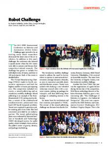
IEEE TRANSACTIONS ON NEURAL SYSTEMS AND REHABILITATION ENGINEERING, VOL. 20, NO. 3, MAY 2012
351
Robot Training of Upper Limb in Multiple Sclerosis: Comparing Protocols With or WithoutManipulative Task Components Ilaria Carpinella, Davide Cattaneo, Rita Bertoni, and Maurizio Ferrarin, Member, IEEE
Abstract—In this pilot study, we compared two protocols for robot-based rehabilitation of upper limb in multiple sclerosis (MS): a protocol involving reaching tasks (RT) requiring arm transport only and a protocol requiring both objects’ reaching and manipulation (RMT). Twenty-two MS subjects were assigned to RT or RMT group. Both protocols consisted of eight sessions. During RT training, subjects moved the handle of a planar robotic manipulandum toward circular targets displayed on a screen. RMT protocol required patients to reach and manipulate real objects, by moving the robotic arm equipped with a handle which left the hand free for distal tasks. In both trainings, the robot generated resistive and perturbing forces. Subjects were evaluated with clinical and instrumental tests. The results confirmed that MS patients maintained the ability to adapt to the robot-generated forces and that the rate of motor learning increased across sessions. Robot-therapy significantly reduced arm tremor and improved arm kinematics and functional ability. Compared to RT, RMT protocol induced a significantly larger improvement in movements involving grasp (improvement in Grasp ARAT sub-score: RMT 77.4%, RT 29.5%, p=0.035) but not precision grip. Future studies are needed to evaluate if longer trainings and the use of robotic handles would significantly improve also fine manipulation. Index Terms—Motor learning, multiple sclerosis, robot-therapy, upper limb function.
I. INTRODUCTION
A
LTHOUGH multiple sclerosis (MS) is the most common cause of chronic neurological disability in young adults, current pharmacological therapies are so far not able to substantially improve motor functionality [1]. Although no firm conclusions can be drawn, a recent review suggests that rehabilitation may be useful to maximize the functional status of these subjects [2]. Most studies about motor rehabilitation in MS are focused on walking and mobility [3], but, as the disease progresses, nearly 75% of MS subjects experience also upper limb dysfunction [4], mainly related to tremor [5], coordination deficit [6] and muscle weakness [7]. Although arm impairment highly contributes to reduce the quality of life of Manuscript received July 14, 2011; revised October 20, 2011; accepted January 29, 2012. Date of current version May 18, 2012. This work was supported in part by FISM—Fondazione Italiana Sclerosi Multipla. I. Carpinella and M. Ferrarin are with the Biomedical Technology Department, Don Carlo Gnocchi Foundation Onlus IRCCS, 20148 Milan, Italy (e-mail:
[email protected]). D. Cattaneo and R. Bertoni are with the Department of Neurorehabilitation, Don Carlo Gnocchi Foundation Onlus IRCCS, 20148 Milan, Italy (e-mail:
[email protected]). Digital Object Identifier 10.1109/TNSRE.2012.2187462
MS subjects [8], only few studies exist about neuromotor rehabilitation of upper limb in MS [7], [9], [10]. The results emerged from these works showed that physical rehabilitation generated an improvement in arm strength [7], in the execution of the activities of daily living (ADL) [10] and a tendency to an improved manual dexterity, although not significant [9]. Given the paucity of existing studies, more research is needed to explore the effects of neuro-rehabilitation on upper limb dysfunction in MS subjects. In the last decade, robotic devices for upper limb motor rehabilitation have been increasingly studied, becoming a promising complement to traditional therapy, as they can provide high-intensity, repetitive and interactive treatment of the impaired upper limb and, inherently, an objective, quantitative measurement of patient’s progress. Although robotic systems are mostly used in rehabilitation of stroke patients [11], they appear good candidates also for the treatment of tremor and incoordination due to MS. At present, three studies have addressed the application of robot-based treatment of upper limb in MS [12]–[14]. Gijbels et al. [14] used a gravity-supporting exoskeleton which allowed the subject to execute 3-D tasks simulated in a virtual environment on a computer screen. Carpinella et al. [12] and Vergaro et al. [13] used a planar robotic system and adaptive training protocols, in which the robot did not assist subjects during the execution of the movement, but, rather it provided unfamiliar dynamic environments to which subjects were required to adapt, by learning to predict the effects of perturbing forces [15], [16]. A common finding of these studies was the post-treatment improvement in the execution of functional tasks implying distal movements not directly involved in the training. In particular, robotic training consistently improved manual dexterity, as shown by the Nine Hole Peg Test score [17] which significantly decreased from 157 to 109 s in severe MS subjects [14] and from nearly 60 to 48 s in mild/moderate patients [12], [13]. These results indicated that, even though the mean improvement was significant, the post-treatment score was still consistently higher than the threshold value typical of healthy adults with comparable age, that is 19 [18]. Starting from these results, one may wonder if the implementation of a functional-based robot training, which involves not only the movement of proximal joints but also the use of distal arm and the manipulation of real objects should even improve the rehabilitation outcome and facilitate the skill transfer from the experimental setting to the ADL [19]. This approach has been recently applied to stroke patients [20], not yet to MS subjects.
1534-4320/$31.00 © 2012 IEEE
352
IEEE TRANSACTIONS ON NEURAL SYSTEMS AND REHABILITATION ENGINEERING, VOL. 20, NO. 3, MAY 2012
In the present study we designed a robot-based functional training which combines a typical adaptive paradigm, previously applied to stroke [21] and MS subjects [12], [13], [22], with two main principles of motor learning: 1) the use of real objects during purposeful and challenging functional tasks involving whole arm, to enhance motor performance and maximize subjects’ active participation [19] and 2) the execution of different types of exercises during each session, that has been shown to promote motor learning [23]. A robot-based protocol incorporating these two concepts was applied to a group of MS subjects and the results were compared to those obtained by a second group of MS patients treated with a more traditional robot-based protocol involving reaching tasks [12], [13]. Goal of this pilot study was to evaluate if a robot therapy approach involving both objects’ reaching and manipulation leads to better outcomes than training involving only the transport of the arm.
TABLE I DEMOGRAPHIC AND CLINICAL CHARACTERISTICS OF TREATED MS SUBJECTS
II. METHODS A. Subjects A consecutive sample of 22 MS subjects were enrolled in the study within a period of one year and a half. Their demographic and clinical characteristics are shown in Table I. All subjects signed an informed consent to the protocol which was conformed to the standards for human experiments set by the Declaration of Helsinki. Subjects fulfilled the following inclusion criteria: a definite diagnosis of MS according to McDonald criteria [24], Expanded Disability Status Scale [25] , Nine Hole Peg Test [17] score between 30 and 300 s, Mini-Mental State Examination [26] , willing to follow the rehabilitation program. Subjects were excluded if they had reduced and not amendable visual acuity and/or ocular motility which interfere with the execution of the training tasks. Each subject was allocated to one of the following groups: a group receiving a robot-based rehabilitation program involving the execution of reaching tasks (RT) and a group receiving a robot-based rehabilitation program involving both objects’ reaching and manipulation (RMT). Group allocation was performed following minimization method [27], that is a widely acceptable alternative approach to randomization in small trials, as it ensures excellent balance between groups, even in small samples. In particular, each subject was allocated to one group or the other, depending on the baseline clinical characteristics of those participants already enrolled, in order to minimize the imbalance between the two groups. As shown in Table I, demographic and clinical characteristics were similar in both groups.
Fig. 1. (a) Experimental set-up. (b) The “traditional” handle. (c) The “functional” handle. (d) Example of a subject executing a functional task, i.e., insert a key in a padlock.
manipulate real objects [Fig. 1(d)]. The “functional” handle is fixed to the robotic arm through a 12-cm-long vertical rod that supports a cylindrical ball bearing which can rotate around the rod and translate in vertical direction with low friction. A forearm splint made of thermoplastic material is fixed to the ball bearing. The inner surface of the splint is covered with foam rubber coat to guarantee subject’s comfort.
B. Experimental Equipment
C. Robot-Based Rehabilitation Protocols
The apparatus consists of a planar robotic manipulandum [Fig. 1(a)] with two back-driveable degrees-of-freedom (Braccio di Ferro, Celin srl, La Spezia, Italy), which has been fully described elsewhere [28]. It has an 80 40 cm elliptic workspace and is smoothly impedance-controlled in order to generate continuous forces up to 25 . Subjects can interact with the robotic arm through two handles: a “traditional” handle grasped by the patient [Fig. 1(b)] and a customized “functional” handle [Fig. 1(c)] wrapped around the forearm of the subject and fastened through velcro stripes leaving the hand free to
Both RT and RMT protocols were composed of eight sessions. The number of sessions was chosen on the basis of the results obtained by Vergaro et al. [13]. Each session consisted of eight epochs of 20 movements each, for a total of 160 movements and a duration of 30–45 min. The most affected arm of each subject was treated. In the RT protocol (see also [12], [13], and [22]), subjects sat on a chair behind a table and grasped the “traditional” handle of the robot [Fig. 1(b)]. A 19-in LCD screen, positioned in front of the subject, was used to display the current position of the
CARPINELLA et al.: ROBOT TRAINING OF UPPER LIMB IN MULTIPLE SCLEROSIS
end-effector and the target (circles with a diameter of 1.5 cm and 3 cm, respectively). Subjects performed center-out reaching movements from the same center position to peripheral targets (directions: 45 and 135 with respect to the horizontal axis) and then they returned to the center (directions: 225 and 315 ). The amplitude of the nominal reaching trajectory was 25 cm. All subjects were able to fulfill the task. The movements were executed at self-selected speed and the targets were presented in a random order. Each training session consisted of two phases [13], [22]: 1) null field phase (2 epochs, 40 movements) and 2) force field phase (6 epochs, 120 movements). During the null field phase, subjects executed the reaching task while the robot did not generate forces. Null field trials had the purpose to monitor the progress of unperturbed reaching movement and the retention of the effects of each training session on arm kinematics. During the force field phase, subjects executed the reaching task while the robot generated a velocity-dependent clockwise force field [12], [13], [15], [22] to which subjects were required to adapt. Perturbing force was perpendicular to the instantaneous movement direction and had a magnitude proportional to the handle speed. This force disturbed the movement executed by the subject by deviating hand’s trajectory from the nominal straight path. During each epoch, force was unexpectedly turned off in 1/5 of the movements (catch trials, 4 per epoch) to monitor the progress of adaptation. In particular, if adaptation is occurring and the subject is learning the appropriate internal model to predict and cancel the perturbations induced by the robot, two effects should be noticed: 1) a gradual reduction of the execution errors in the force trials and 2) a gradual increase of erroneous movements in directions opposite to the perturbations in catch trials, when the force is unexpectedly removed (see also [12], [13], [15], [16], [22]). To increase the difficulty of the exercise, the robot generated also a spring-like resistive force which opposed hand’s movement. In particular, the force was proportional to the distance between the current position of the end-effector and the starting position and was directed along the line which connected the starting point and the instantaneous position of the handle. RMT training sessions also consisted of a null field phase and a force field phase. During the null field phase (2 epochs, 40 movements), subjects executed the reaching task (as in RT protocol), while the robot did not generate forces. At the end of this phase, the “traditional” handle of the robot was replaced with the “functional” handle [Fig. 1(c)] and the splint was worn by the subject. During the force field phase (6 epochs, 120 movements), subjects executed a set of 6 functional tasks (one per epoch) involving reaching and manipulation of “real” objects, built using LEGO bricks (LEGO Group, Billund, Denmark) (e.g., small containers, pegs, beads, coin slots) or taken from typical activities of daily living (e.g., bottles, jars, padlocks, keys). The set of functional tasks was selected on the basis of the specific deficit of each subject, as measured by clinical assessment. The tasks included both grasp (e.g., grasp and release of water bottles or cylindrical jars) and precision grips, mainly key grip (e.g., locking of a padlock or insertion of a coin into a slot) and pinch grip (e.g., putting a bead down a needle or insert a rectangular peg into a squared hole). An example is shown in
353
Fig. 1(d). The objects to be reached and manipulated were fixed on a LEGO supporting base and arranged on the table within the elliptic workspace of the manipulandum. The positions of the end-effector when each object had been reached were calibrated for each patient at the beginning of the first training day and maintained for all sessions. These positions and the instantaneous position of the handle were represented on the screen by circles, as in RT protocol. This allowed the operator to check the correct execution of the task which was performed by the patient by looking at the objects on the table. Subjects were invited to perform the tasks at self-selected speed, trying to counteract the pull of gravity by maintaining the ball bearing in the middle of the handle rod and by avoiding to rest their elbow on the table. During the training, the difficulty of the tasks was increased by varying the size and the weight of the objects to be manipulated. Each functional movement consisted in a transport phase (during which the subject reached the object) and a manipulation phase. During the transport phase the robot generated the same forces and used in the RT protocol. Moreover the same number of catch trials (4 per epoch) were inserted. To analyze the effect of resistive force , all subjects were also required to execute the reaching task (1 epoch, 20 movements) while only was turned on. This test was performed pre- and post-treatment. D. Clinical Assessment All subjects were evaluated pre- and post-treatment by means of three clinical tests administered by a blinded examiner: Nine Hole Peg test (9 HPT) [17], Action Research Arm Test (ARAT) [29], and Tremor Severity Scale (TSS) [30]. The 9HPT evaluates hand dexterity. The test requires the subject to place nine pegs in nine holes. Subjects are scored on the amount of time they take to place and remove all nine pegs. The ARAT evaluates proximal and distal function of upper limb and was used in this study to evaluate the transfer of the training effects on functional tasks involving 3-D movements not executed during the treatment. The ARAT consists of 19 items organized in four sections: Grasp, Grip, Pinch, and Gross. Each item is given an ordinal score of 0, 1, 2, or 3, with higher values indicating better performance. The maximum ARAT score is 57. In the present study, total ARAT score and sub-scores related to the four sections were considered. Moreover, a variable related to the execution time was analyzed. When a subject was not able to perform some ARAT tasks , execution time was used to calculate execution frequency (60/time), that represents the number of times an item was executed in a minute. A value of 0 was assigned when subjects were unable to perform the task. Total ARAT frequency was calculated as the mean frequency of all items. The TSS measures the severity of tremor in four domains: 1) rest tremor: tremor occurring at rest with the body part supported; 2) postural tremor: tremor occurring while maintaining a position against gravity; 3) kinetic tremor: tremor during movement; and 4) intention tremor: pronounced exacerbation of kinetic tremor towards the end of a goal directed movement. Each domain is rated by a 10-point scale, with higher score indicating more severe tremor.
354
IEEE TRANSACTIONS ON NEURAL SYSTEMS AND REHABILITATION ENGINEERING, VOL. 20, NO. 3, MAY 2012
III. RESULTS
E. Kinematic Analysis Handle coordinates were sampled at 100 Hz and low-pass filtered using a sixth-order Savitzky-Golay filter with a 200 ms window and a cutoff frequency of 9.4 Hz [12]. The same filter was used to estimate the subsequent time derivatives of the trajectory. Kinematic data were segmented into separated movements and, for functional tasks, each movement was divided into a reaching phase and a manipulation phase. Then, the following parameters were extracted. • Reaching duration: time between reaching onset and termination (first instants in which handle velocity exceeded and fell below a threshold of 20% of peak speed, respectively [13]). • Manipulation duration: time elapsed between reaching termination and the end of whole task. This parameter was calculated only for functional tasks. • Jerk Index: logarithm of the jerk (norm of the third time derivative of the trajectory), averaged over the movement duration and normalized with respect to the amplitude and duration of the reaching movement [13]. Jerk Index evaluates the smoothness of the trajectories. • Mean and maximum lateral deviation: mean and maximum distance of the actual trajectory from the nominal trajectory (straight line connecting the start and the end points). • Normalized path length: length of the actual trajectory normalized with respect to the length of the nominal trajectory. • Learning Index : calculated to assess the subjects’ ability to adapt to the perturbing force field generated by the robot. In this study we used the definition of learning index proposed by Donchin et al. [15] and expressed by
(1) and are the maximum lateral deviations where in the field and catch trials, respectively. This index compares a signed measure of execution error (here, maximum lateral deviation, positive in the direction of force ) in movements where force is turned on (force trials) and where force is turned off (catch trials). Values of equal to 1 indicate a perfect adaptation, in which errors in force trials tend to 0, while errors in catch trials are large and in the opposite direction (negative values) with respect to force [15]. F. Statistical Analysis Kinematic parameters were analyzed with a two-way mixed ANOVA, with session and group (RT versus RMT protocol) as fixed factors. Considering that clinical data were not normally distributed (Shapiro-Wilks W test, ), nonparametric procedures were used. In particular, comparisons between pre- and post-treatment data were evaluated using Wilcoxon matched pairs test (Wt), while differences between RT and RMT groups were tested by means of Mann-Whitney U test (MWt). Level of significance was set to 0.05.
A. Null Field Trials The results related to the unperturbed reaching movements performed by the treated MS subjects are shown in Fig. 2. A significant effect of session was found for reaching duration , jerk index and mean lateral deviation , which gradually decreased during the training [Fig. 2(a)–(c)], while normalized path length remained almost unchanged [see Fig. 2(d)]. The effect of training protocol was not significant for all parameters. These results indicated that the unperturbed reaching movements of MS subjects became gradually faster, smoother and more linear across sessions, similarly in both groups, as shown in the examples reported in Fig. 2(e)–(h). B. Force Field Trials The subjects’ improvement in counteracting the robot-generated disturbing force is shown in Fig. 3. The amplitude of the perturbing force field [Fig. 3(a)] showed a significant effect of session . No difference was noticed between RT and RMT groups but the significant session group interaction revealed that amplitude increased mainly in RT group, while remained almost stable in RMT group. Despite this difference, the rate of force field adaptation was similar in both groups. As shown in Fig. 3(b), the learning index increased over sessions , similarly in both groups . The increase of was due to a significant decrease of the lateral deviation in the force trials [see Fig. 3(c)] and to a significant increase (in the opposite direction) of the execution error in catch trials [see Fig. 3(d)]. Examples of the trajectories executed during force field trials by two representative MS subjects from RT and RMT group are shown in Fig. 3(e)–(h), respectively. With respect to the first session [Fig. 3(e) and (g)], in the last training day [Fig. 3(f)–(h)] the deviation from the straight line decreased during force field trials (continuous lines) and increased in the opposite direction during catch trials (dashed lines), thus indicating adaptation to the perturbing force. The results related to the trajectories executed pre and post-treatment, when only force was turned on revealed that, after the treatment, MS subjects improved their ability in counteracting the resistive force which opposed the movement. In particular the trajectories performed against resistance showed a significant decrease in duration , jerk index , and lateral deviation , with respect to pretreatment evaluation. No statistically significant difference was noticed between the two groups. Moreover, similarly in both groups, movements executed against were characterized by higher duration and jerk index , with respect to unperturbed reaching.
CARPINELLA et al.: ROBOT TRAINING OF UPPER LIMB IN MULTIPLE SCLEROSIS
Fig. 2. (a)–(d) Quantitative parameters (mean confidence interval) describing null field trials. Significant differences with respect to session 1 are and RMT group . (e)–(h) Unperturbed reaching trajectoreported for RT ries executed during the first and the last treatment sessions by two MS subjects (V09 and F03) treated with the RT and the RMT protocols, respectively. -axis represents the direction of movement.
The results related to the duration of the manipulation tasks executed by the RMT group are reported in Fig. 4. The time required for manipulative tasks involving grasp gradually decreased across sessions , while the time of precision grip tasks (i.e., key or pinch grip) remained almost unchanged . C. Clinical Evaluations At the baseline evaluation (Pre) both groups showed similar clinical characteristics (Table II). The MS subjects showed moderate to severe upper limb dysfunction (ARAT score: ). All subjects showed cerebellar
355
Fig. 3. (a)–(d) Quantitative parameters (mean confidence interval) describing force field trials. Significant differences with respect to session 1 are and RMT group . (e)–(h) Reaching trajectories executed reported for RT during the first and the last treatment sessions by two MS subjects (V06 and F05) treated with the RT and the RMT protocols, respectively. -axis represents the direction of movement. Continuous lines: force field trials; dashed lines: catch trials.
symptoms. In particular, upper limb ataxia (as measured by items of the Ataxia scale [31]) was mild/moderate (Ataxia score: 1–3) in 18 subjects and severe (5–6) in four subjects. Kinetic and intention tremor was mild/moderate (TSS score: 1–5) in 20 patients and severe (7–8) in two patients. As concerns the effects of training on upper limb function (see Table II), both groups significantly improved ARAT total score and Pinch sub-score, while only RMT group consistently increased Grasp and Grip sub-scores. Gross sub-score did not change significantly in both groups because of a high ceiling
356
IEEE TRANSACTIONS ON NEURAL SYSTEMS AND REHABILITATION ENGINEERING, VOL. 20, NO. 3, MAY 2012
Fig. 4. Duration (mean confidence interval) of manipulation tasks, involving grasp and precision grip, executed by MS subjects during the eight sessions of the RMT protocol. ANOVA p-values related to the effect of session and are reported. significant differences with respect to session
TABLE II PRE- AND POST-TREATMENT CLINICAL SCORES FOR RT AND RMT GROUPS
Fig. 5. Post treatment percentage change in ARAT total score and sub-scores. . Column: mean; whisker: standard deviation. P-value from Mann Whitney U test comparing RT and RMT groups are reported.
(15.8%); RMT: 38.4% (21.3%); ], while a similar improvement in both groups was noticed for execution frequency of Grip [RT: 18.1% (17.8%); RMT: 21.6% (19.8%); ] and Pinch items [RT: 17.2% (20.0%); RMT: 17.8% (34.0%); ]. As concerns manual dexterity, both groups significantly improved the 9 HPT score after the treatment (Table II). The percentage change obtained after the training was similar in both groups [RT: 14.1% (16.3%); RMT: 12.1% (19.3%); ]. Three subjects in the RT group and five subjects in the RMT group attained an improvement greater than 20% (the threshold for clinical significance [32]). No subjects showed a significant worsening. As shown in Table II, both groups significantly reduced intention tremor. A significant reduction of postural tremor was noticed in the RMT group. IV. DISCUSSION
effect. In particular, 16/22 patients (eight per group) showed a baseline Gross score equal to the maximum (nine points) and maintained the same score after the treatment. The remaining subjects (three per group) significantly improved their Gross sub-score . A direct comparison of the percentage change obtained by the two groups after the treatment confirmed the above results. As shown in Fig. 5, RMT group obtained a percentage improvement of Grasp sub-score significantly higher than that attained by RT group. Analysis of the execution frequency of ARAT tasks revealed that both groups significantly increased this parameter after the treatment . Again, the percentage change in execution frequency of Grasp items was significantly higher in RMT group [RT: 14.3%
The main goal of the present study was to compare the effects of two protocols for robot-based rehabilitation of upper limb in MS: a protocol involving reaching tasks (RT) and a protocol requiring objects’ reaching and manipulation (RMT). The enrolled patients did not report any adverse event in terms of muscle aches, fatigue or increased muscle stiffness. The results related to the whole sample of treated MS subjects (RT and RMT groups together) confirmed those found in previous studies and added further evidence that robot-based training significantly improved upper limb coordination, functionality and dexterity in people with MS [12]–[14], thus representing a valid complement to traditional rehabilitation approaches. An important question that arises from these results is whether the observed improvements are due to the forces generated by the robot or are just the effect of repeated movements. This pilot study did not analyze this aspect, but a recent work of Vergaro et al. [13] found that, within each session, motor improvements were significant only after robot-assisted trials, whereas mere exercise alone did not show any effect. This result suggests a specific within-session effect of the robot,
CARPINELLA et al.: ROBOT TRAINING OF UPPER LIMB IN MULTIPLE SCLEROSIS
but future studies comparing robot training with unassisted exercise are warranted to explore long term effects. Analysis of instrumental data confirmed that MS subjects maintained the ability to adapt to novel dynamic environments by learning to predict the perturbations induced by the robot. In particular the appearance of erroneous movements in opposite direction of force , when it was unexpectedly turned off (catch trials), suggested that MS subjects reacted to the perturbation by learning a suitable internal model of the disturbing field rather than resisting the perturbation by increasing their arm stiffness through muscles co-contraction, as found also in healthy subjects [16]. The time course of learning index indicated that the capability to adapt was preserved not only within each session, as found by Casadio et al. [22], but also across training days. Similar results were found by Tomassini et al. [33] who analyzed the learning curves of MS subjects during the execution of a simple tracking task. Contrarily to the protocols described by Casadio et al. [22] and Vergaro et al. [13], which forced the subjects to maintain an approximately constant velocity, in the present study MS patients executed the movements at self-selected speed. In particular, MS subjects gradually increased their reaching velocity across sessions, thus inducing a consequent gradual augmentation of the perturbation generated by the robot. Despite this factor, the parameters describing motor learning improved along the treatment, thus suggesting that MS subjects maintain the ability to adapt to the disturbing force, not only when it remains nearly constant [13], [22] but also when it increases across sessions. Analysis of the learning curves obtained in the present study revealed that MS subjects’ adaptation mainly improved in the first part of the treatment, with an increase of of approximately 50% during the first four training days, followed by a further but lower increase of about 15% during the last four sessions. This typical asymptotic behavior of motor learning has been found also in several studies on healthy subjects and has been demonstrated to be associated with different mechanisms of neural plasticity indispensable for the acquisition of new skills (e.g., [34]). This similarity with healthy subjects suggests, in turn, that the mechanisms of force field adaptation and brain plasticity are preserved, at least partly, in MS patients and that, for this reason, these subjects may benefit of adaptive trainings which have been shown to promote neural reorganization [35]. Interestingly, the rate of adaptation was similar in both groups, thus indicating that subjects treated with the RMT protocol adapted to the perturbing force field similarly to patients who underwent RT training, even though the exercises proposed in RMT protocol were more complex and the forces were transmitted to the forearm of the patients and not directly to the hand as in the RT protocol. The only difference found in RMT group was the time course of the perturbing force amplitude, whose increase across sessions was lower with respect to RT group. This could be ascribed to the difficulty of the proposed functional tasks which was gradually augmented during the training, thus limiting the increase of hand velocity and, consequently, of the disturbing force. Despite
357
this difference, direct comparisons between the two groups, performed separately within each training day, revealed that, during each session, both groups were exposed to forces of similar amplitude, thus allowing the comparison of their performances. Analysis of trials executed when only was turned on, revealed that movements against resistance were slower and less smooth than unperturbed reaching. This suggested that adding a spring-like resistive component to the task actually increased the difficulty of the exercise. Moreover, after the treatment both groups of MS subjects improved their ability in counteracting the resistive force . This could be ascribed not only to an improvement in upper limb coordination, but also to an increase in muscle strength. Future studies should include a direct measure of muscle force to test this hypothesis. Analysis of the reaching trajectories performed by the subjects during null field trials clearly showed that kinematics of unperturbed upper limb movements significantly improved over sessions, independently from the training protocol. In particular, arm movements became gradually faster, smoother and more linear, similarly in both groups. According to Fitts’ law [36], an increase in movement speed should reduce the accuracy of the trajectory. The fact that the movements became smoother and more linear despite the increased velocity suggested that the improvements in arm kinematics were actually due to a beneficial effect of the therapy for both groups. Clinical counterpart of this result was the similar improvement attained by the two groups in Gross sub-score of the ARAT, which evaluates the movement of proximal arm only. Taken together, these results suggest that adding a manipulation component to the reaching exercise did not limit the improvements of proximal arm function in MS subjects. An opposite result was found by Krebs et al. [20] on chronic stroke patients. In particular, they found that subjects who underwent a robot-assisted whole-arm training obtained a significantly larger improvement in wrist/hand movement but a consistently lower increase in proximal arm function, with respect to stroke patients treated with a robot training involving arm transport only. The authors speculated that stroke subjects could have focused their attention on the hardest component of the task, that was the manipulation of the object, relying on the assistive force generated by the robot for the transport of the arm to the target. This was not the case of MS patients treated in the present study, as they did not receive an assistive force which helped them to extend the elbow, but, rather, resistive and perturbing forces which disturbed their movement. This training paradigm, in turn, required the subject to actively participate not only in the manipulation phase of the task, but also in the reaching phase, thus promoting the active use of whole arm. Importantly, the applied clinical tests showed that both robotbased protocols significantly reduced intention tremor and improved upper limb functionality and dexterity. A hypothesis about tremor reduction arises from the indication that cerebellar symptoms typical of MS (including coordination deficit and tremor), may partly depend on the alteration of the anticipatory
358
IEEE TRANSACTIONS ON NEURAL SYSTEMS AND REHABILITATION ENGINEERING, VOL. 20, NO. 3, MAY 2012
(feed-forward) component of motor control and, thus, on the reduced, although present, capability to predict the motor commands required to perform a complex task [37]. The fact that force field adaptation exercises specifically train these feed-forward control mechanisms could thus explain tremor reduction. As concerns the improvements of upper limb function, the results related to the RT group confirmed those previously found in [12]–[14] and suggested that the effect of robot-therapy may partly transfer to tasks more related to ADL. This could be ascribed to two factors. Firstly, the reduction of tremor and the improvement of coordination between shoulder and elbow may have improved the control of proximal arm, thus allowing a better orientation of the hand in space, that is a fundamental prerequisite for a correct grip. Secondly, the beneficial effect of the adaptive training on the feed-forward control mechanisms, strongly involved in manipulative tasks [38], could explain the improvement observed also in distal functions not directly involved in the treatment. Interestingly, the comparison between the two groups revealed that MS subjects treated with the RMT protocol obtained a significantly higher improvement in 3-D tasks involving grasp but not precision grip (i.e., key and pinch grip). This finding was confirmed by instrumental results, which revealed a significant decrease in the execution time of grasp tasks only. This could be due to different factors. First of all, precision grip is more complex than grasp, as it requires independent finger movements that involve fine control of the directions and magnitudes of fingertip forces [38]. This aspect has been confirmed by fMRI studies which showed that precision grip engages different and more complex neural circuits with respect to those involved during grasp [39]. Moreover, it has been demonstrated that MS subjects reported more difficulties in pinch than grasp [40]. This could be ascribed not only to motor problems, but also to altered and/or reduced tactile sensibility [41] which can impair the feedback control of fingertip actions mainly during manipulation of small and light objects. On the basis of these considerations, it could be possible that more training sessions are necessary to obtain a further improvement in precision grip. A second hypothetical explanation could be ascribed to the splint of the “functional” handle, which highly reduced forearm prono-supination movements. Possibly, this characteristic may have facilitated the subjects during the training by reducing the degrees of freedom to be controlled and, consequently, by simplifying the whole motor problem to be solved. This, in turn, may have limited the transfer of the acquired skill to the more complex 3-D tasks measured by ARAT. A third hypothesis can also be formulated. In particular, in the investigated protocol the haptic device generated forces only during the reaching phase of the tasks while the patient-robot interaction was indeed null during the manipulation phase. Moreover such forces were applied to the forearm/wrist and not to the fingers. This leaves open the possibility that assistive/resistive/perturbing forces operating directly on the hand and/or fingers (as described for example in [42], [43]) might be a significant factor for a further improvement in manipulation tasks and, in particular, in precision skills.
There are some limitations that need to be addressed regarding the present study. Considering that no correlation was found between the baseline clinical characteristics and the level of post-treatment improvement, studies on a greater number of patients are warranted to identify the MS subjects who would mostly benefit of robot therapy and to make the results generalizable to the entire population with MS. A second limitation concerns the design of the “functional” handle. In particular, future studies should include the use of wearable sensorized robotic handles which can generate forces operating on the distal part of the upper limb (i.e., hand and fingers) and which can provide an objective, quantitative measurement of hand’s movement. The third limitation concerns the parameters and related to the forces generated by the robot, that were maintained fixed for all subjects and during all training sessions. Given the high variability of the symptoms typical of MS, these parameters should be tuned on the basis of the individual capabilities and gradually adjusted to account for the change of performance across the training sessions. Future studies should include automatic procedures for the fine tuning of the exercises. In conclusion the present pilot study confirmed that adaptive robot training may be a useful approach to improve upper limb kinematics and functional ability in subjects with MS. Even though caution must be taken given the small sample size, the present results suggested that the inclusion of a manipulation component to the typical reaching exercise can significantly improve the execution of functional tasks involving grasp. Further larger studies including follow-up evaluations are needed to evaluate if longer treatments, which involve the use of a wearable robotic handle, could induced also significant amelioration of precision grip and if the beneficial effects of robot therapy are maintained long-term. REFERENCES [1] S. J. Pittock and C. F. Lucchinetti, “The pathology of MS: New insights and potential clinical applications,” Neurologist, vol. 13, pp. 45–56, Mar. 2007. [2] F. Khan, L. Turner-Stokes, L. Ng, and T. Kilpatrick, “Multidisciplinary rehabilitation for adults with multiple sclerosis,” Cochrane. Database Syst. Rev., pp. CD006036–CD006036, 2007. [3] E. M. Snook and R. W. Motl, “Effect of exercise training on walking mobility in multiple sclerosis: A meta-analysis,” Neurorehabil. Neural Repair, vol. 23, pp. 108–116, Feb. 2009. [4] S. Johansson, C. Ytterberg, I. M. Claesson, J. Lindberg, J. Hillert, M. Andersson, H. L. Widen, and L. von Koch, “High concurrent presence of disability in multiple sclerosis. Associations with perceived health,” J. Neurol., vol. 254, pp. 767–773, Jun. 2007. [5] P. Feys, F. Maes, B. Nuttin, W. Helsen, V. Malfait, G. Nagels, A. Lavrysen, and X. Liu, “Relationship between multiple sclerosis intention tremor severity and lesion load in the brainstem,” Neurorep., vol. 16, pp. 1379–1382, Aug. 2005. [6] A. J. Bastian, T. A. Martin, J. G. Keating, and W. T. Thach, “Cerebellar ataxia: Abnormal control of interaction torques across multiple joints,” J. Neurophysiol., vol. 76, pp. 492–509, Jul. 1996. [7] N. F. Taylor, K. J. Dodd, D. Prasad, and S. Denisenko, “Progressive resistance exercise for people with multiple sclerosis,” Disabil. Rehabil., vol. 28, pp. 1119–1126, Sep. 2006. [8] N. Yozbatiran, F. Baskurt, Z. Baskurt, S. Ozakbas, and E. Idiman, “Motor assessment of upper extremity function and its relation with fatigue, cognitive function and quality of life in multiple sclerosis patients,” J. Neurol. Sci., vol. 246, pp. 117–122, Jul. 2006.
CARPINELLA et al.: ROBOT TRAINING OF UPPER LIMB IN MULTIPLE SCLEROSIS
[9] L. K. Storr, P. S. Sorensen, and M. Ravnborg, “The efficacy of multidisciplinary rehabilitation in stable multiple sclerosis patients,” Mult. Scler., vol. 12, pp. 235–242, Apr. 2006. [10] V. W. Mark, E. Taub, K. Bashir, G. Uswatte, A. Delgado, M. H. Bowman, C. C. Bryson, S. McKay, and G. R. Cutter, “Constraint-Induced Movement therapy can improve hemiparetic progressive multiple sclerosis. Preliminary findings,” Mult. Scler., vol. 14, pp. 992–994, Aug. 2008. [11] G. Kwakkel, B. J. Kollen, and H. I. Krebs, “Effects of robot-assisted therapy on upper limb recovery after stroke: A systematic review,” Neurorehabil. Neural Repair, vol. 22, pp. 111–121, Mar. 2008. [12] I. Carpinella, D. Cattaneo, S. Abuarqub, and M. Ferrarin, “Robot-based rehabilitation of the upper limbs in multiple sclerosis: Feasibility and preliminary results,” J. Rehabil. Med., vol. 41, pp. 966–970, Nov. 2010. [13] E. Vergaro, V. Squeri, G. Brichetto, M. Casadio, P. Morasso, C. Solaro, and V. Sanguineti, “Adaptive robot training for the treatment of incoordination in Multiple Sclerosis,” J. Neuroeng. Rehabil., vol. 7, pp. 37–37, 2010. [14] D. Gijbels, I. Lamers, L. Kerkhofs, G. Alders, E. Knippenberg, and P. Feys, “The Armeo Spring as training tool to improve upper limb functionality in multiple sclerosis: A pilot study,” J. Neuroeng. Rehabil., vol. 8, pp. 5–5, 2011. [15] O. Donchin, L. Sawaki, G. Madupu, L. G. Cohen, and R. Shadmehr, “Mechanisms influencing acquisition and recall of motor memories,” J. Neurophysiol., vol. 88, pp. 2114–2123, Oct. 2002. [16] J. L. Patton and F. A. Mussa-Ivaldi, “Robot-assisted adaptive training: Custom force fields for teaching movement patterns,” IEEE Trans Biomed. Eng., vol. 51, no. 4, pp. 636–646, Apr. 2004. [17] D. E. Goodkin, D. Hertsgaard, and J. Seminary, “Upper extremity function in multiple sclerosis: Improving assessment sensitivity with boxand-block and nine-hole peg tests,” Arch. Phys. Med Rehabil., vol. 69, pp. 850–854, Oct. 1988. [18] G. K. Oxford, K. A. Vogel, V. Le, A. Mitchell, S. Muniz, and M. A. Vollmer, “Adult norms for a commercially available Nine Hole Peg Test for finger dexterity,” Am. J. Occup. Ther., vol. 57, pp. 570–573, Sep. 2003. [19] J. Carr and R. Shepherd, A Motor Relearning Programme for Stroke, 2 ed. Rockville, MD: Aspen, 1987. [20] H. I. Krebs, S. Mernoff, S. E. Fasoli, R. Hughes, J. Stein, and N. Hogan, “A comparison of functional and impairment-based robotic training in severe to moderate chronic stroke: A pilot study,” NeuroRehabil., vol. 23, pp. 81–87, 2008. [21] J. L. Patton, M. Kovic, and F. A. Mussa-Ivaldi, “Custom-designed haptic training for restoring reaching ability to individuals with poststroke hemiparesis,” J. Rehabil. Res. Develop., vol. 43, pp. 643–656, Aug. 2006. [22] M. Casadio, V. Sanguineti, P. Morasso, and C. Solaro, “Abnormal sensorimotor control, but intact force field adaptation, in multiple sclerosis subjects with no clinical disability,” Mult. Scler., vol. 14, pp. 330–342, Apr. 2008. [23] V. S. Huang and J. W. Krakauer, “Robotic neurorehabilitation: A computational motor learning perspective,” J. Neuroeng. Rehabil., vol. 6, pp. 5–5, 2009. [24] W. I. McDonald, A. Compston, G. Edan, D. Goodkin, H. P. Hartung, F. D. Lublin, H. F. McFarland, D. W. Paty, C. H. Polman, S. C. Reingold, M. Sandberg-Wollheim, W. Sibley, A. Thompson, N. S. van den, B. Y. Weinshenker, and J. S. Wolinsky, “Recommended diagnostic criteria for multiple sclerosis: Guidelines from the International Panel on the diagnosis of multiple sclerosis,” Ann. Neurol., vol. 50, pp. 121–127, Jul. 2001. [25] J. F. Kurtzke, “Rating neurologic impairment in multiple sclerosis: An expanded disability status scale (EDSS),” Neurology, vol. 33, pp. 1444–1452, Nov. 1983. [26] E. Pfeiffer, “A short portable mental status questionnaire for the assessment of organic brain deficit in elderly patients,” J. Am. Geriatr. Soc., vol. 23, pp. 433–441, Oct. 1975. [27] D. G. Altman and J. M. Bland, “Treatment allocation by minimisation,” BMJ, vol. 330, pp. 843–843, Apr. 2005. [28] M. Casadio, V. Sanguineti, P. G. Morasso, and V. Arrichiello, “Braccio di Ferro: A new haptic workstation for neuromotor rehabilitation,” Technol. Health Care, vol. 14, pp. 123–142, 2006. [29] R. C. Lyle, “A performance test for assessment of upper limb function in physical rehabilitation treatment and research,” Int. J. Rehabil. Res., vol. 4, pp. 483–492, 1981.
359
[30] P. G. Bain, L. J. Findley, P. Atchison, M. Behari, M. Vidailhet, M. Gresty, J. C. Rothwell, P. D. Thompson, and C. D. Marsden, “Assessing tremor severity,” J Neurol. Neurosurg. Psychiatry, vol. 56, pp. 868–873, Aug. 1993. [31] S. H. Alusi, J. Worthington, S. Glickman, and P. G. Bain, “A study of tremor in multiple sclerosis,” Brain, vol. 124, pp. 720–730, Apr. 2001. [32] S. R. Schwid, A. D. Goodman, M. P. McDermott, C. F. Bever, and S. D. Cook, “Quantitative functional measures in MS: What is a reliable change?,” Neurology, vol. 58, pp. 1294–1296, Apr. 2002. [33] V. Tomassini, H. Johansen-Berg, L. Leonardi, L. Paixao, S. Jbabdi, J. Palace, C. Pozzilli, and P. M. Matthews, “Preservation of motor skill learning in patients with multiple sclerosis,” Mult. Scler., vol. 17, pp. 103–115, Jan. 2011. [34] L. Ma, B. Wang, S. Narayana, E. Hazeltine, X. Chen, D. A. Robin, P. T. Fox, and J. Xiong, “Changes in regional activity are accompanied with changes in inter-regional connectivity during 4 weeks motor learning,” Brain Res., vol. 1318, pp. 64–76, Mar. 2010. [35] R. J. Nudo, “Adaptive plasticity in motor cortex: Implications for rehabilitation after brain injury,” J. Rehabil. Med., pp. 7–10, May 2003. [36] P. M. Fitts, “The information capacity of the human motor system in controlling the amplitude of movement. 1954,” J. Exp. Psychol. Gen., vol. 121, pp. 262–269, Sep. 1992. [37] V. Sanguineti, P. G. Morasso, L. Baratto, G. Brichetto, M. G. Luigi, and C. Solaro, “Cerebellar ataxia: Quantitative assessment and cybernetic interpretation,” Hum. Mov Sci., vol. 22, pp. 189–205, Apr. 2003. [38] J. R. Flanagan, M. C. Bowman, and R. S. Johansson, “Control strategies in object manipulation tasks,” Curr. Opin. Neurobiol., vol. 16, pp. 650–659, Dec. 2006. [39] H. H. Ehrsson, A. Fagergren, T. Jonsson, G. Westling, R. S. Johansson, and H. Forssberg, “Cortical activity in precision- versus power-grip tasks: An fMRI study,” J. Neurophysiol., vol. 83, pp. 528–536, Jan. 2000. [40] C. C. Chen, N. Kasven, H. I. Karpatkin, and A. Sylvester, “Hand strength and perceived manual ability among patients with multiple sclerosis,” Arch. Phys. Med Rehabil., vol. 88, pp. 794–797, Jun. 2007. [41] V. Iyengar, M. J. Santos, M. Ko, and A. S. Aruin, “Grip force control in individuals with multiple sclerosis,” Neurorehabil. Neural Repair, vol. 23, pp. 855–861, Oct. 2009. [42] A. E. Fiorilla, F. Nori, L. Masia, and G. Sandini, “Finger impedance evaluation by means of hand exoskeleton,” Ann. Biomed. Eng., Aug. 2011. [43] L. Masia, H. I. Krebs, P. Cappa, and N. Hogan, “Design and characterization of hand module for whole-arm rehabilitation following stroke,” IEEE/ASME Trans. Mechatron., vol. 12, pp. 399–407, Aug. 2007.
Ilaria Carpinella was born in Vigevano, Italy, in 1977. She received the M.Sc. degree in biomedical engineering from thr Polytechnic of Milan, Milan, Italy, in 2002, with a thesis developed at the University of Glasgow, Glasgow, U.K. Since 2003, she has been a Researcher with the Biomedical Technology Department, Don C. Gnocchi Foundation Research Hospital, Milan, and from 2005 to 2007 she was a Professor of electric and electronic measures at the University of Milan. Her main research interests include rehabilitation engineering and robotics, motor control, and movement analysis.
Davide Cattaneo was born in Milan, Italy, in 1970. He received the M.Sc. degree in rehabilitation science in 2007 and the Ph.D. degree in biomedical engineering in 2010. He is Research Coordinator at the Larice Lab, Don C. Gnocchi Foundation Research Hospital, Milan. He has published papers on the assessment and treatment of functional disorders in subjects with neurological diseases using traditional and innovative instruments. He is a reviewer for several peer-reviewed international journals and international research institutions. Dr. Cattaneo is a member of the European Group for Rehabilitation in Multiple Sclerosis.
360
IEEE TRANSACTIONS ON NEURAL SYSTEMS AND REHABILITATION ENGINEERING, VOL. 20, NO. 3, MAY 2012
Rita Bertoni was born in Milan, Italy, in 1982. She graduated as a Sport Scientist from the Università Cattolica del Sacro Cuore in 2005 and received the B.S. degree in physical therapy, in 2008, from the University of Milan with a thesis on the robotic rehabilitation in people with multiple sclerosis. Since 2010, she has worked as a Physical Therapist and researcher at Don Carlo Gnocchi Foundation Onlus IRCCS, Milan.
Maurizio Ferrarin (M’97) received the M.Sc. and Ph.D. degrees in biomedical engineering. He is Research Coordinator with the Biomedical Technology Department, Don C. Gnocchi Foundation Research Hospital, Milan, Italy. He was Professor of rehabilitation engineering at the Polytechnic of Milan (1997–2003) and, since 2003, has been a Professor of electronic bioengineering at the University of Milan. His research interests include movement analysis, FES, motor control, rehabilitation robotics. He authored 60 full papers in peer-reviewed journals and two books. He is member of IFESS and a Founding Member of the Italian Society of Clinical Movement Analysis. He served in the board of directors of IFESS (2003–2006) and SIAMOC (2009–present).