Introduction. Inorganic arsenic determination is based on bioluminescence produced by the bioreporter cells in response to arsenite exposure.
CONffIDENCE: Contaminants in food and feed: Inexpensive detection for control of exposure
Robust methods for detecting inorganic arsenic with biosensor bacteria using luminescence measurement and fish imaging Anne Rantala, Daniel Paul and Matti Karp Department of Chemistry and Bioengineering, Tampere University of Technology, P.O.Box 541, 33101, Tampere, FINLAND Visualization
Introduction Inorganic arsenic determination is based on bioluminescence
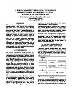
An agar diffusion assay (ADA) was performed for the comparison of
produced by the bioreporter cells in response to arsenite exposure.
arsenic detection on plate. Logarithmically grown cells (E. coli XL-1
bacterium
parsRluxCDABE) were added to soft agar supplemented with
Escherichia coli XL-1 (parsRluxCDABE) (Hakkila, 2004) was used
appropriate antibiotic, mixed gently and poured on top of the LA
A
bioluminescence-producing
arsenic-inducible
in this study as the reporter organism. These sensor cells express
agar plates containing fish filet (Baltic herring, Clupea harengus
luciferase upon exposure to arsenite, the activity of which was
membras) samples soaked in 5 ml of 30 µM arsenic (Fig. 2a) and 5
detected by measurement of cellular bioluminescence. Arsenate (V)
ml of milliQ water (Fig. 2b) for 24 hours. Biophotonic imaging station
is spontaneously reduced by the cells to arsenite (III) and hence can
(IVIS Xenogen, Caliper life sciences) was used to visualize the
also indirectly cause luciferase synthesis. The data obtained by the
arsenic detection. Exposure time at each measurement point was
two
methods,
instrumental
luminescence
measurement
and
visualization with CCD camera were used to show the performance
30 sec. The more red intensity the higher is the concentration around the filet.
on the biosensor cells.
Method Fig.2a. Comparison of arsenic using fish sample immersed in 5 ml of 30 µM Na2HAsO4
Luminescence measurement Freeze-dried E. coli XL-1 (parsRluxCDABE) were rehydrated and
(left spots) and NaAsO2 (right spots) for 24 hours and exposed to E. coli XL1(parsRluxCDABE) for up to 3 hours
used as fresh cells in arsenic analysis to produce a standard curve. Assay mixtures were prepared directly in the 96-well microtiter plate containing equal volumes of each As standard and reconstituted biosensor cell suspension. E.Coli XL-1 parsRluxCDABE in LB 100 80
0h
Fig.2b. Fish sample immersed in 5 ml of milli-Q water for 24 hours and exposed to E. coli XL-1(parsRluxCDABE) for up to 3 hours.
IC
60 1h
40 20 0 0,03
0,1
0,3
1
3
10
30
2h
Conclusion
3h
The intensity of the bioluminescence is proportional to the arsenite
100
NaAsO2 (III) concentration (µM)
concentration in the luminescence assay measurements; detection limit being 0.3 µM. Theoretical explanation was confirmed with the
Fig.1. Induction coefficiencies from the E. coli XL-1 (parsRluxCDABE) cells in LB is plotted as a function of the arsenite concentration in the assay, ranging from 0 to 100 µM As(III).The detection limit is shown with the arrow.
visualization method: the higher is the arsenic concentration, the higher the luminescence is seen in biophotonic imaging. Response intensities by the sensor cells on inorganic arsenic demonstrate the
Assays were incubated with shaking (300 rpm) at 37 °C for 180 min. Luminescence was measured once an hour (Chameleon multilabel Detection Platform luminometer, Hidex Oy, Turku, Finland). Induction coefficients were calculated using the formula IC = Li/Lb, where IC is the induction coefficient, Li is the luminescence value of
correlation of the two methods used in this study (data not shown). Also the
use
of
reagent-like freeze-dried
bacteria
in
the
luminescence measurement make the sensors available as robust detectors, which can simply be reconstituted and used, thus enabling rapid and simple analysis of inorganic arsenic.
the sample, and Lb is the luminescence value of a blank.
Reference: Hakkila K., Green T., Leskinen P., Ivask A., Marks R., Virta M. (2004) Detection of bioavailable heavy metals in EILATox-Oregon samples using whole-cell luminescent bacterial sensors in suspension or immobilized onto fibre-optic tips. J.Appl Toxicol. 24(5):333–42.
The research leading to these results has received funding from the European Community's Seventh Framework Programme (FP7/2007-2013) under grant agreement n° 211326: CONffIDENCE project (www.conffidence.eu). The research leading to these results has received funding from the European Community's Seventh Framework Programme (FP7/2007-2013) under grant agreement n° 211326: CONffIDENCE project (www.conffidence.eu).