PHYSIOLOGICAL REVIEWS Vol. 81, No. 1, January 2001 Printed in U.S.A.
Role of Alternative Splicing in Generating Isoform Diversity Among Plasma Membrane Calcium Pumps EMANUEL E. STREHLER AND DAVID A. ZACHARIAS Department of Biochemistry and Molecular Biology, Mayo Graduate School, Mayo Clinic/Foundation, Rochester, Minnesota; and Howard Hughes Medical Institute, Cell and Molecular Medicine, University of California San Diego, La Jolla, California
I. II. III. IV. V.
VI.
VII. VIII.
IX.
Introduction Overview of Structural and Regulatory Characteristics of Plasma Membrane Calcium Pumps Nomenclature of Plasma Membrane Calcium Pump Isoforms Four Genes and Alternative Ribonucleic Acid Splicing Generate a Multitude of Plasma Membrane Calcium Pump Isoforms Alternative Splicing Options of Mammalian Plasma Membrane Calcium Pumps A. Splice site A: highly variable complexity among different PMCA genes B. Splice site B: a splicing artifact? C. Splice site C: a multitude of options with some conserved principles Tissue Distribution of Plasma Membrane Calcium Pump Isoforms and Splice Variants A. Differential expression of the four PMCA genes in adult tissues B. Differential expression of the four PMCA genes during development C. Differential expression of alternative splice variants Regulation of Alternative Splicing in the Plasma Membrane Calcium Pump Family Physiological Significance of Alternative Splicing in the Plasma Membrane Calcium Pump Family A. Experimental challenges in determining the function of individual PMCA isoforms B. Splicing at site A: differential phospholipid sensitivity and/or differential interaction with regulatory proteins? C. Splicing at site C: multiple effects on the complex and modular COOH-terminal regulatory domain Conclusions and Future Perspectives
22 22 25 26 26 26 26 28 29 29 29 31 32 35 35 37 39 45
Strehler, Emanuel E., and David A. Zacharias. Role of Alternative Splicing in Generating Isoform Diversity Among Plasma Membrane Calcium Pumps. Physiol Rev 81: 21–50, 2001.—Calcium pumps of the plasma membrane (also known as plasma membrane Ca2⫹-ATPases or PMCAs) are responsible for the expulsion of Ca2⫹ from the cytosol of all eukaryotic cells. Together with Na⫹/Ca2⫹ exchangers, they are the major plasma membrane transport system responsible for the long-term regulation of the resting intracellular Ca2⫹ concentration. Like the Ca2⫹ pumps of the sarco/endoplasmic reticulum (SERCAs), which pump Ca2⫹ from the cytosol into the endoplasmic reticulum, the PMCAs belong to the family of P-type primary ion transport ATPases characterized by the formation of an aspartyl phosphate intermediate during the reaction cycle. Mammalian PMCAs are encoded by four separate genes, and additional isoform variants are generated via alternative RNA splicing of the primary gene transcripts. The expression of different PMCA isoforms and splice variants is regulated in a developmental, tissue- and cell type-specific manner, suggesting that these pumps are functionally adapted to the physiological needs of particular cells and tissues. PMCAs 1 and 4 are found in virtually all tissues in the adult, whereas PMCAs 2 and 3 are primarily expressed in excitable cells of the nervous system and muscles. During mouse embryonic development, PMCA1 is ubiquitously detected from the earliest time points, and all isoforms show spatially overlapping but distinct expression patterns with dynamic temporal changes occurring during late fetal development. Alternative splicing affects two major locations in the plasma membrane Ca2⫹ pump protein: the first intracellular loop and the COOH-terminal tail. These two regions correspond to major regulatory domains of the pumps. In the first cytosolic loop, the affected region is embedded between a putative G protein binding sequence and the site of phospholipid sensitivity, and in the COOH-terminal tail, splicing affects pump regulation by calmodulin, phosphorylation, and differential interaction with PDZ domain-containing anchoring and signaling proteins. Recent evidence demonstrating differential distribution, dynamic regulation of expression, and major functional differences between alternative splice variants suggests that these transporters play a more dynamic role than hitherto assumed in the spatial and http://physrev.physiology.org
0031-9333/01 $15.00 Copyright © 2001 the American Physiological Society
21
22
EMANUEL E. STREHLER AND DAVID A. ZACHARIAS
Volume 81
temporal control of Ca2⫹ signaling. The identification of mice carrying PMCA mutations that lead to diseases such as hearing loss and ataxia, as well as the corresponding phenotypes of genetically engineered PMCA “knockout” mice further support the concept of specific, nonredundant roles for each Ca2⫹ pump isoform in cellular Ca2⫹ regulation.
I. INTRODUCTION Plasma membrane calcium pumps are now well recognized as a primary system for the specific expulsion of Ca2⫹ from eukaryotic cells. Together with Ca2⫹-specific ion channels and exchangers, these pumps are largely responsible for the regulated transport of Ca2⫹ between the intracellular and the extracellular milieu. Since their original identification in mammalian erythrocyte membranes in the mid 1960s (141), the calcium pumps have gained increasing attention as a ubiquitous mechanism for high-affinity calcium extrusion across the cell membrane. Due to the emergence of sophisticated protein biochemical and molecular techniques, tremendous progress has been made over the last two decades in elucidating their enzymatic properties, biochemical regulation, gross functional domain structure, and primary amino acid sequences. Accordingly, several reviews have been published on these transporters during the last few years, including contributions by Carafoli (24, 25), Carafoli and Stauffer (27), Monteith and Roufogalis (119), Lehotsky (111), Guerini (71), and Penniston and Enyedi (129). Several overviews comparing the calcium pumps of the plasma and the organellar membranes have also been published (128, 158), including one on the different calcium pumps in plants (54). In addition, the structural organization and mechanistic properties of P-type primary ion pumps (which include the plasma membrane calcium pumps) have received in-depth treatment in two excellent recent reviews by Andersen and Vilsen (5) and Møller et al. (118). Lastly, the molecular, functional, and physiological properties of Na⫹/Ca2⫹ exchangers, the major alternative to the calcium pumps for Ca2⫹ removal from a cell, have recently been reviewed in a comprehensive manner by Blaustein and Lederer in this journal (11). The biochemical characteristics and overall domain structure of the plasma membrane calcium pumps have been comprehensively discussed in a review by Carafoli (24) published almost 10 years ago in this journal. Since then, rapid progress has been made in the identification of multiple isoforms of the pump and in the elucidation of their unique regulatory and functional properties. Of particular interest were findings showing that much of the isoform diversity among plasma membrane calcium pumps is due to complex alternative RNA splicing. This in turn has raised questions concerning the regulation of splicing, the cell and tissue specificity of isoform expression, and the functional significance of calcium pump isoforms differing only in a small and defined portion of
the molecule. This review does not recapitulate the earlier findings on the characteristics of the calcium pump discussed extensively by Carafoli in 1991 (24) but, rather, continues where the previous review left off. The focus is on the unique aspect of the isoform complexity among plasma membrane calcium pumps, in particular as it relates to structural and functional differences among isoforms generated via alternative mRNA splicing. We begin with a brief overview of the general structural and regulatory properties of the plasma membrane calcium pumps and a section on the nomenclature of the different isoforms. We then compile the information concerning alternative splicing of the mammalian plasma membrane calcium pumps in a manner that will provide a comprehensive catalog of the splice variants, where and when they are expressed, and what consequences the alternate splices may have on the structural and functional properties of the encoded isoforms. This is followed by a discussion concerning the physiological significance of alternative splicing as it occurs in the plasma membrane calcium pump gene family. We conclude the review with a brief outlook into promising future developments, emphasizing the importance of transgenic animal models to study the physiological consequences of the selective ablation (“knockout”) of specific plasma membrane calcium pump isoforms, as well as of investigations into naturally occurring diseases linked to mutations in specific calcium pump genes. II. OVERVIEW OF STRUCTURAL AND REGULATORY CHARACTERISTICS OF PLASMA MEMBRANE CALCIUM PUMPS The plasma membrane calcium pumps, also known as plasma membrane Ca2⫹-ATPases (PMCAs), belong to the P2 (subtype 2B) subfamily of P-type primary ion transport ATPases (8, 115, 118), which are characterized by the formation of an aspartyl phosphate intermediate as part of their reaction cycle. The PMCAs appear to be ubiquitous in eukaryotic cells where they are thought to be the major high-affinity transporter for Ca2⫹ in the plasma membrane. A “generic” schematic model of a PMCA is shown in Figure 1. The PMCAs are predicted to contain 10 membrane-spanning segments, and the NH2 and COOH termini are both located on the cytosolic side of the membrane. As shown in Figure 1B, the bulk of the protein mass is facing the cytosol and consists of three major parts: the intracellular loop between transmembrane segments 2 and 3, the large unit between membrane-spanning
January 2001
ISOFORMS OF THE PLASMA MEMBRANE CALCIUM PUMP
23
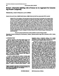
2⫹ FIG. 1. Models of the plasma membrane Ca -ATPase (PMCA). A: linear representation of a generic PMCA showing the major domain substructure. The 10 putative transmembrane regions (TM) are numbered and indicated by shaded boxes. The NH2-terminal phospholipid-sensitive region (PL) preceding transmembrane domain 3 and the calmodulin binding domain (CaM-BD) are shown as black boxes. P is the site of the obligatory aspartyl-phosphate formed during pump function. The regions of highest sequence divergence among PMCA isoforms and splice variants are denoted by black bars above the model. The sites where alternative RNA splicing affects the protein are indicated below the model and are labeled A, B, and C. Site B may represent a splicing artifact and is shown in parentheses. B: two-dimensional model of the PMCA in its autoinhibited form (left) and upon stimulation by Ca2⫹-calmodulin (right). The domains are labeled as in A, and the location where the protein is affected by splicing at sites A and C is emphasized. At site A, the short peptide segment encoded by the alternatively spliced exon(s) is indicated by a hatched box. The PDZ domain binding/putative COOH-terminal targeting domain is shown as a shaded box at the extreme COOH terminus (C). N is the NH2 terminus. Note that the bulk of the protein mass is located on the cytosolic face of the membrane. Also note that the first cytosolic loop and the major catalytic loop are drawn differently in the inhibited (left) and stimulated (right) state to indicate the conformational changes that are likely to accompany the binding of Ca2⫹-calmodulin (Ca2⫹-CaM; hatched oval) to the autoinhibitory calmodulin binding site.
domains 4 and 5, and the extended “tail” following the last transmembrane domain (25, 71, 129). The first intracellular loop region between membrane-spanning domains 2 and 3 corresponds to the “transduction domain” thought to play an important role in the long-range transmission of conformational changes occurring during the transport cycle. The large cytosolic region of ⬃400 residues between membrane-spanning segments 4 and 5 contains the major catalytic domain including the ATP binding site and the invariate aspartate residue that forms the acyl phosphate intermediate during ATP hydrolysis. Finally, the extended COOH-terminal tail corresponds to the major regulatory domain of the PMCAs (25, 24, 129, 157). On the basis of computer modeling and sequence comparisons, the overall structure of the PMCAs closely resembles that of other P2-type ATPases, notably that of the Ca2⫹
ATPases of the sarco/endoplasmic reticulum (SERCAs) (5, 118, 189). Indeed, the major “global” difference between the two types of calcium pumps is confined to the COOH-terminal tail, which is generally much smaller in the SERCAs (ranging from ⬍20 to ⬃50 residues) than in the PMCAs (70 up to 200 residues). On the other hand, the global structural arrangement of the membrane-spanning domains and of the bulky intracellular loop regions appears to be surprisingly similar in different P2-type ion pumps (140, 154), although differences in the size, structure, and relative spatial orientation of subdomains are obviously present and likely related to the specific cation(s) transported and the regulatory mechanisms operating on each transporter. A particularly distinctive feature of the PMCAs is the number of different regulatory mechanisms that alter
24
EMANUEL E. STREHLER AND DAVID A. ZACHARIAS
their functionality (reviewed in Refs. 24, 119, 128, 129). Of interest to the topic of this review is how alternative splicing impacts the regulation of the PMCAs, primarily their activation by Ca2⫹-calmodulin, acidic phospholipids, serine/threonine phosphorylation, and the possibility that they are regulated by heterotrimeric G proteins. Ca2⫹calmodulin binds to a region in the COOH-terminal portion of the PMCAs located ⬃40 residues downstream of the last transmembrane domain (90). In the absence of Ca2⫹-calmodulin, this sequence acts as an “autoinhibitory” domain; cross-linking studies using labeled peptides demonstrated that the calmodulin binding domain interacts intramolecularly with two separate regions of the pump, one located in the first cytosolic loop and the other in the major catalytic unit between the phosphorylation and the ATP binding site (53, 55, 56) (see Fig. 1B). In the absence of Ca2⫹-calmodulin, the autoinhibitory COOHterminal domain is thought to prevent catalytic turnover, keeping the pump in an inactive state (Fig. 1B, left). An elevation in the cytoplasmic Ca2⫹ results in an increase in Ca2⫹-calmodulin, which then binds with high affinity to the autoinhibitory domain of the PMCA, thereby releasing the inhibition and stimulating pump activity to near-maximal potential (Fig. 1B, right). This regulatory mechanism is similar to that of other Ca2⫹-calmodulin-dependent enzymes such as smooth muscle myosin light-chain kinase or Ca2⫹/calmodulin-dependent protein kinase I (69, 97). In the latter case, a recent study of the crystal structure of the enzyme in its autoinhibited state showed convincingly that the COOH-terminal regulatory domain (which overlaps with the calmodulin binding domain) interacts with multiple sites in the catalytic core, preventing substrate access and obstructing ATP binding in the absence of Ca2⫹-calmodulin (69). Interestingly, alternative splicing affects the affinity of the PMCAs for Ca2⫹ and calmodulin as well as the maximum velocity (Vmax) of the activated enzyme (47, 52, 129, 142). Because the Ca2⫹-calmodulin affinity of some isoforms of the PMCA is in the low nanomolar range (30, 47, 80), at least two to three orders of magnitude below calmodulin concentrations in tissues like the brain (23), it has been predicted that calmodulin may act as a pseudosubunit for some of the PMCAs (171). In addition to mediating regulation by Ca2⫹-calmodulin, the COOH-terminal region of the calcium pumps has also been shown to be the target of phosphorylation by protein kinases A and C (reviewed in Refs. 25, 119, 129, 173, 181), to be affected by proteases such as calpain (25, 173), to contain structural as well as potentially regulatory Ca2⫹ binding sites (84), and to mediate interactions with PDZ domains (100). The PMCAs are also activated by self-association (104), and although they are still able to bind Ca2⫹-calmodulin in the oligomerized state (105), there is no further activation by calmodulin. These and additional studies using synthetic peptides and COOHterminally truncated PMCAs have shown that the dimer-
Volume 81
ization (oligomerization) of the PMCAs is mediated via the calmodulin binding domain itself (104, 105, 172). Finally, the calmodulin binding domain of the PMCAs also binds to phospholipids and may thereby participate in the known stimulation of the pumps by acidic phospholipids (20, 59). Taken together, the above data strongly suggest that alternative splicing affecting the COOH-terminal sequence of PMCA isoforms could have profound effects on the regulatory and protein interaction properties of the different splice variants. These are discussed in more detail in the appropriate sections below. Acidic phospholipids, polyphosphoinositides in particular, are the most potent stimulators of the PMCA (117, 124, 125). They reduce the Michaelis constant for Ca2⫹ [Km(Ca2⫹)] of the enzyme to ⬃0.3 M compared with 0.4 – 0.7 M in the case of calmodulin stimulation (117, reviewed in Refs. 111, 128, 173). Activation by acidic phospholipids renders the PMCA insensitive to calmodulin activation, i.e., Ca2⫹-calmodulin and phospholipids are not additive although acidic phospholipids can further reduce the Km(Ca2⫹) of the Ca2⫹-calmodulin-stimulated pump (50, 173). Data showing that the fully Ca2⫹-calmodulin-stimulated pump (or a truncated pump lacking the COOH-terminal autoinhibitory, calmodulin binding domain) could be further stimulated by phospholipids indicated that a region(s) other than the COOH-terminal domain must also be involved in the lipid stimulation (50). Work with different proteolytic fragments of the PMCA and with synthetic peptides revealed that there are indeed two separate phospholipid binding regions in the pump: one corresponding to the previously mentioned COOH-terminal calmodulin binding domain and the other situated in the first cytosolic loop immediately preceding the third membrane-spanning domain (20, 59, 190) (Fig. 1). The mechanism by which different phospholipids activate the PMCAs is not known. However, given the location of the lipid-binding sequences in the pump, one may speculate that the interaction of acidic phospholipids (presumably via their charged headgroups) with the calmodulin binding domain leads to some structural rearrangement that weakens the autoinhibitory intramolecular interactions formed by the COOH-terminal tail (Fig. 1B). How phospholipid interactions with the first cytosolic loop further stimulate PMCA activity in a fully Ca2⫹-calmodulin-activated pump (corresponding to the model in Fig. 1B, right) is less obvious. Structural rearrangements of the transduction and catalytic domains could be invoked that may facilitate access of Ca2⫹ to its high-affinity transport sites and/or positively influence a rate-limiting step in the catalytic cycle. Regardless, it is intriguing that alternative splicing at the A site (Fig. 1) affects the first cytosolic loop in a region located between the phospholipid binding sequence and the sequence involved in autoinhibitory
January 2001
25
ISOFORMS OF THE PLASMA MEMBRANE CALCIUM PUMP
interactions (Fig. 1B). Alternative splicing may thus affect the phospholipid regulation of PMCA isoforms by altering the accessibility of the phospholipid binding domain to the acidic phospholipids. Similarly, alternative splice variants may show different levels of autoinhibition due to structural differences in their first cytosolic loop caused by the variable sequence insertions. In addition to protein-lipid and intramolecular protein interactions, the same alternate splice could also potentially affect interactions with external proteins. For example, the possibility that members of the heterotrimeric G protein family directly regulate the PMCA has been raised (94, 119). The amino acid sequence immediately preceding alternative splice site A displays sequence and structural similarity with G protein /␥ binding regions in other effector proteins (33, 109), rendering this region of the PMCA well suited to participate in G protein interactions (see sect. VIIIA).
lowercase letter designating the splice variant is replaced by a capital letter indicating the splice site (A, B, and C going from the NH2-terminal to the COOH-terminal splices; see Fig. 1) followed by a Roman numeral (I, II, III. . .) denoting which exon(s) is inserted. An advantage of the newer system is that it explicitly specifies the splice site concerned. On the other hand, the addition of Roman numerals to indicate whether or not a specific exon is included at a given site and to designate variants with different combinations of multiple exons at the same site is still not intuitive. A comparison of the two systems as applied to the currently known PMCA splice variants is shown in Table 1. Given that the alternative system is not free of drawbacks, and to maintain consistency with the large number of earlier publications on the PMCA isoforms, we use the old system of nomenclature throughout this review.
III. NOMENCLATURE OF PLASMA MEMBRANE CALCIUM PUMP ISOFORMS
TABLE
In order that the reader understands the nomenclature used throughout this review, some history must be reviewed and some rules must be established. Shortly after the publication of the first full-length PMCA cDNAs (144, 169), others were cloned and a consensus on isoform naming was reached by comparing the sequences and by naming the isoforms numerically (1, 2, 3. . .) in the order of their discovery. A lowercase letter is added as a prefix to designate the species (e.g., h for human, r for rat, m for mouse, etc.). Currently, the cDNA sequences of four different PMCA isoforms are known in humans (hPMCA1, hPMCA2, hPMCA3, hPMCA4) and rats (rPMCA1, rPMCA2, rPMCA3, rPMCA4). The naming of the alternative splice variants has been a bit more controversial. A scheme for splice site designation was introduced shortly after the first description of alternative splicing in hPMCA1 (70, 162). In this scheme, the alternate splice variants are designated by lowercase letters following the isoform number (e.g., hPMCA1a, hPMCA1b, etc.) where the letters at the beginning of the alphabet (a, b, c. . .) are used to designate the historically earlier identified splices at the COOH terminus of the pumps, and those at the end of the alphabet (w, x, y, z) indicate the splices at the NH2-terminal region (95, 128, 153, 156). This scheme of nomenclature has been used and extended in many subsequent publications concerning alternative splice variants, and although it is not without its problems, it is still widely used to maintain consistency with the earlier literature. An alternative system for the nomenclature of PMCA splice forms was later proposed by Carafoli (25). In this system, the
1. Nomenclature for known alternative splice variants of the PMCAs Isoform
Splice Site
Original Nomenclature
Alternative Nomenclature
PMCA1
A B B C C C C C A A A A C C C A A C C C C C C A A B B C C
x* ⫺† ⫺† a b c d e w x y z a b c x z a b c d e f x z (g)† ⫺† a b
AII* (BI)† (BII)† CII CI CIII CIV CV AIII AII AIV AI CII CI CIII AII AI CII CI CIII CIV CV CVI AII AI (BI)† (BII)† CII CI
PMCA2
PMCA3
PMCA4
For a schematic overview of the exon configuration for each splice variant, see Figures 2 and 3. For original nomenclature, see References 95, 153. For alternative nomenclature, see Reference 25. * This site in plasma membrane Ca2⫹-ATPase (PMCA) 1 does not appear to be alternatively spliced; on the basis of the exon structure, all currently known PMCA1 splice variants correspond to the “x” or “AII” type. † Alternative splicing at site B has only been observed in a few intestinal tissues (see Ref. 86) and may represent aberrant splicing. In the alternative nomenclature, the “normal” variant would be called BII because it includes the alternatively spliced exon.
26
EMANUEL E. STREHLER AND DAVID A. ZACHARIAS
IV. FOUR GENES AND ALTERNATIVE RIBONUCLEIC ACID SPLICING GENERATE A MULTITUDE OF PLASMA MEMBRANE CALCIUM PUMP ISOFORMS The mammalian PMCAs are encoded by a gene family composed of at least four nonallelic members. Complete cDNA-derived coding sequences have been determined for all four PMCA isoforms in rats and humans, the two organisms in which the majority of the work on the genes and their products has been performed. The chromosomal loci for the human genes have been determined; they are 12q21-q23 for PMCA1, 3p25-p26 for PMCA2, Xq28 for PMCA3, and 1q25-q32 for PMCA4 (15, 110, 126, 175). Because of the large size of the genes, information on the gene structures of the different PMCA isoforms is still scarce. In most cases, only fragments of the genes have been characterized; however, these generally correspond to the areas of interest with respect to the alternative splicing options. Complete genomic structures have so far been reported for the rat PMCA3 (22) and the human PMCA1 gene (81), whereas partial information is available on the rat PMCA1, -2, and -4 genes (95, 96); the human PMCA2, -3, and -4 genes (17, 21, 77, 108); and the mouse PMCA genes (43, 106; Zacharias and Strehler, unpublished results). With the rapid progress of the Human Genome Project, complete sequence and structural information for all human genes should become available within a few years, and the corresponding information from other species will likely follow in rapid succession from many ongoing and future whole genome sequencing efforts. The mammalian PMCA genes appear to be very closely related in their exon-intron structure, as demonstrated by the almost perfect conservation of intron locations in all currently known human, rat, and mouse PMCA genes and gene fragments. The currently known genes are large, ranging from ⬃70 kb for the rat PMCA3 gene (22) to well over 100 kb for the human PMCA1 and -2 genes (81, 108). The rat PMCA3 and the human PMCA1 genes contain 24 and at least 22 exons, respectively, and several of these exons specify only 5⬘-untranslated sequences or are subject to alternative splicing (22, 81). Each of the genes’ primary transcripts is subject to alternative splicing. The occurrence of alternative splicing in PMCA transcripts was first suggested by Shull and Greeb (144) and first demonstrated for hPMCA1 by Strehler et al. (162). The sites where alternative RNA splicing may occur in the protein-coding sequence of the PMCA transcripts are indicated in Figure 1 and are termed A, B, and C (157, 160). Because of the combinatorial potential of the various splice options at each site, a large number of splice variants are theoretically possible (see also Table 1), of which over 20 have been detected at the RNA (cDNA) level. In the following section, we systematically survey the different splice options that have so far
Volume 81
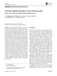
been observed for each PMCA isoform at each of the three splice “hot spots” A, B, and C. V. ALTERNATIVE SPLICING OPTIONS OF MAMMALIAN PLASMA MEMBRANE CALCIUM PUMPS A. Splice Site A: Highly Variable Complexity Among Different PMCA Genes The alternative splice options at site A (Fig. 2) have been extensively characterized in all four human and rat PMCA isoforms (22, 96, 153, 183). The splicing affects a small exon of 36 – 42 nucleotides (nt) present in all PMCA genes and coding for a short segment of the first intracellular loop of the pump molecule. This exon can be optionally inserted or excluded in the mature transcripts from all PMCA genes, with the apparent exception of PMCA1. Despite intense efforts to detect alternative usage of the corresponding 39-nt exon in PMCA1, this exon has always been found to be included in every PMCA1 transcript. Thus all PMCA1 variants are of the x splice type (see Table 1 and Fig. 2). The situation for PMCA2 at splice site A is more complex. There are three exons of 33, 60, and 42 nt (see Fig. 2) whose alternative usage theoretically gives rise to eight splice variants at this site (77). This is possible because all exons contain integral multiples of three nucleotides, and any combination of their insertion or exclusion will maintain an open protein reading frame. However, only four of the eight possible combinations have so far been detected in mRNAs from a variety of tissues. These are the variants w (with all 3 exons included), z (all 3 exons excluded), x (only the 42-nt exon included), and y (inclusion of the 33- and 60-nt exons). In humans, only variants w, x, and z have been detected, whereas in rat, variant y also has been found (2). Compared with the z and x variants, PMCA2 splice variants of the “w” type contain an additional 45 and 31 amino acid residues, respectively, in their first cytosolic loop (see Fig. 1). In PMCAs 3 and 4, there is a single exon (exon 8 in the rat PMCA3 gene) that may be included or excluded in the mature mRNA (yielding the x and z splice variants, respectively; see Fig. 2). In both human and rat, the size of this exon is 42 bp in the PMCA3 and 36 bp in the PMCA4 gene. B. Splice Site B: A Splicing Artifact? The occurrence in vivo of alternative splicing at site B has been a point of some controversy. The isolation of a hPMCA4 cDNA that lacked a 108-nt segment in the
January 2001
ISOFORMS OF THE PLASMA MEMBRANE CALCIUM PUMP
27
FIG. 2. Observed alternative splicing options at site A of the PMCAs. The exon structure of the region affected by alternative splicing at site A is shown for each of the 4 PMCA genes. The constitutively spliced exons flanking site A are represented by hatched boxes; the alternatively spliced exons are displayed as open boxes with their size indicated in nucleotides (nt). Introns are shown by solid lines connecting the exons; exon and intron sizes are not to scale. The different splice options are indicated for each PMCA, and the resulting splice variants are labeled by their lowercase symbol. Note that the 39-nt exon in PMCA1 transcripts corresponds to alternatively spliced exons of 42, 42, and 36 nt, respectively, in transcripts of the genes for PMCAs 2, 3, and 4. This exon has never been found to be excluded from any PMCA1 mRNA; hence, all PMCA1 variants are of the “x” splice type. y*, splicing of this type has so far only been observed in the rat.
COOH-terminal coding region led Strehler et al. (156, 159) to postulate that alternative splicing may occur at this site. This was supported by the finding that the 108-nt sequence is encoded in a single exon in the hPMCA1 and rPMCA3 genes (22, 81). Furthermore, as in the case of the alternatively spliced exons at site A, the 108-nt exon encodes an integral multiple of codons, and its exclusion does not alter the reading frame of the remainder of the protein. Independent cDNA clones and PCR products corresponding to PMCA1 and PMCA4 transcripts alternatively spliced at this site were subsequently isolated. However, they appear to represent only an extremely minor proportion of the total message and have only been detected in the intestine and liver (85, 87). Several studies by other laboratories have found no indication that the 108-nt exon at splice site B is ever removed in any of the tissues analyzed (95, 153). The failure by most researchers to positively identify this splice variant, and/or its very low abundance in those cases where it has been detected, support the view that mRNAs lacking the 108-nt exon are possibly the products of aberrant splicing (87, 95, 153). If translated into protein, the physiological significance of such splice site B variants would also be questionable. Exclusion of the 108-nt exon sequence leads to a pump protein lacking the tenth putative transmembrane domain (see Fig. 1A). Numerous studies have shown that the long COOH-terminal tail of the
PMCA is cytosolic (reviewed in Ref. 25). Given the proposed 10-transmembrane domain model, splicing at site B would alter the topology of the PMCA such that the COOH terminus would protrude into the extracellular space. It should be noted, however, that the precise number of membrane-spanning domains of the PMCAs has not yet been experimentally determined. In particular, the number and arrangement of membranespanning domains in the COOH-terminal half of the molecule are still a matter of debate, although the recent determination of the closely related SERCA Ca2⫹ pump (189) and Neurospora plasma membrane H⫹ pump structures (7) at a resolution of 8 Å leaves little doubt that the PMCAs will also contain 10 transmembrane domains. Interestingly, when a recombinant hPMCA4 splice variant lacking the exon at site B was expressed in insect Sf9 cells, it showed no Ca2⫹-dependent ATPase activity but was still able to form the phosphoenzyme intermediate from inorganic phosphate. When it was expressed in COS cells, it was retained in the endoplasmic reticulum with the COOH terminus apparently in the cytosol, indicating that an even number of transmembrane domains had been eliminated (142). Considering the activity data reported by Seiz-Preiano et al. (142), however, the physiological significance of such splice site B variants remains doubtful regardless of their COOH-terminal transmembrane topology.
28
EMANUEL E. STREHLER AND DAVID A. ZACHARIAS
C. Splice Site C: A Multitude of Options With Some Conserved Principles Alternative splicing at site C occurs in all isoforms, albeit in varying degrees of complexity. Generally, however, similar structural options have been found in all PMCA isoforms at this site (Fig. 3). The variations, as they occur in rat, have all been examined in a single comprehensive work by Keeton et al. (95), whereas those for the human PMCAs were most thoroughly analyzed by Stauffer et al. (153). For PMCA1, five different variants (a, b, c, d, e) have been shown to arise by inclusion or exclusion of a single 154-nt exon, and/or by utilization of multiple internal donor sites within that exon (95, 153, 162). In PMCA2, an exon of 172 nt corresponds to the
Volume 81
154-nt alternatively spliced exon found in PMCA1. However, the most frequent insertion at this site in PMCA2 is a sequence of 227 nt consisting of the 172-nt exon and an additional 55-nt exon (95, 153). In the rat PMCA2 gene, these two exons are separated by an intronic sequence of ⬃3.5 kb (95). There have been several reports of the insertion of the 172-nt exon independently of the smaller 55-nt exon, resulting in a variant designated “2c” (74, 95). In contrast, the splice variant produced from mRNA containing both exons is known as “2a.” However, based on COOH-terminal amino acid sequence similarities, we propose that the 2c variant should be more appropriately named 2a (see Figs. 3 and 6). This would also be consistent with the mechanism for the generation of the 1a, 3a, and 4a splice variants that all arise from the insertion of a single large exon (see also below).
FIG. 3. Alternative splicing options at site C of the PMCAs. The exon structure of the region affected by alternative splicing at site C is shown for each of the 4 PMCA genes. The constitutively spliced exons are represented by hatched boxes, and the alternatively spliced exons are displayed as open boxes. The sizes of the alternatively spliced exons are indicated in nucleotides (nt) and are those for the human genes. Introns are shown by solid lines connecting the exons; exon and intron sizes are not to scale. The different splice options are indicated for each PMCA gene, and the resulting splice variants are labeled by their lowercase symbol. Note that the “b” splice variants exclude the alternatively spliced exon(s), whereas the “a” variants include an entire large exon of 154, 172, 154, and 175 nt, respectively, in PMCAs 1, 2, 3, and 4. In PMCA2, splice variant a was originally proposed to arise from insertion of both the 172- and 55-nt exons; however, based on exon utilization and amino acid comparisons, this variant should be renamed “c,” and the former c should henceforth be called variant a. This change is indicated by asterisks in the splicing diagram for PMCA2. In PMCA3, splice variant “e” results from a read-through of the 154-nt exon into the following intron (indicated as thin open box). The position of the translation stop codon for each splice form is indicated by a corresponding capital letter.
January 2001
ISOFORMS OF THE PLASMA MEMBRANE CALCIUM PUMP
In PMCA3, which appears to be expressed at high levels only in skeletal muscle and brain, two exons are subject to alternative splicing at site C (Fig. 3). These are a 68-nt exon (exon 22 in the rat gene) followed by a 154-nt exon (exon 23 in the rat gene) that contains multiple internal donor sites (22) and is analogous to the 154- and 172-nt exons found at splice site C in PMCA1 and -2, respectively, and the 175/178-nt exon in isoform 4 (see below). As in all other PMCA isoforms, the splice variant lacking any of the alternatively inserted exons is the “b” variant (Fig. 3). The splice variant including the 154-nt exon is named 3a. Variants 3c and 3d utilize internal splice donor sites at positions 87 and 114, respectively, within the 154-nt exon. Splice variant 3e is generated when a “read through” of the 154-nt exon occurs, adding 88 nt of the following intron and a poly(A) tail (22). This read through leads to a short alternate COOH-terminal sequence in this variant compared with variant 3a because the open reading frame terminates just three codons into the intron. Finally, the inclusion of the 68-nt exon located 5⬘ to the 154-nt exon results in variant 3f. Insertion of this exon introduces an in-frame translation-termination codon after 45 nt, thereby generating a PMCA3 variant with the shortest COOH-terminal tail of all known PMCAs (22, 95). Insertion of the 68-nt exon generates variant 3f irrespective of the splice options utilized further downstream (e.g., whether or not the following 154-nt exon is inserted in the mRNA). In PMCA4, alternative splicing at site C is relatively simple. In humans, cows, and rats, there is a single alternatively spliced exon of 178, 181, or 175 nt, respectively, first described by Brandt et al. (17) and Keeton et al. (95). As in the case of variants 1a, 2a, and 3a, the inclusion of this exon (generating variant 4a) causes a truncation of the open reading frame that results in a protein with a shorter regulatory COOH-terminal domain that differs significantly from that of the b variant. VI. TISSUE DISTRIBUTION OF PLASMA MEMBRANE CALCIUM PUMP ISOFORMS AND SPLICE VARIANTS A. Differential Expression of the Four PMCA Genes in Adult Tissues Many studies have documented the tissue- and developmental-specific pattern of expression of several PMCAs. These studies have also shown that the brain expresses by far the greatest abundance and diversity of isoforms and splice variants (17, 95, 149, 153, 183; reviewed in Refs. 27, 111). PMCA isoforms 1 and 4 are, in general, expressed throughout most tissues, whereas PMCA isoforms 2 and 3 are expressed in a much more restricted manner and, in the adult, are found predomi-
29
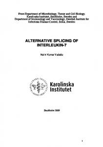
nantly in the brain and striated muscle. Within the brain, PMCA2 is primarily expressed within specialized cell types such as cerebellar Purkinje cells (152) and cochlear hair cells (64, 155). However, significant amounts of PMCA2 are also present in tissues such as uterus, liver, and kidney, and very high local levels of PMCA2 are apparently found in lactating mammary glands (132). Expression of PMCA3 appears to be even more restricted, and in the brain is particularly high in the choroid plexus (149, 152). In rats, high levels of PMCA3 expression are also found in skeletal muscle (70), whereas in humans, PMCA3 expression in skeletal muscle appears to be transient and is essentially undetectable in neonatal and adult skeletal muscle (153). Recently, isoform-specific, monoand polyclonal antibodies have been produced against peptide epitopes corresponding to small, nonconserved portions of the NH2 termini of each PMCA isoform. Using these as well as other independently generated antibodies to detect PMCA expression at the protein level, several studies have shown that there is generally a good correlation between the proteins and the previously demonstrated mRNAs in terms of their localization and relative abundance (31, 44, 58, 151, 152). B. Differential Expression of the Four PMCA Genes During Development Very few studies have appeared in the literature on the developmental pattern of expression of the different PMCA isoforms. Such information is crucial, however, if we are to understand the potential involvement of the different PMCAs in specific processes of organ and tissue development. It will also be essential for proper interpretation of the phenotypic consequences of PMCA knockout mice that are certain to be generated in the very near future or, as for PMCA2, have already been reported (106). The most comprehensive analysis of the expression of the four PMCA genes during development was recently published in a study using in situ hybridization on tissue sections of mouse embryos between days 9.5 and 18.5 in utero (186) (see Fig. 4). In this study, PMCA1 expression was detected throughout the embryo from the earliest time point analyzed, and although there appear to be some fluctuations in the level of expression in different tissues during development, the data confirm that PMCA1 is ubiquitously expressed and may be considered a “house-keeping” isoform of the pump. All other PMCA isoforms were first detectable by in situ hybridization around day 12.5, and all showed pronounced changes in the level and/or tissue distribution during further development (186). PMCA2 expression was essentially confined to the developing nervous system and reached high levels in the dorsal root ganglia, the retinoblasts of the
30
EMANUEL E. STREHLER AND DAVID A. ZACHARIAS
Volume 81
FIG. 4. Temporal and spatial expression of the 4 PMCA genes during mouse development. Contact autoradiograms are shown of in situ hybridizations to sagittal/parasagittal sections of embryos isolated from 9.5 to 18.5 days postcoitum (dpc). Sections on the left were stained with Alcian blue (AB) and Nuclear Fast Red (NFR) to illustrate overall anatomy and morphology for each stage (scale bar, 0.5 cm). PMCA1 is expressed from at least 9.5 dpc on. Signal is distributed throughout the embryo but is greatest in the nervous system, heart, skeletal muscle, and intestine. PMCA2 is first detected at 12.5 dpc. Its expression is confined to the nervous system (central nervous system, dorsal root ganglia, and pituitary) throughout development. PMCA3 is first detected at 12.5 dpc. At the early stages, expression is greatest in the nervous system but is also evident transiently in skeletal muscle. Onset of PMCA4 expression is also at ⬃12.5 dpc, although expression levels seem to remain low throughout development. PMCA4 expression is highest in the liver but decreases by 18.5 dpc. [From Zacharias and Kappen (186) with permission from Elsevier Science.]
developing eye, and the central nervous system, where the external granular layer of the developing cerebellum showed the most intense labeling. Interestingly, PMCA3, which shows a very restricted tissue distribution in the adult, was widely expressed at the transcript level early in development (between 12.5 and 15.5 days). Only at later time points (starting at around 16.5 days) did the expression of this isoform become more restricted and was high in the nervous system, the developing limb skeletal muscles, and the lung. This finding is of interest as it suggests that “knocking out” the PMCA3 gene may not merely affect specific areas of the brain (such as the choroid plexus and habenula) but could have severe, potentially lethal consequences during early embryonic development due to the possible importance of this pump isoform in the development of vital organs such as the lung. Finally, PMCA4 was generally found to be expressed at much lower levels than the other isoforms throughout mouse development, which is in contrast to the situation in adult human tissues where PMCA4 is almost as abundant and
ubiquitous as PMCA1. An exception appears to be the liver, where PMCA4 gene expression was relatively high during early time points (12.5–16.5 days) but then decreased at 18.5 days. Significant levels of PMCA4 expression were detected at the later time points in the brain, dorsal root ganglia, heart, and intestine (186). The developmental regulation of PMCA isoform and COOH-terminal splice variant expression has also been studied by PCR amplification of reverse-transcribed mRNA in the developing rat brain (16). As expected, PMCA1 is expressed at all time points from embryonic day 10 to postnatal day 30. PMCA1b is the predominant splice form early on but slowly decreases as expression of the 1a variant gradually increases until it reaches a steadystate level around embryonic day 18. Clearly, a splice shift from variant 1b to the “brain-specific” 1a variant occurs during maturation of the neuronal system. In the study by Brandt and Neve (16), PMCA2 mRNA expression in the brain was first detected around day 18, and both PMCA2a/c and 2b appeared to be about equally abundant
January 2001
ISOFORMS OF THE PLASMA MEMBRANE CALCIUM PUMP
at all time points analyzed. During postnatal development, significant changes in the relative expression of PMCA2a/c were apparent, however, in several subregions of the brain. For example, expression of PMCA2a/c appears to be upregulated in the developing cerebellum, whereas this splice variant decreases in septum, caudate, and hypothalamus during the same time period (postnatal days 2–30) (16). PMCA3 transcripts showed a similar pattern of expression in the developing rat brain as PMCA1: PMCA3b was detected from embryonic day 10, whereas expression of PMCA3a did not become apparent until embryonic day 18. As in the case of PMCA1, the emergence of the PMCA3a splice form thus appears to accompany neuronal maturation. In accordance with the in situ hybridization study on mouse embryos (186), and previous indications of relatively low expression of PMCA4 in rat brain (95, 96), PMCA4 mRNAs were not detected in the developing rat brain by the methods used in the study by Brandt and Neve (16). C. Differential Expression of Alternative Splice Variants The problem of the tissue distribution and relative abundance of the various alternative splice variants has been more difficult to address. At the mRNA level, this issue has been examined extensively by Northern blots, reverse transcriptase (RT)-PCR, ribonuclease protection assays (RPA), and in situ hybridization, with the bulk of these studies being performed on rat and human tissues. In the following sections, we review each of these techniques with respect to their relative merit and potential problems as they relate to the characterization of different PMCA isoforms and alternative splice variants. The collected set of data on the expression of PMCA splice variants is then summarized in tabular form at the end. 1. Northern blots Northern blots have been extensively used to determine the overall size, tissue distribution, and relative abundance of PMCA gene transcripts. The most detailed studies have been performed in the rat by Shull and co-workers (22, 70, 96). The major disadvantage of this method is its relative insensitivity, requiring large amounts of high-quality RNA as starting material. The problem is exacerbated in the case of the PMCAs because of their apparently low abundance in most tissues. Therefore, it has been generally necessary to analyze the poly(A) RNA fraction to detect the PMCA transcripts with the required sensitivity. This fact, combined with the often limited amount of a specific tissue source, severely limits the usefulness of the Northern technique to determine the expression pattern of the various PMCA isoforms and subtle changes in alternative splice forms.
31
However, an advantage of this method is that it allows the determination of the full-length transcript size and that relative steady-state transcript levels of specific PMCAs can be directly compared. Northern blots have been used to show, for example, that several PMCA3 transcripts of widely divergent size (7.5 and 4.5 kb) are expressed in brain and skeletal muscle and that they are generated by alternative splicing (22, 70). 2. RT-PCR Once a site for alternative splicing has been characterized, RT-PCR is the easiest method to examine the expression of differently spliced variants. However, its major advantage, exquisite sensitivity, may also be one of its main problems. PCR artifacts such as the creation of heteroduplex molecules and spurious amplification of very minor transcripts (including aberrant splice products) may lead to erroneous conclusions concerning the presence of “novel” splice variants (95, 183, 185). This problem may have arisen in a number of studies on the expression of (rare) PMCA alternative splice variants in different cell types and tissues. For example, the sensitivity of the RT-PCR approach may be responsible for the detection in some tissues of alternative splice products of PMCAs 1 and 4 at splice site B (87) and may have led to an overestimate of the diversity of alternative splice options utilized in some cell types (74). The importance of a careful analysis of all RT-PCR products by nucleotide sequencing and the inadequacy of agarose gel electrophoresis and Southern blotting alone to determine alternative splice variants have been pointed out by Zacharias et al. (185) and White et al. (180). Information concerning the exact cellular distribution of the various splice variants is generally not obtainable by RT-PCR because all tissue structure and cellular identity is destroyed in the process of extracting the RNA. There have been some attempts at microdissection of various subregions from tissues such as the kidney, but the potential for obtaining a mixed population of cell types is still very high (28, 29, 116). This problem is exemplified in the human brain; a study in which 14 regions of the brain were carefully dissected and assayed for the expression of the various splice variants (183) was not able to define which of the different cell types expressed the different spliced variants, although it was still possible to detect gross differences among the brain regions in overall splice variant expression. Single-cell RT-PCR approaches are able to solve this problem, but they are technically demanding and not always feasible in highly complex tissues. 3. RPA Although this method is labor intensive, technically demanding, and more expensive than most of the other methods, it also has several advantages. 1) RPA is usually
32
EMANUEL E. STREHLER AND DAVID A. ZACHARIAS
sensitive enough to detect even very rare messages. 2) The data produced are not prone to the same artifacts that occur in RT-PCR analyses (see sect. VIC2). 3) It can be quantitative. If one uses known quantities of radiolabeled antisense probes for an mRNA of interest and an internal standard such as glyceraldehyde-3-phosphate dehydrogenase, each with a known specific activity, then the signal obtained by detection of the protected fragments by an instrument such as the PhosphorImager (Molecular Dynamics) theoretically provides a highly quantitative measure of a given mRNA species in a mixed population. 4) RPA can detect alternatively spliced mRNA species. RPA methods were first used to detect the alternative splice options at site C in human PMCA1 transcripts from skeletal muscle (162) and have also been successfully employed to determine changes in the expression of PMCA2 splice site A variants in human IMR32 neuroblastoma cells (187). 4. In situ hybridization The major advantage of this method is that it allows the direct visualization of the transcripts of interest in specific cell types. However, for low-abundance transcripts, sensitivity issues and problems with background signals may arise. The method is also technically challenging and time consuming, and the interpretation of results is not always straightforward, particularly when complex tissues with multiple cell types are being studied. The detection of alternatively spliced products by in situ hybridization can also be difficult, if not impossible, if the alternatively spliced sequence is too short to generate sufficiently specific probes. To date, there are no in situ hybridization studies that have specifically examined the location of alternatively spliced variants of the PMCAs, only those in which probes common to most or all splice variants of an isoform were utilized. The most comprehensive studies examining the cellular distribution of the PMCA transcripts are those by Stahl and co-workers (44, 149, 150) in which the gene products for all four isoforms were localized in the rat brain, and the recent publication by Zacharias and Kappen (186) where the four PMCA gene transcripts were localized in the developing mouse embryo. These studies have shown, for example, that PMCA mRNAs are primarily expressed in neurons and at much lower levels in glial cells and astrocytes (however, see Ref. 61) and that dramatic differences exist in the distribution of the different isoform transcripts in various brain areas and cell layers. In situ hybridization has also revealed species differences in the distribution of some PMCA transcripts. PMCA4 was found to be highly expressed in granule cells of the dentate gyrus and in the CA2 region of the human hippocampus (184), whereas studies in the rat brain showed little, if any, PMCA4 in the hippocampus. In the rat, PMCA4 transcripts were de-
Volume 81
tected at high levels in the piriform cortex, the amygdaloid nucleus, and several cortical layers (150). In situ hybridization has also been used to detect temporal changes in PMCA transcript expression in the rat hippocampus following kainic acid-induced seizure activity (65). Lastly, in situ hybridization has recently been used in an elegant study to detect the cellular distribution of the four PMCA isoform transcripts in the developing and adult rat cochlea (64) as well as to study their appearance and tissue localization during mouse embryonic development (186) (see sect. VIB). 5. Summary of the tissue and cell type expression of the PMCA isoforms and their splice variants Taken together, the data from the above approaches provide a fairly detailed picture of the identity, expression level, and tissue (in some cases even cell type) distribution of the various PMCA transcripts and their splice variants. The studies at the mRNA level are being complemented at the protein level by a growing number of studies using PMCA isoform- and splice variant-specific antibodies. Tables 2– 6 summarize the data on the currently known PMCA splice variants with respect to their tissue distribution and abundance. Because the vast majority of these studies were performed in the rat and human systems, our current knowledge of PMCA isoforms and splice variants in other mammals (and definitely in nonmammalian species) is more limited. Where possible, however, data from other mammalian species have been included in Tables 2– 6. VII. REGULATION OF ALTERNATIVE SPLICING IN THE PLASMA MEMBRANE CALCIUM PUMP FAMILY There are numerous examples of specific expression of the PMCA splice variants in different cells or tissues and at different time points during development. In addition, there are an increasing number of reports showing the plasticity of PMCA splice variant expression, i.e., the shift from the expression of a particular variant to the expression of additional or different splice forms. Although the mechanism(s) affecting such splice shifts are virtually unknown, it is becoming evident that rapid signaling events, for example, in response to growth factors, can induce these shifts. Throughout a body, whether rat or human, both the PMCA isoforms as well as their individual splice variants have a relatively unique distribution. Isoforms 1 and 4 are expressed in virtually all tissues, but not all the known splice variants of these isoforms are expressed in all tissues. PMCA isoforms 2 and 3 are expressed almost exclusively in excitable tissues, although certain cell types in other tissues such as kidney, liver, or mammary gland may also express significant
TABLE
33
ISOFORMS OF THE PLASMA MEMBRANE CALCIUM PUMP
January 2001
2. Tissue distribution of PMCA1 Splice Variants at Site C
Tissue
Brain Spinal cord Skeletal muscle Heart Aorta Lung Liver Spleen Pancreas Stomach Smooth muscle Mucosa Small intestine Colon Kidney Adrenal gland Thymus Uterus Testis Mammary gland Placenta Erythrocytes Human platelets
Site A (1x)
⫹⫹ ⫹⫹ ⫹⫹ ⫹⫹ ⫹⫹ ⫹⫹ ⫹⫹ ⫹⫹ ⫹⫹ ⫹⫹ ⫹⫹ ⫹⫹ ⫹⫹ ⫹⫹ ⫹⫹ ⫹⫹ ⫹⫹ ⫹⫹ ⫹⫹ ⫹ ⫹⫹
Site B (⌬exon)
1a
1b
1c
1d
1e
⫹⫹ ⫹⫹ ⫹ ⫹ ⫹ ⫺ ⫺ ⫺ ⫺ ⫺ ⫹
⫹ ⫹ ⫹⫹ ⫹⫹ ⫹⫹ ⫹⫹ ⫹⫹ ⫹⫹ ⫹⫹ ⫹⫹ ⫹⫹
⫹ ⫹ ⫹⫹ ⫹⫹ ⫹/⫺ ⫺ ⫹/⫺† ⫺ ⫺ ⫺ ⫹/⫺
⫹/⫺ ⫺ ⫹ ⫹ ⫺ ⫺ ⫺ ⫺ ⫺ ⫺ ⫺
⫹ ND ⫺ ⫺ ND ⫺ ⫺ ⫺ ⫺ ⫺ ND
⫺ ⫺ ⫺ ⫹/⫺† ⫺ ⫺ ⫺ ⫺ ⫺ ⫹ (by Northern) ⫺ ⫺ (by Western)
⫹⫹ ⫹⫹ ⫹⫹ ⫹⫹ ⫹⫹ ⫹⫹ ⫹⫹ ⫹⫹ ⫹
⫺ ⫺ ⫺ ⫹/⫺† ⫺ ⫺ ⫺ ⫺ ⫺
⫺ ⫺ ⫺ ⫹/⫺† ⫺ ⫺ ⫺ ⫺ ⫺
ND ⫺ ⫺ ⫺ ND ND ⫺ ⫺ ND
⫹⫹
⫺
⫺
ND
⫹/⫺*
⫹/⫺* ⫹/⫺*
The relative abundance of the different splice variants is given as follows: ⫹⫹, abundant; ⫹, present; ⫹/⫺, rare; ⫺, absent; ND, not determined. ⌬Exon refers to optional exclusion of a 108-nt exon; in the default splice pathway, this exon is included. * Very low abundance, detected only by sensitive PCR (85, 87). † Only detected by PCR in a single study in hamster (139). The data were collected from the following references: human (17, 85– 87, 127, 151, 153), rat (87, 95, 132, 151), gerbil (35), hamster (139), rabbit (99), mouse (37), and pig (38).
amounts of either or both of these isoforms (see Tables 3 and 4). It should be noted, however, that most of these studies rely on data obtained by RT-PCR and that isoformspecific antibodies have so far failed to detect either isoform outside of the brain by Western blotting (151). The data accumulated over the last few years clearly demonstrate that the process of alternative splicing in the PMCA family is a regulated event. For example, some of the variants of PMCA1 appear to be specific to excitable tissue (1a, 1c, 1d, 1e) (17, 95, 153), and their expression is regulated during embryonic development and during differentiation in vivo and in vitro (16, 18, 37, 74). In the case of muscle, the alternative splicing events that occur as a result of myogenic differentiation may be mimicked by the application of the muscle differentiation factor myogenin (74). Although this does not establish a causative link between the splicing event and the differentiation agent, it is an interesting finding and certainly fits with in vitro data showing the induction of the 1c splice form upon myotube formation (18, 37). Nerve growth factor treatment of PC12 pheochromocytoma cells also leads to the appearance of the “differentiation-specific” splice variants of PMCAs 1, 2, and 4 (i.e., 1c, 2a, 4a) (74), suggesting a link between nerve growth factor signaling and PMCA splice variant expression.
Recent reports have shown that the mRNAs (and the corresponding proteins) of several PMCA isoforms and splice variants are regulated by changes in Ca2⫹ itself (26, 72, 107, 187). For example, rat cerebellar granule cells kept under depolarizing conditions for several days (leading to increased Ca2⫹ influx) showed a marked upregulation of PMCA1a as well as of PMCA2 and PMCA3 at the mRNA and protein level (72). In contrast, elevation of intracellular Ca2⫹ resulted in a rapid (within hours) and specific downregulation of the PMCA4a splice variant. This downregulation likely occurs at the transcriptional level and has very recently been shown to be mediated by the Ca2⫹/calmodulin-sensitive phosphatase calcineurin (73). In a study in the human neuroblastoma cell line IMR32, we showed that splicing at site A of hPMCA2 can be regulated by a second messenger signaling pathway elicited by a rise in intracellular Ca2⫹ (187). These data also illustrated that at least some changes in the expression of mRNA alternative splice variants of the PMCA occur rapidly (within minutes) and independently of new protein synthesis. In the IMR32 cells, differentiation is accompanied by a marked upregulation of PMCA isoforms 2 and 4 (and to a lesser extent, of PMCA1) which in turn leads to an improved Ca2⫹ extrusion efficiency of these cells (Y. M. Usachev, S. L. Toutenhoofd, E. E.
34
EMANUEL E. STREHLER AND DAVID A. ZACHARIAS
TABLE
Volume 81
3. Tissue distribution of PMCA2 Splice Variants at Site A
Site C
Tissue
2w
2x
2y
2z
2a
2b
2c
Brain Spinal cord Skeletal muscle Heart Lung Liver Spleen Pancreas Stomach Small intestine Colon Kidney Adrenal gland Thymus Uterus Testis Mammary gland (lactating) Placenta Erythrocytes Human platelets
⫹ ND ⫹/⫺ ⫹ ⫹/⫺ ⫹/⫺ ND
⫹ ND ⫹/⫺ ⫹ ⫹/⫺ ⫹/⫺ ND
⫹/⫺* ND ⫺ ⫹/⫺* ⫺ ⫺ ND
⫹⫹ ND ⫹/⫺ ⫹ ⫺ ⫺ ND
⫹⫹ ⫹ ⫹/⫺ ⫹/⫺ ⫹/⫺ ⫹ ⫹/⫺
⫹/⫺ ⫹/⫺ (?) ⫺ ⫺ ⫺ ⫹/⫺ (?) ⫺
ND
ND
ND
ND
⫹/⫺
⫺
⫹ ND
⫹/⫺ ND
⫺ ND
⫺ ND
⫹ ⫹
⫹/⫺ (?) ⫺
⫹ ND ND
⫺ ND ND
⫺ ND ND
⫺ ND ND
⫹ ⫹ ⫹/⫺ ⫹ ⫺ ⫺ ⫺ ⫺ (by PCR) ⫺ (by Western and PCR) ⫹/⫺ ⫺ (by PCR) ⫺ ⫺ ⫺ (by PCR) ⫹ ⫺ ⫺ ⫺ (by PCR) ⫺ (by Western) ⫺ (by Western)
⫹ ⫹ ⫹⫹
⫹/⫺ (?) ⫹ ⫺
The relative abundance of the different splice variants is given as follows: ⫹⫹, abundant; ⫹, present; ⫹/⫺, rare; ⫺, absent; ND, not determined. * Detected only by sensitive PCR in a single study in the rat (2). (?), Found only in some cases and/or questionable due to lack of sequence identification. Data were collected from the following references: human (17, 85, 86, 127, 151, 153), rat (2, 70, 95, 132, 151), and gerbil (35).
TABLE
4. Tissue distribution of PMCA3 Splice Variants at Site A
Site C
Tissue
3x
3z
3a
3b
3c
3d
3e
3f
Brain Spinal cord Skeletal muscle* Heart Lung Liver Spleen Pancreas Stomach Small intestine Colon Kidney Adrenal gland Thymus Uterus Testis Placenta Erythrocytes Human platelets
⫹ ND ⫹
⫹/⫺ ND ⫺
⫹ ⫹ ⫹
⫹/⫺(?) ND ⫹/⫺(?)
⫹/⫺(?) ND ⫺
⫹ ND ⫺
⫹/⫺(?) ND ⫹
ND
ND
⫺
⫺
⫺
ND
⫺
ND ND ND ND ND
ND ND ND ND ND
⫺ ⫺ ⫺ ⫹/⫺ ⫹⫹
⫺ ⫺ ⫺ ⫹/⫺ ND
⫺ ⫺ ⫺ ⫺ ND
ND ND ND ND ND
⫺ ⫺ ⫺ ⫺ ND
ND ND
ND ND
⫹⫹ ⫹⫹ ⫹/⫺ ⫺ (by PCR) ⫹/⫺ ⫺ (by PCR) ⫺ (by PCR) ⫺ (by PCR) ⫹/⫺ ⫹/⫺ ⫹/⫺ ⫹/⫺ ⫺ ⫺ (by PCR) ⫹/⫺ ⫹ ⫺ (by PCR) ⫺ (by Western) ⫺ (by Western)
⫺ ⫹/⫺
⫺ ⫹/⫺
⫺ ⫺
ND ND
⫺ ⫺
The relative abundance of the different splice variants is given as follows: ⫹⫹, abundant; ⫹, present; ⫹/⫺, rare; ⫺, absent; ND, not determined. * Data for rat skeletal muscle; in humans, PMCA3 is expressed only in fetal skeletal muscle and is almost undetectable in neonatal and adult skeletal muscle (17, 153). (?), Found only in some cases and/or questionable due to lack of sequence identification. The data were collected from the following references: human (17, 86, 127, 151, 153), rat (22, 29, 70, 95, 151), and gerbil (35).
ISOFORMS OF THE PLASMA MEMBRANE CALCIUM PUMP
January 2001 TABLE
5. Tissue distribution of PMCA4 Splice Variants at Site C
Site A Tissue
4x
4z
Brain Spinal cord Skeletal muscle Heart Lung Liver* Spleen Pancreas Stomach Small intestine Colon Kidney Adrenal gland Thymus Uterus Testis Mammary gland (lactating) Placenta Erythrocytes Platelets
⫹⫹ ND ⫹⫹ ⫹ ⫹⫹ ⫹⫹ ⫹⫹ ND ⫹⫹ ⫹⫹ ⫹⫹ ⫹⫹ ND ND ⫹⫹ ⫹ ND ND ND ND
⫺ ND ⫺ ⫹⫹ ⫺ ⫺ ⫺ ND ⫺ ⫺ ⫺ ⫺ ND ND ⫺ ⫹⫹ ND ND ND ND
Site B (⌬exon)
⫹/⫺†
⫹/⫺† ⫹/⫺†
4a
4b
⫹⫹ ⫹⫹ ⫹ ⫹⫹ ⫹/⫺ ⫺ ⫺ ⫹/⫺ ⫹⫹ ⫹⫹ ⫹⫹ ⫺ ⫺ ⫺ ⫹⫹ ⫹⫹ ⫺ ⫺ ⫺ ⫺
⫹⫹ ⫹⫹ ⫹⫹ ⫹⫹ ⫹⫹ ⫹⫹ ⫹⫹ ⫹⫹ ⫹ ⫹ ⫹ ⫹⫹ ⫹⫹ ⫹⫹ ⫹ ⫹ ⫹ ⫹⫹ ⫹⫹ ⫹/⫺
The relative abundance of the different splice variants is given as follows: ⫹⫹, abundant; ⫹, present; ⫹/⫺, rare; ⫺, absent; ND, not determined. * Data for human liver; in rat liver, PMCA4 is expressed at very low levels (85, 96). † Very low abundance, detected only by sensitive PCR (85, 87). Data were collected from the following references: human (17, 85– 87, 127, 151, 153), rat (87, 95, 96, 151), and gerbil (35).
Strehler, and S. A. Thayer, unpublished data). Very recent studies also indicate that PMCA1 expression can be altered (repressed) on a relatively rapid time scale by glucocorticoids in the rat hippocampus (10) or by c-mybmediated transcriptional repression during the cell cycle in vascular smooth muscle cells (4). These findings are of interest not only because of their implications for PMCA expression but more generally as a potential mechanism of inducing rapid changes in protein (isoform) components in response to specific signaling events. Such signals are likely part of the molecular events leading to specific biological outcomes such as synaptic plasticity in neurons. VIII. PHYSIOLOGICAL SIGNIFICANCE OF ALTERNATIVE SPLICING IN THE PLASMA MEMBRANE CALCIUM PUMP FAMILY A. Experimental Challenges in Determining the Function of Individual PMCA Isoforms For a number of reasons, determining the physiological role of each PMCA isoform and splice variant in the context of an intact cell has proven to be extremely
35
difficult. One significant reason is that there are no specific pharmacological inhibitors for any of the PMCA isoforms or splice variants. This barrier has arguably been the greatest impediment to a complete understanding of the function of these pumps in a living cell or tissue. The PMCAs are believed to have turnover rates on the order of only 10 –50 Ca2⫹ translocated/s (138). This rate is considerably below that which is necessary to make successful measurements of pump activity by standard methodologies like patch clamping, which have proven so useful in the ion channel field. Another confounding factor is that in most cell lines suitable for calcium imaging, more than one PMCA isoform is expressed (most cells express at least PMCA1 and PMCA4), and multiple splice variants may be present simultaneously. Therefore, assignment of calcium extrusion characteristics to a particular isoform or splice variant is not readily possible in vivo. Because of these technical limitations, our knowledge of the specific physiological role of each PMCA isoform, and of each of the multiple alternative splice variants, in a specific cell type and under dynamic Ca2⫹ loads is still in its infancy. The best strategies currently available for determining the in vivo participation of the PMCAs in calcium regulation attempt to isolate their contribution from that of other calcium regulatory processes by selectively inhibiting major alternative Ca2⫹ transporting pathways. Generally, this involves blocking the SERCA pumps of the endoplasmic reticulum with thapsigargin, “poisoning” mitochondrial calcium transport by reagents such as the protonophore carbonyl cyanide m-chlorophenylhydrazone, and blocking Na⫹/Ca2⫹ exchange by replacing extracellular Na⫹ with N-methyl-D-glucamine or tetraethylammonium ions (9, 79, 177, 178). Combined with genetic manipulations, i.e., selective “knock down” of specific PMCA isoforms via antisense strategies or overexpression of specific PMCA isoforms and splice variants from recombinant expression vectors, the contributions of PMCA isoforms in shaping intracellular Ca2⫹ signals and maintaining resting Ca2⫹ levels are beginning to be unraveled (19, 67, 75, 76, 112, 166). Presently, the best estimations of functional differences among isoforms and splice variants stem from in vitro assays measuring the uptake of 45Ca2⫹ into reconstituted microsomal vesicles (prepared from cells overexpressing recombinant pump isoforms) and/or from the ATPase activity of specific PMCA isoforms purified from eukaryotic overexpression systems such as recombinant baculovirus-infected insect Sf9 cells (3, 30, 52, 78, 80). These methods are valid to determine the isolated functional characteristics of the isoforms and their splice variants with respect to enzyme kinetics [Vmax, Km(Ca2⫹)] and regulation by calmodulin, phosphorylation, and phospholipids. However, they provide only limited information concerning the true physiological properties of the iso-
2z
⫹* ⫺ ND ND ND ND ND ND
2x
ND ND ND ND ND ND ND ND ND
⫹ ND ND ND
2w
⫺ ⫺ ⫺ ⫺ ⫺ ⫺ ⫺ ⫺ ⫺ ⫹ ⫺
⫺ ⫺ ⫹
2b
2c
3x
3z
3a
3b
3c
3d
4x
4z
⫺ ⫹ ⫺ ⫹/⫺ ⫺ ⫹/⫺ ⫹/⫺ ⫺ ⫺ ⫹ ⫺ ⫺ ⫹/⫺ ⫺ ND ND ⫹/⫺ ⫹/⫺? ⫹/⫺? ⫺ ND ND ⫹/⫺ ⫹ ⫹/⫺? ND ND ⫹/⫺ ⫹/⫺? ⫹/⫺? ⫺ ND ND ⫺ ⫹ ⫺ ND ND ⫺ ⫺ ⫺ ⫺ ND ND (by PCR) ⫺ (by PCR) ND ND (by PCR) ⫺ (by PCR) ND ND (by PCR) ⫹ (by PCR) ⫹ (by ND ND ND ND ND ND ND ND ND ND ND ND ND ND ND ND ND ND ND ND ND ND ⫹/⫺ ⫹ ⫹/⫺? ⫺ (by PCR) ND ND (by PCR) ⫺ (by PCR) ND ND (by PCR) ⫺ (by PCR) ND ND (by PCR) ND ND ⫹/⫺ ⫹/⫺ ⫹/⫺ ⫺ ND ND (by Western) ⫺ (by Western) ⫺ (by (by Western) ⫺ (by Western) ND ND (by Western) ⫺ (by Western) ND ND (by PCR) ⫺ (by PCR) ND ND (by PCR) ⫺ (by PCR) ND ND (by PCR) ⫺ (by PCR) ⫺ (by (by PCR) ⫺ (by PCR) ⫹ (by (by Western) ⫺ (by Western) ND ND
2a
PMCA Isoform/Splice Variant 4b
⫺ ⫹ ⫺ ⫹ ⫹/⫺ ⫹ ⫺ ⫹ ⫺ ⫹ ⫹/⫺ ⫹ PCR) ND ND ND ND ⫹/⫺ ⫹ ⫺ ⫹ ⫺ ⫹ ⫹ ⫹ Western) ⫺ ⫹ ⫺ ⫹ ⫺ ⫹ ⫺ ⫹ PCR) PCR) ⫺ ⫹
4a
The relative abundance of the different splice variants is given as follows: ⫹, present; ⫹/⫺, rare; ⫺, absent; ND, not determined. * Expression regulated in a calcium-dependent manner (187). Data were collected from the following references: human cell lines (127, 133, 151, 187), rat primary cells and cell lines (1, 61, 74, 136), mouse cell lines (37, 186), and monkey COS-7 cells (58, 127).
ND
⫹ ⫹ ⫹ ⫹ ⫹ ND ND ND ND ⫹ ⫹
⫺ ⫹ ⫹/⫺ ⫹/⫺ ⫺ ⫹ ⫺ ⫺ ⫺ ⫹ ⫹/⫺ ⫺ ⫺ ⫹ ⫺ ⫺ ⫺ ⫹ ⫺ ⫺ ⫺ ⫹ ⫹ ⫹/⫺ ⫹ (by PCR) ⫺ ⫹ ⫺ ⫺ ⫺ ⫹ ⫹ ⫺ ⫺ ⫹ ⫹ ⫹/⫺ ⫺ ⫹ ⫺ ⫺ ⫺ ⫹ ⫺ ⫺ ⫺ ⫹ ⫺ ⫺ ⫺ ⫹ (by Western) ⫺ ⫹ (by Western) ⫺ ⫹ (by Western) ⫺ ⫹ ⫺ ⫺ ⫺ ⫹ ⫺ ⫺ ⫹ (by PCR) ⫹ (by PCR) ⫺ ⫹/⫺ (by Western)
1d
⫹ ⫹ ⫹ ND ⫹ ⫹
1c
Human IMR32 neuroblasts Rat PC12 neuroblasts Rat NGF-treated PC12 cells Rat cortical astroctes Rat L6 myoblasts Rat L6 myotubes Mouse embryonic stem cells Mouse BC3H1 myoblasts Mouse BC3H1 myotubes Rat adult cardiomyocytes Rat aortic smooth muscle cells Rat aortic endothelial cells Rat mesangial cells Monkey COS cells Human HeLa cells Human HEK 293 cells Human WI38 fibroblasts Rat fetal skin fibroblasts Rat ROS17/2.8 osteosarcoma cells Rat UMR-106 osteosarcoma cells Human DAMI megakaryoblastoid cells
1b
1a
1x
6. PMCA expression in various cell types
Cell Type
TABLE
36 EMANUEL E. STREHLER AND DAVID A. ZACHARIAS Volume 81
January 2001
ISOFORMS OF THE PLASMA MEMBRANE CALCIUM PUMP
forms and splice variants because in these systems, the PMCAs are separated from the entire network of endogenous regulatory factors of which many are likely to exist. Despite these caveats, a number of distinct differences among the major splice variants of several PMCA isoforms have been established. In the following sections, we discuss these findings as well as speculate on additional or alternative roles of alternative splicing in providing specific functionalities to the various pumps. We first consider potential consequences of alternative splicing at site A, followed by those at site C. We do not discuss any further the potential consequences of alternative splicing at site B because all available evidence suggests that such splicing events happen only in a small proportion of the PMCA transcripts and can only be detected by sensitive PCR methods in a few tissues (see Tables 2 and 5).
B. Splicing at Site A: Differential Phospholipid Sensitivity and/or Differential Interaction With Regulatory Proteins? 1. Differential interactions with phospholipids and with the intramolecular autoinhibitory domain may contribute to functional differences among splice variants The functional consequence of alternative splicing at site A is still unknown, but a few speculations on its possible impact on pump regulation and/or localization may appropriately be made here. The splicing event that occurs at site A affects the sequence immediately 5⬘ to the region that encodes a phospholipid-sensitive portion of the PMCA (see Fig. 1). Acidic phospholipids, in particular polyphosphoinositides, are potent activators of the PCMA (reviewed in Refs. 24, 128, 131, 173). Splice site A is also situated between the phospholipid binding region and a sequence further upstream that appears to be involved in an intramolecular inhibitory interaction with the COOHterminal calmodulin binding domain (56) (see also sect. II). Insertions of different length and sequence at site A are thus likely to alter the overall conformation of the second cytosolic loop of the pump and change the spatial connectivities between the phospholipid binding and upstream autoinhibitory domain. To date, only one study has attempted to measure functional differences between site A alternative splice variants of the PMCA (80). In this study, the three site A splice variants of human PMCA2b (2z/b, 2x/b, and 2w/b) were overexpressed in recombinant baculovirus-infected Sf9 cells, and the ATP-dependent production of the phosphorylated intermediate as well as the Ca2⫹-dependent ATPase activity were determined directly on membrane preparations from these cells. The data were compared
37
with those obtained with the hPMCA4x/b isoform that has become the standard comparator for these studies because it was the first PMCA to be overexpressed in eukaryotic cell systems (3, 78). All PMCA2 splice variants had a higher affinity for ATP than hPMCA4x/b and showed a much higher basal ATPase activity in native membranes than hPMCA4x/b. This activity could be stimulated only marginally by the addition of calmodulin. Calmodulin stimulation of all PMCA2b variants was observed, however, in EDTA-washed membranes, suggesting that calmodulin had not been completely removed from the native membranes. The study also showed that PMCA2b (all site A variants) has a 5- to 10-fold higher affinity for calmodulin than PMCA4b (80). This has been confirmed more recently in an independent study that also indicated that the high basal activity may be an intrinsic, calmodulin-independent property of all PMCA2 splice variants (47). In a comparison of the three site A splice variants, a decreased maximal activity was found for 2z compared with 2x and 2w in both native and EDTA-washed membranes (80). This difference was particularly obvious in the EDTA-washed membranes where both the basal and the calmodulin-stimulated activity were reduced to ⬃50% of the activity of the other two splice variants. As pointed out by the authors (80), the potential difference in activity between PMCA2z/b and the other splice forms must be interpreted with caution, however, because of potential problems with calmodulin release from the EDTA-washed membranes and the limited amounts of material available for these studies. The latter may also be responsible for the fact that no reliable data are so far available on the phospholipid sensitivity of the splice forms. Phosphatidylserine was found to be a more potent activator than phosphatidylinositol for both hPMCA2w/b and hPMCA4x/b when added exogenously to the PMCA preparations (80). Unfortunately, however, no comprehensive comparison of the effects of different phospholipids on the different site A splice variants has yet been performed, leaving open the possibility that they show major functional differences as a consequence of differential lipid regulation. The first two alternatively spliced exons of PMCA2 at site A encode a relatively hydrophobic stretch of amino acids that is positioned amidst a highly charged region. This could have significant consequences for the interaction of the first intracellular loop with the membrane environment (2). Alternative splice variants may well differ in their ability to interact optimally with different phospholipids containing different head groups (with respect to both size and charge). With the development of improved expression systems for individual PMCAs (e.g., the baculovirus system), these issues will hopefully soon be addressed in more detail.
38
EMANUEL E. STREHLER AND DAVID A. ZACHARIAS
2. Differential interactions with regulatory proteins: G protein subunits as potential candidates? The region of the protein around and including alternative splice site A is predicted to form an amphipathic, ␣-helical structure. This particular feature may indicate that this region of the PMCA is involved in intra- or intermolecular protein-protein interactions that may be responsible for some of its observed regulatory properties. A notable feature of the sequence encoded by the site A exons is the occurrence of a KxxDG motif at the beginning of each insert. In PMCA2, this motif is thus found only once in the z splice variant, duplicated in the x variant, and repeated three times in the w variant (2). Repeated elements of defined structure are often involved in specific molecular interactions. There is evidence that the PMCAs are regulated by both the ␣- and the ␥-subunits of heterotrimeric G proteins (93, 94, 113). However, it is not yet clear if these regulatory phenomena reflect a membrane-delimited event, meaning that the effects observed on the PMCA molecules could be occurring circuitously via stimulation of other signaling or regulatory pathways that also interact on the PMCA. Direct regulation of several membranebound effector proteins (e.g., ion channels) by the ␣- or the ␥-G protein subunits occurs via interactions with specific cytosolic loops in these proteins (e.g., the linker loop between membrane domains I and II in the ␣1-
Volume 81
subunit of voltage-gated Ca2⫹ channels, Refs. 40, 188). The alternatively spliced region at site A of the PMCAs would seem to be an ideal candidate domain for the interaction with regulatory proteins such as G protein subunits. Indeed, the sequence immediately preceding splice site A shows significant similarity to a consensus motif for G protein ␥ binding identified earlier in a variety of membrane receptors and channels (33). Figure 5 shows that the putative ␥-interacting consensus sequence is conserved in all PMCA isoforms and not directly affected by alternative splicing. However, alternative splicing will change the immediate COOH-terminal extension of this sequence. Because the extent and overall structure of the complete ␥-interacting sequence is not known, alternative splicing would be expected to have significant consequences for a possible G protein-PMCA interaction in this region. Although purely speculative at this time, the above hypothesis for a direct G protein interaction of the PMCAs should be fairly easy to address experimentally. Alternative splicing affecting a limited region within an intracellular loop has also been shown to be responsible for the differential interaction of the ␣-subunit of brain Ca2⫹ channels with the presynaptic proteins syntaxin and SNAP-25 (134). Similar differential protein interactions of the alternatively spliced region in the first cytosolic loop of the PMCAs could allow differential regulation of the
FIG. 5. A potential G protein ␥ binding site is present in the first intracellular loop of the PMCA. The location of a possible G protein interaction site (black oval) immediately NH2 terminal to the alternative splice site A is indicated in a model of the PMCA similar to that in Fig. 1B. Alternative splice site A is shown for PMCA2, i.e., containing 3 separate exons (striped boxes; see Fig. 2). The position of alternative splice site C is also indicated. The domains are labeled as in Fig. 1. The putative G protein binding sequences in all 4 human PMCA isoforms are shown and have been aligned with the G protein ␥ binding sequences identified in a number of other G protein-regulated proteins (33). Conserved acidic and basic residues are indicated in bold type. The position of alternative splice site A is indicated by a vertical line in the PMCA sequences. The amino acid number of the last residue shown (AA#) is also indicated for each protein. ARK, -adrenergic receptor kinase; AC, adenylyl cyclase; GIRK, G proteingated inwardly rectifying potassium channel.
January 2001
ISOFORMS OF THE PLASMA MEMBRANE CALCIUM PUMP
splice variants (e.g., by G proteins as noted above) and/or their differential recruitment to specific membrane domains. Differential regulation by phospholipids, heterotrimeric G proteins, and additional protein interactions need not be mutually exclusive. In an interesting recent report, Huang et al. (88) showed that inward rectifier K⫹ channels were activated by phosphatidylinositol 4,5-bisphosphate (PIP2) (as are the PMCAs) and that this lipid interaction was modified (stabilized) by G protein ␥. In the absence of a sufficiently high concentration of PIP2, the channel could not be stimulated by the G protein. Thus there appeared to be synergism between the G protein ␥-subunits and PIP2 in effector activation. Conversely, inhibition of an effector via G protein regulation of the local phospholipid concentration could also be envisaged. In the case of the PMCA, agonist-stimulated, G proteindependent activation of phospholipase C may decrease the basal Ca2⫹ pump activity due to PIP2 hydrolysis (119). Effective control of local PMCA activity could be exerted if the phospholipase were held in close proximity of the PMCAs. Scaffolding protein(s), perhaps the G protein subunits themselves, may recruit the phospholipase to the PMCA to ensure local control of signal transmission to the pump. Given the mounting evidence that short variable sequences generated by alternative splicing serve to provide alternative protein interaction interfaces within otherwise well-conserved protein isoforms, we predict that splicing at site A in the PMCAs may fulfill such a function. A systematic search for differential interacting partners, e.g., by using the yeast two-hybrid interaction trap, may well lead to exciting new insights into the functional role of this variable region in the PMCAs. C. Splicing at Site C: Multiple Effects on the Complex and Modular COOH-Terminal Regulatory Domain 1. Calmodulin regulation A) SEQUENCE DIFFERENCES BETWEEN “A” AND “B” SPLICE VARIANTS IN THEIR CALMODULIN BINDING DOMAIN.
The functional implications of alternative splicing at site C have been characterized to a much greater degree than those at sites A and B. This is due in large part to the early realization that much of the regulation of the PMCAs is mediated via their COOH-terminal cytosolic tail (reviewed in Refs. 24, 128, 129). This tail is unique to the PMCAs in that it is much longer (⬃100 residues in the “a” splice forms to over 150 residues in the “b” splice forms) than any of the COOH-terminal extensions in the other P2-type ion pumps. Significantly, the amino acid sequence around splice site C had previously been shown to correspond to the calmodulin binding domain (90, 169). The amino acid sequence of each of the four isoforms is completely conserved immediately preceding the location of the alterna-
39
tive splice site (see Fig. 6). Following the conserved IQTQ, the sequence varies depending on whether a large, conserved alternatively spliced exon (154 –175 nt) is included (a splice variants) or excluded (b splice variants) (Fig. 3). Inclusion of the large exon in all isoforms effectively alters the COOH-terminal half of the calmodulin binding domain and changes the reading frame for the entire remaining COOH-terminal tail. The frame shift also introduces an earlier translation stop site than in the b splice variant, resulting in the PMCA a variants being smaller by ⬃5 kDa than the b splice variants. In isoforms 1 and 3, the 154-bp alternatively spliced exon contains internal donor sites, adding to the degree of splice form diversity (see Fig. 3). The second half of the calmodulin binding domain is similarly changed in splice variants 1c, 1d, and 1e as in 1a, although variants 1c and 1d continue further downstream with the same reading frame as in 1b (162). PMCA1e is identical to 1a except for the last three residues (Fig. 6). An analogous situation is present in PMCA3 for the splice variants 3c, 3d, and 3e. B) DIFFERENCES IN RESPONSIVENESS TO CALMODULIN AND RESULTING EFFECTS IN CALCIUM SENSITIVITY. In PMCA1, alternative splicing at site C was found to confer pH sensitivity to the calmodulin binding properties of the different splice variants (98). hPMCA1b (which contains the “canonical” calmodulin binding domain) bound Ca2⫹-calmodulin at pH 7.2 and pH 5.9 with equal affinity, whereas variants 1a, 1c, and 1d that contain histidine-rich inserts bound Ca2⫹calmodulin with higher affinity at the lower pH. It has thus been suggested that the activation of PMCA variants 1a, 1c, and 1d could be pH sensitive. At acidic pH, these splice variants would bind calmodulin (and thus be activated) more readily than at neutral pH. It is of interest that acidic conditions (pH below 6.5) can (temporarily) exist in certain tissues such as skeletal muscle (147). Perhaps it is no coincidence that this is also one of the tissues with the highest abundance of PMCA splice forms 1c and 1d (see Tables 2 and 6). The impact of alternative splicing on the calmodulin binding and activation properties of the PMCA has been studied in most detail on the human 4a and 4b variants. The exon insertion responsible for generating the a variant results in a pump with a reduced affinity for calmodulin [measured as mean affinity constant (K1/2) for halfmaximal stimulation of the pump] and, as a result, an apparent reduced affinity for Ca2⫹ (52, 142). At 1.5 M calmodulin, the K1/2 for Ca2⫹ was determined to be 0.25 M for hPMCA4b versus 0.54 M for hPMCA4a (52). These values are relevant because they are well within the physiological range of free Ca2⫹ observed in living cells. The difference in calmodulin affinity between the PMCA4a and -4b splice variants is explained by sequence and structural differences in the COOH-terminal portion of their calmodulin binding domains. Using different COOH-terminal truncation mutants of PMCA4a and
40
EMANUEL E. STREHLER AND DAVID A. ZACHARIAS
Volume 81
FIG. 6. COOH-terminal sequence comparison of human PMCA isoforms. Top: alignment of the sequences of the 4 human PMCA isoforms, starting after the last (10th) putative membrane-spanning domain and ending at splice site C. The exon-intron boundary is indicated by a slash, and the last residue (a Gln) before the splice junction is numbered in parentheses for each of the PMCAs. Residues conserved in all PMCAs are marked with a caret (∧). Middle: alignment of the COOH-terminal sequences of the major alternative splice variants containing exon insertions at site C. Exon-intron boundaries are indicated by a slash; the COOH-terminal residue (marked by an asterisk) is numbered for each splice variant. #, Note that the last few amino acids of PMCA2a and 2c have been switched with respect to the previous nomenclature; 2a now ends with the sequence . . .HPRREGVP*, which is closely related to the sequences of 1a, 3a, and 4a and is consistent with the general splicing pattern leading to the “a” splice forms (see Fig. 3). Residues conserved in all a splice forms are indicated with a caret (∧). Bottom: alignment of the COOH-terminal sequences of the “b” splice variants following splice site C. The exon-intron boundary is indicated by a slash, and the COOH-terminal residue is numbered for each splice variant. Conserved amino acids in all 4 b splice variants are marked by a caret (∧).
PMCA4b, Verma et al. (167) demonstrated that the calmodulin binding domain of PMCA4b is entirely contained within a 28-residue sequence consisting of 18 residues upstream and 10 residues downstream of the splice site. In contrast, in PMCA4a, the calmodulin binding domain is bipartite, spanning ⬃49 residues, including 18 residues upstream of the splice site but extending at least 31 residues downstream of it (168). As mentioned in section II and shown in Figure 2B, the calmodulin binding sequence of the PMCA acts as an autoinhibitory domain that keeps the pump in a relatively inactive state in the absence of Ca2⫹ and calmodulin (25, 128). The detailed studies on PMCA4a and -4b have shown that the calmodulin binding domain in PMCA4b does not correspond to the entire autoinhibitory domain and that additional, downstream sequences are necessary to confer full autoinhibition on the pump. In contrast, in PMCA4a, the extended calmodulin binding domain of ⬃49 residues also corresponds to the entire autoinhibitory domain (167, 168). Lower Ca2⫹-calmodulin affinity of the a compared with the b splice form also characterizes PMCA2 (47) and is probably a general finding for all a versus b comparisons (49). Another finding of general relevance in a comparison of the a and the b splice forms is that the a forms show a significantly elevated basal pumping activity (47, 142, 168). Because the a forms are
more restricted in their expression pattern than the b forms and appear to be prevalent in excitable cells where elevated free Ca2⫹ levels and frequent Ca2⫹ fluctuations may prevail, they may be adapted to Ca2⫹ handling under these specialized conditions. A higher basal activity and hence a lesser dependence on calmodulin may enable these splice forms to be active at moderately elevated Ca2⫹ even when calmodulin is limiting. C) CALMODULIN ACTIVATION OF PMCA4 IS SLOW AND IS INFLUENCED BY ALTERNATIVE SPLICING. Very recently, an improved in vitro reconstitution assay was developed for measuring PMCA activity in a continuous manner, allowing for the first time the precise determination of calmodulin association and dissociation rates (30). Surprisingly, this study revealed that activation of PMCA4b by calmodulin appears to be very slow with a half time (t1/2) of almost 1 min at a calmodulin concentration of 235 nM and at 0.5 M Ca2⫹. In contrast, activation of PMCA4a was significantly faster, with a t1/2 of ⬃12 s. The t1/2 for the rate of inactivation upon removal of calmodulin was ⬍1 min for PMCA4a but extremely long (over 20 min!) for PMCA4b, consistent with the much higher Ca2⫹-calmodulin affinity of isoform 4b than of 4a (30). Recent structural studies using NMR and fluorescence spectroscopy have shown that the COOH-terminal half of calmodulin binds with much higher affinity to calmodulin binding peptides of the
January 2001
ISOFORMS OF THE PLASMA MEMBRANE CALCIUM PUMP
PMCA than the NH2-terminal half and that a stable calmodulin-PMCA peptide complex may be formed that utilizes the COOH-terminal half of calmodulin alone (46, 163). It is conceivable, therefore, that once formed, such a strong interaction between the PMCA and the COOHterminal portion of calmodulin may be difficult to disrupt, leading to the observed slow off rates. The determination of on rates and off rates for calmodulin activation allowed calculations of the true calmodulin affinity of the PMCA4 splice variants, yielding dissociation constant (Kd) values of 53 and 7.6 nM for PMCA4a and -4b, respectively (30). Compared with several other calmodulin-regulated enzymes, both PMCA4a and -4b show a much slower on rate (by 1–3 orders of magnitude), indicating a significant lag time for activation of the pump by calmodulin. Considering the high affinity of PMCA4 for calmodulin, a possible interpretation of these findings is that the calmodulin binding domain of the (inactive) pump is “occluded,” thereby decreasing its accessibility to calmodulin (30). Conversely, once bound to calmodulin, the pump (particularly PMCA4b) may remain active for prolonged periods of time. However, as pointed out by Caride et al. (30), caution should be used in extrapolating from the in vitro data to the physiological situation in a living cell (30). Of note, the a splice form of PMCA4 is mainly found in cells with fast response times to Ca2⫹ transients, such as in muscle and nerve (see Tables 5 and 6). D) PMCA2A AND -2B ARE EXTREMELY SENSITIVE TO CALMODULIN AND SHOW ELEVATED BASAL ACTIVITY. A surprising finding was the discovery of a much (5- to 10-fold) higher calmodulin affinity in PMCA2b than in PMCA4b (47, 80). The sequence of the first 18 residues of the calmodulin binding domain is identical in all PMCA isoforms (see Fig. 6), and only 2 conservative differences (Arg for Lys and Arg for His) are found in the 10 following residues in PMCA2b versus PMCA4b. The immediate conclusion is that sequences outside of the 28-residue calmodulin binding domain must be responsible for this large effect on calmodulin affinity. This is supported by results showing that the calmodulin affinity of PMCA2a, while smaller than that of PMCA2b, is still higher than that of either PMCA4b or PMCA4a (47). Thus PMCA2 (splice variants a and b) remains activated at low Ca2⫹-calmodulin levels (in the nanomolar range) and will be able to efficiently extrude Ca2⫹ even when free Ca2⫹ drops to ⬍100 nM (47, 129). This has led to the speculation that PMCA2 is constitutively active because the concentration of cytosolic calmodulin in the brain (where PMCA2 is most abundant) may be as high as 50 M (23). This is supported by the finding that the basal activity of both PMCA2a and -2b in the complete absence of calmodulin is remarkably high, reaching Vmax values of 47 and 72%, respectively, of the maximal Ca2⫹-calmodulin-stimulated activity (47). E) SIMILARITY OF THE CALMODULIN BINDING SEQUENCE OF PMCAS TO THE IQ DOMAIN. It has recently been pointed out
41
(129) that the calmodulin binding domain of the PMCA bears some resemblance to the IQ motif originally identified as a light chain/calmodulin binding sequence in unconventional myosins (34). The IQ motif takes its name from its core consensus sequence IQxxxRGxxxR, although a more extended consensus is represented by the sequence IQxxxRGxxxRRxxL, where x stands for any amino acid (see Ref. 135 for a recent review of calmodulin binding sequence motifs). The IQ-like sequence in the PMCAs begins a few residues upstream of splice site C, and the change in sequence affected by the splice leads to a “weakening” in the IQ consensus in the a compared with the b variants. Significantly, the nonconservative change at the second position after the splice from a basic residue (R, K) in the b variants to an acidic residue (D, E) in the a variants replaces an important conserved residue in the IQ consensus sequence (see Fig. 7). If this sequence is indeed involved as an IQ motif in calmodulin binding, the noted sequence difference may help explain the observed difference in affinity between the a and the b variants of the PMCA. Moreover, if the presence of an arginine residue at the second and eighth position after the splice is crucial for high-affinity calmodulin binding, the lower calmodulin affinity of PMCA4b compared with PMCA2b becomes plausible. As shown in Figure 7, these two positions are occupied by a Lys and a His residue in PMCA4b. F) PMCA3F: A CONSTITUTIVELY ACTIVE, VIRTUALLY CALMODULININDEPENDENT PUMP. The splicing that occurs at site C of PMCA3 is complex (see Fig. 3). Again, the theme of exchange of a portion of the calmodulin regulatory domain by inclusion of a large exon (154 bp as in PMCA1) remains consistent. Presumably, the overall calmodulin regulatory properties of 3a and 3b differ similarly as those in other a versus b splice pairs, although the lessons learned with the PMCA2 suggest caution in further speculation until experimental evidence has been obtained. A unique situation is presented by PMCA3f, where the insertion of a 68-nt exon before (or instead of) the 154-nt exon at site C creates a pump with yet a different COOH terminus. Not only is the second half of the calmodulin binding domain replaced by a sequence completely different from that in the a and the b splice variants, but in addition, the pump is truncated just 15 residues downstream of the splice (see Fig. 6). PMCA3f is thus by far the smallest PMCA isoform, and it lacks the entire downstream regulatory region. A peptide corresponding to the 28-residue calmodulin binding sequence of rat PMCA3f displayed an over 50-fold reduced affinity for calmodulin (Kd of 15 nM) compared with a corresponding peptide of PMCA2b (Kd of 0.2 nM) (60). A chimera of hPMCA4 carrying the COOH-terminal tail sequence of rPMCA3f showed full activity in the absence of calmodulin, although it was still able to bind calmodulin. Similarly, the full-length rPMCA3f bound to, but was only minimally
42
EMANUEL E. STREHLER AND DAVID A. ZACHARIAS
Volume 81
FIG. 7. Alignment of the IQ domain consensus sequence with the IQ-like sequence at splice site C of the PMCAs. The core of the IQ consensus sequence is shown on top and has been aligned with the sequence of the PMCA splice variants starting at their common IQTQ sequence just upstream of the splice and extending 11 residues beyond the splice junction (indicated by a slash). Residues matching the IQ consensus sequence are printed in bold (perfect matches) or underlined (conserved substitutions). Note the better match of the “b” splice forms to the IQ consensus sequence compared with the “a” (and 3f) splice variants. x, Any amino acid.
stimulated by, calmodulin (60). In the context of the resemblance of the PMCA calmodulin binding sequence to the IQ motif, the PMCA3f sequence would be expected to be a much poorer calmodulin binding domain than the b splice sequences (see Fig. 7). Because the expression of rPMCA3f is essentially confined to skeletal muscle (22, 60, 95), it is tempting to speculate that its calmodulin-independent “constitutive” activity may be a physiological adaptation to the unique Ca2⫹ fluxes occurring in skeletal muscle. 2. Regulation by phosphorylation A) OVERVIEW AND DATA ON PHOSPHORYLATION BY PROTEIN KINASE A.
Phosphorylation of the PMCAs by a variety of serine/threonine kinases, most notably by protein kinase A (PKA) and protein kinase C (PKC), has been demonstrated for several isoforms and splice variants (reviewed in Refs. 119, 129, 131). In addition, tyrosine phosphorylation has recently been shown to occur on the PMCA in human platelets and to be responsible for downregulation of PMCA activity (36). In all cases, phosphorylation is thought to influence the regulation of the pumps either directly (e.g., by deinhibiting the PMCA) or indirectly (e.g., via interference with calmodulin regulation). Because the most striking differences among isoforms and splice variants reside in their COOH-terminal tails, the tail sequences are likely the relevant targets of differential phosphorylation. Indeed, the entire COOH-terminal sequence of the PMCAs following splice site C is very rich in Ser and Thr residues (see Fig. 6 and Ref. 129), but the distribution and absolute position of many of these residues vary greatly between isoforms and splice variants. PKA, for example, has been shown to phosphorylate Ser1178 in hPMCA1b within the sequence context KRNSS
(91). A comparable (but not identical) substrate sequence (KQNSS) is found in only one other PMCA isoform (PMCA2b), suggesting that PKA phosphorylation may be specific for, and confined to, a certain subset of the PMCAs. Stimulation of PKA in heart sarcolemma has been shown to activate the PMCA (32, 41, 123), and PKAmediated phosphorylation of Ser-1178 of PMCA1b in vitro leads to an increase in the pump’s Ca2⫹ affinity and Vmax (91). Because only some of the PMCA isoforms/splice variants appear to be targets of PKA phosphorylation, great variability exists in the potential for regulation of Ca2⫹ extrusion by cAMP-dependent protein kinase, depending on the pattern of PMCA isoform expression in the tissue/cell type of interest. It should also be noted that induction of PKA activity need not lead to obligatory activation of the PMCA. Most tissues express PMCA1b, yet PKA stimulation does not activate pump-mediated Ca2⫹ extrusion in all of them. Additional regulatory mechanisms likely exist that determine whether the PMCA will be phosphorylated by PKA; these may include the regulated targeting of the kinase to the pump via anchoring proteins (see Ref. 39 for a review). B) PKC. Regulation of the PMCA by PKC has been reported in a variety of cell types (see Refs. 119, 129 for recent reviews). In general, stimulation of PKC leads to increased Ca2⫹ extrusion by the PMCA. As in the case of PKA, residues in the COOH-terminal tail region of the PMCAs have been shown to be targets of direct phosphorylation by PKC (51, 174). Although an early in vitro study of the purified erythrocyte PMCA identified the conserved threonine within the calmodulin binding domain (just before the splice site) as a major phosphorylated residue (174), later studies showed that residues in the downstream region are more readily phosphorylated by PKC
January 2001
ISOFORMS OF THE PLASMA MEMBRANE CALCIUM PUMP
(51). Indeed, differential effects of PKC on PMCA activity could not be explained easily if the conserved threonine residue were the major site of phosphorylation. Using the COS cell expression system and different COOH-terminal truncation mutants of PMCA4b, Enyedi et al. (51) found that phosphorylation by PKC affected a region of ⬃20 residues located downstream of the calmodulin binding domain. This region overlapped with the downstream portion of the autoinhibitory domain of the pump. Phosphorylation by PKC partially stimulated the PMCA by releasing the inhibition conferred by that region; however, full activation of the pump was only achieved when the inhibitory effect of the calmodulin binding domain was also relieved. Thus, in PMCA4b studied in a native membrane environment, PKC phosphorylation does not interfere with Ca2⫹-calmodulin binding, and its positive effect on pump activity appears to be confined to releasing a part of the autoinhibition. These findings are in agreement with in vivo data demonstrating that PKC stimulation only partially activates the pump and that the effects of PKC and Ca2⫹-calmodulin are additive (145). In contrast, phosphorylation of the conserved Thr residue in the calmodulin binding domain interferes with calmodulin binding and is expected to substantially activate the pump in a calmodulin-independent way (83). In accordance with the considerable COOH-terminal sequence variation among isoforms and splice variants, recent studies have shown that PKC affects the different PMCAs in very different ways. PKC was found to readily phosphorylate PMCA2a and -3a in microsomal membranes, whereas PMCA2b and -3b were very poor substrates for the kinase (48). Contrary to the results with PMCA4b, PKC-mediated phosphorylation of PMCA2a and -3a did not change their basal activity; rather, it inhibited calmodulin binding and thus prevented Ca2⫹-calmodulin stimulation of pump activity. On the other hand, prior incubation with Ca2⫹-calmodulin inhibited subsequent phosphorylation by PKC. Taken together, these data show that PMCA2a and -3a are phosphorylated by PKC in a downstream region that overlaps with the extended calmodulin binding domain in these variants. The finding by Enyedi et al. (48) that PMCA2b and -3b were not significantly phosphorylated by PKC provides further evidence that the conserved Thr residue present in all PMCA isoforms is not a likely site for PKC phosphorylation in vivo. Whereas the precise residues phosphorylated by PKC in PMCA2a, -3a, and -4b are not yet known, a recent study identified Ser-1115 in the downstream part of the bipartite calmodulin binding domain of PMCA4a as the likely site of PKC phosphorylation (170). As was the case for PMCA2a and -3a, PKC phosphorylation of PMCA4a did not activate the pump’s basal activity, and prior incubation with Ca2⫹-calmodulin inhibited phosphorylation. In contrast to the results with PMCA2a and -3a, however,
43
phosphorylation by PKC did not affect calmodulin binding or Ca2⫹-calmodulin activation of PMCA4a (170). In conclusion, the picture that emerges from these studies indicates that the different isoforms and splice variants are very differently regulated by PKC. PMCA2a, -3a, -4a, and -4b are readily phosphorylated by PKC, whereas PMCA2b and -3b are poor substrates for this kinase. The basal activity of the a splice forms is not affected by PKC phosphorylation, whereas PMCA4b gets partially activated through the release of downstream inhibition. The Ca2⫹-calmodulin affinity and activating properties are not influenced by PKC in PMCA4a and -4b, whereas they are severely compromised in PMCA2a and -3a. Although data on the in vivo effects of PKC stimulation on individual PMCA isoforms and splice variants are still lacking (due mainly to the complex pattern of expression of multiple isoforms in most cells and tissues), the above studies argue convincingly for the differential regulation of these pumps by PKC. 3. Differential targeting and assembly into multiprotein signaling complexes A) THE PMCA B SPLICE VARIANTS BIND TO PDZ DOMAIN PROTEINS. Recent information on a new role for alternative splicing at site C has come from the realization that the last four COOH-terminal residues of the b splice forms conform to the minimal consensus sequence for binding to the PDZ protein interaction domain (Fig. 8). With the use of yeast two-hybrid interaction assays, biochemical analyses, and coimmunoprecipitation experiments, hPMCA4b was shown to interact via its COOH-terminal sequence with the PDZ1⫹2 domains of several members of the PSD-95 family of membrane-associated guanylate kinases (MAGUKs) (100). The PMCA4b COOH-terminal sequence shows a perfect match to the E(T/S)XV* sequence that had previously been found to be critical for the interaction of the Kv1.4 potassium channel and the NR2 subunit of the N-methyl-D-aspartate receptor with the PDZ domains of MAGUKs (42, 101, 102, 146). In contrast, the PMCA4a splice variant, which contains a different COOH terminus (Fig. 6), is unable to interact with the PDZ domain (100). The COOH-terminal sequence of the other PMCA b splice variants (ETSL*) also fits the broad consensus sequence for class I PDZ ligands (146 and see Fig. 8). Accordingly, a recent yeast two-hybrid screen using the COOH-terminal tail of PMCA2b as bait identified a number of PDZ domain containing proteins as potential binding partners of this pump (161). Among these are the known MAGUK SAP97/hDlg (114, 121), 2-syntrophin (68), and a protein called NHERF for Na⫹/H⫹ exchanger regulatory factor (176). Subsequent studies in our laboratory have shown that both PMCA2b and -4b interact with high affinity with several members of the PSD-95 family of MAGUKs (such as SAP90/PSD95, SAP97/hDlg, and
44
EMANUEL E. STREHLER AND DAVID A. ZACHARIAS
Volume 81
PSD93/Chapsyn-110), whereas the interaction with syntrophin is much weaker (S. J. DeMarco and E. E. Strehler, unpublished data). Although all b splice variants of the PMCA are predicted to be able to interact with class I PDZ domains, there are clearly variant-specific differences in the preferred PDZ domain proteins. For example, the interaction of hPMCA2b with NHERF appears to be much more robust than that of hPMCA4b, suggesting that residues upstream of the COOH-terminal consensus PDZ binding sequence are involved in conferring selectivity to the interaction. Thus, although there will almost certainly be some promiscuity among the PMCA b splice variants in their PDZ protein partners, a search for specific partners may yield interesting candidates with the requisite binding selectivity. B) CLUSTERING/RETENTION OF PMCA B SPLICE VARIANTS IN SPECIFIC MEMBRANE DOMAINS MAY BE MEDIATED BY PDZ PROTEIN INTERACTIONS.
FIG. 8. The COOH-terminal sequences of the PMCA “a” and “b” splice variants feature consensus motifs suggesting their involvement in protein-protein interactions. Top: alignment of the COOH-terminal sequences of the known b splice variants from different mammalian species. For each PMCA, the human sequence is shown in the first line, and residues identical in the other species are indicated by a dash. The COOH-terminal residues are marked by asterisks. The derived PMCA “b splice” consensus sequence is also shown and compared with the consensus sequence of ligands for the class I PDZ domain. Lowercase letters in the PMCA consensus sequence indicate residues less frequently found at a given position. stands for a hydrophobic amino acid, and X stands for any residue. Bottom: alignment of the COOH-terminal sequences of the known a splice variants, and comparison of the PMCA “a splice” consensus sequence to the COOH-terminal sequence of an alternatively spliced isoform of G␣i-2, which localizes to the Golgi apparatus (120). All symbols are as for the alignment on the top.
All the PDZ proteins mentioned above as potential binding partners of the PMCA b splice variants share the property of linking membrane channels, receptors, and transporters to the underlying membrane cytoskeleton. MAGUKs are thought to be involved in clustering and anchoring channels and receptors at sites of membrane specialization such as the postsynaptic density (see Refs. 57, 103, 130, 137, 143 for reviews). In addition, because most MAGUKs contain multiple PDZ domains as well as other protein interaction modules (e.g., SH3 and guanylate kinase-like domains), they serve as scaffolds for the assembly of multiprotein complexes linking membrane proteins to diverse signaling pathways (122, 130, 165). This may include the recruitment of specific kinases (PKA, PKC) to a particular target protein such as a Ca2⫹ channel, N-methyl-D-aspartate receptor, or PMCA. Specific PMCA-PDZ protein interactions may be responsible for the known concentration of the pump at subcellular sites such as in caveolae (63), basolateral membranes of Ca2⫹-transporting epithelia (12, 13), synaptic spines of cerebellar Purkinje cells (82, 152), or the stereociliary tips of auditory hair cells (6, 182). In addition, clustering of PMCAs via PDZ domain containing scaffolding proteins could lead to high local concentrations of the pump and thus allow the formation of pump dimers. As mentioned in section II, dimerization (oligomerization) of the PMCA via its calmodulin binding COOH-terminal region has previously been shown to be a mechanism for calmodulinindependent pump activation (104, see Ref. 128 for review). C) DIFFERENTIAL TARGETING OF COOH-TERMINAL SPLICE FORMS OF THE PMCA VIA DIFFERENTIAL PROTEIN-PROTEIN INTERACTIONS? These recent findings indicate a new role for alternative splicing at site C of the PMCA: differential localization of pump variants to sites of membrane specialization where they may participate in local Ca2⫹ signaling as part of a multifunctional protein complex. The interaction with PDZ domains is apparently limited to the b splice forms;
January 2001
ISOFORMS OF THE PLASMA MEMBRANE CALCIUM PUMP
the a forms end in a completely different COOH-terminal sequence that does not fit any known PDZ binding consensus. We note, however, that there is significant similarity among the COOH-terminal sequences of the a forms and that their last four residues conform to a consensus motif ES(V/I)(P/S)* (Fig. 8). Of interest, the COOH-terminal sequence DSVP*, which closely matches this consensus motif, has previously been identified in an alternatively spliced G␣i-2 ␣-subunit of heterotrimeric G proteins (120). In this G protein, alternative splicing analogous to that at site C of the PMCAs results in ␣i-2 subunits with completely different COOH-terminal tails. As a consequence, the splice forms show different subcellular localization. Specifically, the variant with the DSVP* COOH terminus has been shown to be targeted to the Golgi apparatus, where it may interact with compartment-specific receptors (120). Based on these data and on similar studies demonstrating that alternative splicing frequently affects protein interaction domains, we predict that the COOH-terminal tails of the PMCA a splice forms also interact with specific target proteins. As in the case of the b splice forms, these interactions may allow the specific targeting of the a variants to subcellular membrane compartments. It will be interesting to see if these include the Golgi or some other vesicular compartment. Retention of specific PMCA splice variants in nonplasma membrane compartments via novel protein interactions may also explain the observation of strong PMCA immunoreactivity in cholinergic synaptic vesicles (62). Of note, the a splice forms of the PMCA are most abundant in excitable cells and appear relatively late in neuronal development (16). IX. CONCLUSIONS AND FUTURE PERSPECTIVES Four nonallelic PMCA genes are expressed in rats and humans and, presumably, in all other mammals. When rat and human protein sequences are compared, the corresponding isoforms are ⬃99% identical. A sequence comparison between protein isoforms within a species shows only 75– 85% identity and 85–90% similarity (157). The existence of four PMCA isoforms performing essentially the same function (i.e., expulsion of Ca2⫹ from the cell against a large concentration gradient) might suggest that they are largely redundant. However, given the high degree of corresponding isoform homology between species and the lower degree of similarity between isoforms within a species, there appears to be selective pressure to maintain all four isoforms in their present form. This indicates that the differences between the isoforms are significant and that all four are somehow necessary for the success of the organism. Moreover, the amino acid sequences of the four PMCAs include regions of variable
45
or lower homology. Regions that are conserved between the isoforms almost certainly represent domains that are essential to the basic structure as well as catalytic and transport functions of the pumps. The regions of higher diversity are likely to reflect isoform-specific regulatory and functional specializations that allow each pump to fulfill a unique role in the specific cell or tissue in which it is expressed. This is particularly obvious for the differences resulting from alternative splicing where otherwise identical PMCA variants are created that differ only in a small, precisely defined region of their primary sequence. The differences among splice variants include differential protein-protein interactions with scaffolding and/or regulatory proteins (e.g., via differential binding to PDZ motifs), differential binding to and regulation by Ca2⫹-calmodulin, as well as differential regulation by kinases. In recent years, several other examples have been reported where alternative splicing profoundly alters a protein’s spatial distribution and/or regulation by differential protein interaction: localization of NMDA receptor subunits NR1A and NR1D to the postsynaptic density (45), targeting of multifunctional Ca2⫹/calmodulin kinase to the nucleus (148), interaction between neuroligin-I and -neurexins (89), or the functional identity (P type vs. Q type) of voltage-gated Ca2⫹ channels (14). Thus it has become clear that inclusion/exclusion or exchange of a protein module or domain as a result of alternative RNA splicing can have profound functional consequences for the encoded protein variants, a situation reflected in an exemplary manner in the PMCA family. It is still possible that the function and specific membrane localization of some of the PMCA isoforms and splice variants overlap enough that the malfunction or absence of one isoform may be compensated for, at least in part, by another expressed in the same cell. One potentially effective way to address the issue of redundancy is to create mice carrying a null mutation for one or more of the PMCA isoforms or splice variants. This strategy has already been successfully used for the recently reported PMCA2 knockout mice (106). These mice survive into adulthood but are deaf and severely impaired in maintaining their balance. Interestingly, mice of the spontaneous “deafwaddler” and “wriggle mouse Sagami” mutant strains carry specific mutations in the PMCA2 gene that either abort the production of the protein or impair its function due to nonconserved amino acid substitutions. As their name suggests, these mice are deaf and display abnormal movements (155, 164). These data thus strongly argue for a specific role of PMCA2 in particular cells and tissues and against the general redundancy of the PMCAs (recently reviewed by Garcia and Strehler, Ref. 66). In the near future, similar studies on the other PMCAs are expected to shed light on their respective roles in tissue and cell function. However, the functional significance of a given PMCA isoform or splice variant in a particular tissue
46
EMANUEL E. STREHLER AND DAVID A. ZACHARIAS
or cell type may be difficult to discern if the expression of that isoform is required in some early developmental stage. For example, the phenotypic consequence of a lack of PMCA3 in adult brain or skeletal muscle may be “masked” by a developmental defect occurring during fetal development where PMCA3 expression is widespread and strong in tissues such as the lung (186). In these cases, null mutants may show embryonic lethality. To address the specific function of isoforms, and particularly of specific alternative splice variants, a conditional “knock-out” or specific “knock-in” approach may be more promising (179). The use of the cre-lox system for conditional gene ablation in combination with tissue-specific promoters (92) promises to yield the most definitive information on the physiological role of the PMCA isoforms and alternative splice variants. For example, specifically “knocking out” the expression of PMCA2 in the adult cerebellum (but not elsewhere in the body or during development) or disabling the production of the b but not the a splice form of a specific pump isoform may ultimately allow us to fully appreciate the significance of the complexity of the PMCA family and the dynamic role played by the various isoforms in local Ca2⫹ handling and Ca2⫹ signaling. We thank the members of the Strehler lab, both past and present, for many fruitful discussions and experimental contributions. Special thanks go to Billie Jo Brown, Steven J. DeMarco, and Michael S. Rogers for their continuous experimental and intellectual input. Thanks are also due to Dr. John T. Penniston and members of his group, especially Dr. A´gnes Enyedi, Adelaida G. Filoteo, and Dr. Anil K. Verma, for stimulating discussions and long-term collaborations. This work was supported by National Institute of General Medical Sciences Grant GM-58710 (to E. E. Strehler) and the Mayo Foundation for Medical Education and Research. Address for reprint requests and other correspondence: E. E. Strehler, Dept. of Biochemistry and Molecular Biology, Mayo Clinic/Foundation, 200 First Street SW, Rochester, MN 55905 (E-mail:
[email protected]).
7.
8. 9. 10.
11. 12.
13.
14.
15.
16.
17.
18.
19.
20.
21.
REFERENCES 22. 1. ABRAMOWITZ J, GONZALES J, ROUSE D, AND SUKI WN. Differential expression of plasma membrane calcium pump mRNA isoforms in rat osteoblast-like cells. Miner Electrolyte Metab 21: 367–374, 1995. 2. ADAMO HP AND PENNISTON JT. New Ca2⫹ pump isoforms generated by alternative splicing of rPMCA2 mRNA. Biochem J 283: 355–359, 1992. 3. ADAMO HP, VERMA AK, SANDERS MA, HEIM R, SALISBURY JL, WIEBEN ED, AND PENNISTON JT. Overexpression of the erythrocyte plasma membrane Ca2⫹ pump in COS-1 cells. Biochem J 285: 791–797, 1992. 4. AFROZE T AND HUSAIN M. c-myb-binding sites mediate G1/S-associated repression of the plasma membrane Ca2⫹-ATPase-1 promoter. J Biol Chem 275: 9062–9069, 2000. 5. ANDERSEN JP AND VILSEN B. Primary ion pumps. In: Principles in Medical Biology. Cell Chemistry and Physiology: Part III. Greenwich, CT: JAI, 1996, p. 1– 66. 6. APICELLA S, CHEN S, BING R, PENNISTON JT, LLINAS R, AND HILLMAN DE.
23. 24. 25. 26.
27.
28.
Volume 81
Plasmalemmal ATPase calcium pump localizes to inner and outer hair bundles. Neuroscience 79: 1145–1151, 1997. ¨ W. Three-dimensional AUER M, SCARBOROUGH G, AND KUHLBRANDT map of the plasma membrane H⫹-ATPase in the open conformation. Nature 392: 840 – 843, 1998. AXELSEN KB AND PALMGREN MG. Evolution of substrate specificities in the P-type ATPase superfamily. J Mol Evol 46: 84 –101, 1998. BABCOCK DF AND HILLE B. Mitochondrial oversight of cellular Ca2⫹ signaling. Curr Opin Neurobiol 8: 398 – 404, 1998. BHARGAVA A, MEIJER OC, DALLMAN MF, AND PEARCE D. Plasma membrane calcium pump isoform 1 gene expression is repressed by corticosterone and stress in rat hippocampus. J Neurosci 20: 3129 – 3138, 2000. BLAUSTEIN MP AND LEDERER WJ. Sodium/calcium exchange: its physiological implications. Physiol Rev 79: 763– 854, 1999. BORKE JL, CARIDE A, VERMA AK, PENNISTON JT, AND KUMAR R. Plasma membrane calcium pump and 28-kDa calcium binding protein in cells of rat kidney distal tubules. Am J Physiol Renal Fluid Electrolyte Physiol 257: F842–F849, 1989. BORKE JL, CARIDE A, VERMA AK, PENNISTON JT, AND KUMAR R. Cellular and segmental distribution of Ca2⫹-pump epitopes in rat intestine. Eur J Physiol 417: 120 –122, 1990. BOURINET E, SOONG TW, SUTTON K, SLAYMAKER S, MATHEWS E, MONTEIL A, ZAMPONI GW, NARGEOT J, AND SNUTCH TP. Splicing of alpha 1A subunit gene generates phenotypic variants of P- and Q-type calcium channels. Nature Neurosci 2: 407– 415, 1999. BRANDT P, IBRAHIM E, BRUNS GAP, AND NEVE RL. Determination of the nucleotide sequence and chromosomal localization of the ATP2B2 gene encoding human Ca2⫹-pumping ATPase isoform PMCA2. Genomics 14: 484 – 487, 1992. BRANDT P AND NEVE RL. Expression of plasma membrane calciumpumping ATPase mRNAs in developing rat brain and adult brain subregions: evidence for stage-specific expression. J Neurochem 59: 1566 –1569, 1992. BRANDT P, NEVE RL, KAMMESHEIDT A, RHOADS RE, AND VANAMAN TC. Analysis of the tissue-specific distribution of mRNAs encoding the plasma membrane calcium-pumping ATPases and characterization of an alternately spliced form of PMCA4 at the cDNA and genomic levels. J Biol Chem 267: 4367– 4385, 1992. BRANDT P AND VANAMAN TC. Splicing of the muscle-specific plasma membrane Ca2⫹-ATPase isoform PMCA1c is associated with cell fusion in C2 myocytes. J Neurochem 62: 799 – 802, 1994. BRANDT PC, SISKEN JE, NEVE RL, AND VANAMAN TC. Blockade of plasma membrane calcium pumping ATPase isoform I impairs nerve growth factor-induced neurite extension in pheochromocytoma cells. Proc Natl Acad Sci USA 93: 13843–13848, 1996. BRODIN P, FALCHETTO R, VORHERR T, AND CARAFOLI E. Identification of two domains which mediate the binding of activating phospholipids to the plasma-membrane Ca2⫹ pump. Eur J Biochem 204: 939 –946, 1992. BROWN BJ, HILFIKER H, DEMARCO SJ, ZACHARIAS DA, GREENWOOD TM, GUERINI D, AND STREHLER EE. Primary structure of human plasma membrane Ca2⫹-ATPase isoform 3. Biochim Biophys Acta 1283: 10 –13, 1996. BURK SE AND SHULL GE. Structure of the rat plasma membrane Ca2⫹-ATPase isoform 3 gene and characterization of alternative splicing and transcription products. J Biol Chem 267: 19683–19690, 1992. CARAFOLI E. Intracellular calcium homeostasis. Annu Rev Biochem 56: 395– 433, 1987. CARAFOLI E. Calcium pump of the plasma membrane. Physiol Rev 71: 129 –153, 1991. CARAFOLI E. Biogenesis: plasma membrane calcium ATPase: 15 years of work on the purified enzyme. FASEB J 8: 993–1002, 1994. CARAFOLI E, GENAZZANI A, AND GUERINI D. Calcium controls the transcription of its own transporters and channels in developing neurons. Biochem Biophys Res Commun 266: 624 – 632, 1999. CARAFOLI E AND STAUFFER T. The plasma membrane calcium pump: functional domains, regulation of the activity, and tissue specificity of isoform expression. J Neurobiol 25: 312–324, 1994. CARIDE AJ, CHINI EN, HOMMA S, PENNISTON JT, AND DOUSA TP. mRNA encoding four isoforms of the plasma membrane calcium pump
January 2001
29.
30.
31.
32.
33.
34. 35.
36.
37.
38.
39. 40.
41.
42.
43.
44.
45.
46.
47.
48.
ISOFORMS OF THE PLASMA MEMBRANE CALCIUM PUMP
and their variants in rat kidney and nephron segments. J Lab Clin Med 132: 149 –156, 1998. CARIDE AJ, CHINI EN, YAMAKI M, DOUSA TP, AND PENNISTON JT. Unique localization of mRNA encoding plasma membrane Ca2⫹ pump isoform 3 in rat thin descending loop of Henle. Am J Physiol Renal Fluid Electrolyte Physiol 269: F681–F685, 1995. CARIDE AJ, ELWESS NL, VERMA AK, FILOTEO AG, ENYEDI A, BAJZER Z, AND PENNISTON JT. The rate of activation by calmodulin of isoform 4 of the plasma membrane Ca2⫹ pump is slow and is changed by alternative splicing. J Biol Chem 274: 35227–35232, 1999. CARIDE AJ, FILOTEO AG, ENYEDI A, VERMA AK, AND PENNISTON JT. Detection of isoform 4 of the plasma membrane calcium pump in human tissues by using isoform-specific monoclonal antibodies. Biochem J 316: 353–359, 1996. CARONI P AND CARAFOLI E. Regulation of the Ca2⫹-ATPase of heart sarcolemma by a phosphorylation/dephosphorylation process. J Biol Chem 256: 9371–9373, 1981. CHEN J, DEVIVO M, DINGUS J, HARRY A, LI J, SUI J, CARTY DJ, BLANK JL, EXTON JH, STOFFEL RH, INGLESE J, LEFKOWITZ RJ, LOGOTHETIS DE, HILDEBRANDT JD, AND IYENGAR R. A region of adenylyl cyclase 2 critical for regulation by G protein ␥ subunits. Science 268: 1166 – 1169, 1995. CHENEY RE AND MOOSEKER MS. Unconventional myosins. Curr Opin Cell Biol 4: 27–35, 1992. CROUCH JJ AND SCHULTE BA. Identification and cloning of site C splice variants of plasma membrane Ca-ATPase in the gerbil cochlea. Hear Res 101: 55– 61, 1996. DEAN WL, CHEN D, BRANDT PC, AND VANAMAN TC. Regulation of platelet plasma membrane Ca2⫹-ATPase by cAMP-dependent and tyrosine phosphorylation. J Biol Chem 272: 15113–15119, 1997. DE JAEGERE S, WUYTACK F, DESMEDT H, VANDENBOSCH L, AND CASTEELS R. Alternative processing of the gene transcripts encoding a plasma membrane and a sarco/endoplasmic reticulum Ca2⫹ pump during differentiation of BC3H1 muscle cells. Biochim Biophys Acta 1173: 188 –194, 1993. DE JAEGERE S, WUYTACK F, EGGERMONT JA, VERBOOMEN H, AND CASTEELS R. Molecular cloning and sequencing of the plasma-membrane Ca2⫹ pump of pig smooth muscle. Biochem J 271: 655– 660, 1990. DELL’ACQUA ML AND SCOTT JD. Protein kinase A anchoring. J Biol Chem 272: 12881–12884, 1997. DE WAARD M, LIU H, WALKER D, SCOTT VES, GURNETT CA, AND CAMPBELL KP. Direct binding of G-protein ␥ complex to voltagedependent calcium channels. Nature 385: 446 – 450, 1997. DIXON DA AND HAYNES DH. Kinetic characterization of the Ca2⫹pumping ATPase of cardiac sarcolemma in four states of activation. J Biol Chem 264: 13612–13622, 1989. DOYLE DA, LEE A, LEWIS J, KIM E, SHENG M, AND MACKINNON R. Crystal structures of a complexed and peptide-free membrane protein-binding domain: molecular basis of peptide recognition by PDZ. Cell 85: 1067–1076, 1996. DU Y, CARLOCK L, AND KUO TH. The mouse plasma membrane Ca2⫹ pump isoform 1 promoter: cloning and characterization. Arch Biochem Biophys 316: 302–310, 1995. EAKIN TJ, ANTONELLI MC, MALCHIODI EL, BASKIN DG, AND STAHL W. Localization of the plasma membrane Ca2⫹-ATPase isoform PMCA3 in rat cerebellum, choroid plexus and hippocampus. Mol Brain Res 29: 71– 80, 1995. EHLERS MD, TINGLEY WG, AND HUGANIR RL. Regulated subcellular distribution of the NR1 subunit of the NMDA receptor. Science 269: 1734 –1737, 1995. ¨ ELSHORST B, HENNIG M, FORSTERLING H, DIENER A, MAURER M, SCHULTE P, SCHWALBE H, GRIESINGER C, KREBS J, SCHMID H, VORHERR T, AND CARAFOLI E. NMR solution structure of a complex of calmodulin with a binding peptide of the Ca2⫹ pump. Biochemistry 38: 12320 –12332, 1999. ELWESS NL, FILOTEO AG, ENYEDI A, AND PENNISTON JT. Plasma membrane Ca2⫹ pump isoforms 2a and 2b are unusually responsive to calmodulin and Ca2⫹. J Biol Chem 272: 17981–17986, 1997. ENYEDI A, ELWESS NL, FILOTEO AG, VERMA AK, PASZTY K, AND PENNISTON JT. Protein kinase C phosphorylates the “a” forms of plasma membrane Ca2⫹ pump isoforms 2 and 3 and prevents binding of calmodulin. J Biol Chem 272: 27525–27528, 1997.
47
49. ENYEDI A, FILOTEO AG, GARDOS G, AND PENNISTON JT. Calmodulinbinding domains from isozymes of the plasma membrane Ca2⫹ pump have different regulatory properties. J Biol Chem 266: 8952– 8956, 1991. 50. ENYEDI A, FLURA M, SARKADI B, GARDOS G, AND CARAFOLI E. The maximum velocity and the calcium affinity of the red cell calcium pump may be regulated independently. J Biol Chem 262: 6425– 6430, 1987. 51. ENYEDI A, VERMA AK, FILOTEO AG, AND PENNISTON JD. Protein kinase C activates the plasma membrane Ca2⫹ pump isoform 4b by phosphorylation of an inhibitory region downstream of the calmodulinbinding domain. J Biol Chem 271: 32461–32467, 1996. 52. ENYEDI A, VERMA AK, HEIM R, ADAMO HP, FILOTEO AG, STREHLER EE, 2⫹ AND PENNISTON JT. The Ca affinity of the plasma membrane Ca2⫹ pump is controlled by alternative splicing. J Biol Chem 269: 41– 43, 1994. 53. ENYEDI A, VORHERR T, JAMES P, MCCORMICK DJ, FILOTEO AG, CARAFOLI E, AND PENNISTON JT. The calmodulin binding domain of the plasma membrane Ca2⫹ pump interacts both with calmodulin and with another part of the pump. J Biol Chem 264: 12313–12321, 1989. 54. EVANS DE AND WILLIAMS LE. P-type calcium ATPases in higher plants: biochemical, molecular and functional properties. Biochim Biophys Acta 1376: 1–25, 1998. 55. FALCHETTO R, VORHERR T, BRUNNER J, AND CARAFOLI E. The plasma membrane Ca2⫹ pump contains a site that interacts with its calmodulin-binding domain. J Biol Chem 266: 2930 –2936, 1991. 56. FALCHETTO R, VORHERR T, AND CARAFOLI E. The calmodulin binding site of the plasma membrane Ca2⫹ pump interacts with the transduction domain of the enzyme. Protein Sci 1: 1612–1621, 1992. 57. FANNING AS AND ANDERSON JM. PDZ domains and the formation of protein networks at the plasma membrane. In: Protein Modules in Signal Transduction, edited by Pawson AJ. Berlin: Springer-Verlag, 1998, p. 209 –233. 58. FILOTEO AG, ELWESS NL, ENYEDI A, CARIDE A, AUNG HH, AND PENNIS2⫹ TON JT. Plasma membrane Ca pump in rat brain. Patterns of alternative splices seen by isoform-specific antibodies. J Biol Chem 272: 23741–23747, 1997. 59. FILOTEO AG, ENYEDI A, AND PENNISTON JT. The lipid-binding peptide from the plasma membrane Ca2⫹ pump binds calmodulin and the primary calmodulin-binding domain interacts with lipid. J Biol Chem 267: 11800 –11805, 1992. 60. FILOTEO AG, ENYEDI A, VERMA AK, ELWESS NL, AND PENNISTON JT. Plasma membrane Ca2⫹ pump isoform 3f is weakly stimulated by calmodulin. J Biol Chem 275: 4323– 4328, 2000. 61. FRESU L, DEHPOUR A, GENAZZANI AA, CARAFOLI E, AND GUERINI D. Plasma membrane calcium ATPase isoforms in astrocytes. Glia 28: 150 –155, 1999. 62. FUJII JT, SU FT, WOODBURY DJ, KURPAKUS M, HU XJ, AND POURCHO R. Plasma membrane calcium ATPase in synaptic terminals of chick Edinger-Westphal neurons. Brain Res 734: 193–202, 1996. 63. FUJIMOTO T. Calcium pump of the plasma membrane is localized in caveolae. J Cell Biol 120: 1147–1157, 1993. 64. FURUTA H, LUO L, HEPLER K, AND RYAN AF. Evidence for differential regulation of calcium by outer versus inner hair cells: plasma membrane Ca-ATPase gene expression. Hear Res 123: 10 –26, 1998. 65. GARCIA ML, MURRAY KD, GARCIA VB, STREHLER EE, AND ISACKSON PJ. Seizure induced alterations of plasma membrane calcium ATPase isoforms 1, 2, and 3 mRNA and protein in rat hippocampus. Mol Brain Res 45: 230 –238, 1997. 66. GARCIA ML AND STREHLER EE. Plasma membrane calcium ATPases as critical regulators of calcium homeostasis during neuronal cell function. Front Biosci 4: 869 – 882, 1999. 67. GARCIA ML, STREHLER EE, AND WINDEBANK AJ. The plasma membrane calcium ATPase is an important determinant of survival for PC12 cells incubated with A23187. Soc Neurosci Abstr 25: 238, 1999. 68. GEE SH, MADHAVAN R, LEVINSON SR, CALDWELL JH, SEALOCK R, AND FROEHNER SC. Interaction of muscle and brain sodium channels with multiple members of the syntrophin family of dystrophinassociated proteins. J Neurosci 18: 128 –137, 1998. 69. GOLDBERG J, NAIRN AC, AND KURIYAN J. Structural basis for the autoinhibition of calcium/calmodulin-dependent protein kinase I. Cell 84: 875– 887, 1996.
48
EMANUEL E. STREHLER AND DAVID A. ZACHARIAS
70. GREEB J AND SHULL GE. Molecular cloning of a third isoform of the calmodulin-sensitive plasma membrane Ca2⫹-transporting ATPase that is expressed predominantly in brain and skeletal muscle. J Biol Chem 264: 18569 –18576, 1989. 71. GUERINI D. The significance of the isoforms of plasma membrane calcium ATPase. Cell Tissue Res 292: 191–197, 1998. 72. GUERINI D, GARCIA-MARTIN E, GERBER A, VOLBRACHT C, LEIST M, GUTIERREZ MERINO C, AND CARAFOLI E. The expression of plasma membrane Ca2⫹ pump isoforms in cerebellar granule neurons is modulated by Ca2⫹. J Biol Chem 274: 1667–1676, 1999. 73. GUERINI D, WANG X, LI L, GENAZZANI A, AND CARAFOLI E. Calcineurin controls the expression of isoform 4CII of the plasma membrane Ca2⫹ pump in neurons. J Biol Chem 275: 3706 –3712, 2000. 74. HAMMES A, OBERDORF S, STREHLER EE, STAUFFER T, CARAFOLI E, VETTER H, AND NEYSES L. Differentiation-specific isoform mRNA expression of the calmodulin-dependent plasma membrane Ca2⫹ATPase. FASEB J 8: 428 – 435, 1994. 75. HAMMES A, OBERDORF-MAASS S, JENATSCHKE S, PELZER T, MAASS A, GOLLNICK F, MEYER R, AFFLERBACH J, AND NEYSES L. Expression of the plasma membrane Ca2⫹-ATPase in myogenic cells. J Biol Chem 271: 30816 –30822, 1996. 76. HAMMES A, OBERDORF-MAASS S, ROTHER T, NETHING K, GOLLNICK F, LINZ KW, MEYER R, HU K, HAN H, GAUDRON P, ERTL G, HOFFMANN S, GANTEN U, VETTER R, SCHUH K, BENKWITZ C, ZIMMER HG, AND NEYSES L. Overexpression of the sarcolemmal calcium pump in the myocardium of transgenic rats. Circ Res 83: 877– 888, 1998. 77. HEIM R, HUG M, IWATA T, STREHLER EE, AND CARAFOLI E. Microdiversity of human-plasma-membrane calcium-pump isoform 2 generated by alternative RNA splicing in the N-terminal coding region. Eur J Biochem 205: 333–340, 1992. 78. HEIM R, IWATA T, ZVARITCH E, ADAMO HP, RUTISHAUSER B, STREHLER EE, GUERINI D, AND CARAFOLI E. Expression, purification, and properties of the plasma membrane Ca2⫹ pump and of its N-terminally truncated 105-kDa fragment. J Biol Chem 267: 24476 –24484, 1992. 79. HERRINGTON J, PARK YB, BABCOCK DF, AND HILLE B. Dominant role of mitochondria in clearance of large Ca2⫹ loads from rat adrenal chromaffin cells. Neuron 16: 219 –228, 1996. 80. HILFIKER H, GUERINI D, AND CARAFOLI E. Cloning and expression of isoform 2 of the human membrane Ca2⫹ ATPase. J Biol Chem 269: 26178 –26103, 1994. 81. HILFIKER H, STREHLER-PAGE MA, STAUFFER TP, CARAFOLI E, AND STREHLER EE. Structure of the gene encoding the human plasma membrane calcium pump isoform 1. J Biol Chem 268: 19717–19725, 1993. 82. HILLMAN DE, CHEN S, BING R, PENNISTON JT, AND LLINAS R. Ultrastructural localization of the plasmalemmal calcium pump in cerebellar neurons. Neuroscience 72: 315–324, 1996. 83. HOFMANN F, ANAGLI J, CARAFOLI E, AND VORHERR T. Phosphorylation of the calmodulin binding domain of the plasma membrane Ca2⫹ pump by protein kinase C reduces interaction with calmodulin and with its pump receptor site. J Biol Chem 269: 24298 –24303, 1994. 84. HOFMANN F, JAMES P, VORHERR T, AND CARAFOLI E. The C-terminal domain of the plasma membrane Ca2⫹ pump contains three high affinity Ca2⫹ binding sites. J Biol Chem 268: 10252–10259, 1993. 85. HOWARD A, BARLEY NF, LEGON S, AND WALTERS JRF. Plasma-membrane calcium-pump isoforms in human and rat liver. Biochem J 303: 275–279, 1994. 86. HOWARD A, LEGON S, AND WALTERS JRF. Plasma membrane calcium pump expression in human placenta and small intestine. Biochem Biophys Res Commun 183: 499 –505, 1992. 87. HOWARD A, LEGON S, AND WALTERS JRF. Human and rat intestinal plasma membrane calcium pump isoforms. Am J Physiol Gastrointest Liver Physiol 265: G917–G925, 1993. 88. HUANG CL, FENG S, AND HILGEMANN DW. Direct activation of inward rectifier potassium channels by PIP2 and its stabilization by G␥. Nature 391: 803– 806, 1998. 89. ICHTCHENKO K, HATA Y, NGUYEN T, ULLRICH B, MISSLER M, MOOMAW C, AND SUDHOF TC. Neuroligin 1: a splice site-specific ligand for betaneurexins. Cell 81: 435– 43, 1995. 90. JAMES P, MAEDA M, FISCHER R, VERMA AK, KREBS J, PENNISTON JT, AND CARAFOLI E. Identification and primary structure of a calmodulin binding domain of the Ca2⫹ pump of human erythrocytes. J Biol Chem 263: 2905–2910, 1988.
Volume 81
91. JAMES PH, PRUSCHY M, VORHERR TE, PENNISTON JT, AND CARAFOLI E. Primary structure of the cAMP-dependent phosphorylation site of the plasma membrane calcium pump. Biochemistry 28: 4253– 4258, 1989. 92. JIANG R AND GRIDLEY T. Gene targeting: things go better with Cre. Curr Biol 7: R321–R323, 1997. 93. JOUNEAUX C, AUDIGIER Y, GOLDSMITH P, PECKER F, AND LOTERSZTAJN S. Gs mediates hormonal inhibition of the calcium pump in liver plasma membranes. J Biol Chem 268: 2368 –2372, 1993. 94. JOUNEAUX C, MALLAT A, SERRADEIL-LEGAL C, GOLDSMITH P, HANOUNE J, AND LOTERSZTAJN S. Coupling of endothelin B receptors to the calcium pump and phospholipase C via Gs and Gq in rat liver. J Biol Chem 269: 1845–1851, 1994. 95. KEETON TP, BURK SE, AND SHULL GE. Alternative splicing of exons encoding the calmodulin-binding domains and C-termini of plasma membrane Ca2⫹-ATPase isoforms 1, 2, 3, and 4. J Biol Chem 268: 2740 –2748, 1993. 96. KEETON TP AND SHULL GE. Primary structure of rat plasma membrane Ca2⫹-ATPase isoform 4 and analysis of alternative splicing patterns at splice site A. Biochem J 306: 779 –785, 1995. 97. KEMP BE, PEARSON RB, GUERRIERO V JR, BAGCHI IC, AND MEANS AR. The calmodulin binding domain of chicken smooth muscle myosin light chain kinase contains a pseudosubstrate sequence. J Biol Chem 262: 2542–2548, 1987. 98. KESSLER F, FALCHETTO R, HEIM R, MEILI R, VORHERR T, STREHLER EE, AND CARAFOLI E. Study of calmodulin binding to the alternatively spliced C-terminal domain of the plasma membrane Ca2⫹ pump. Biochemistry 31: 11785–11792, 1992. 99. KHAN I AND GROVER AK. Expression of cyclic-nucleotide-sensitive and -insensitive isoforms of the plasma membrane Ca2⫹ pump in smooth muscle and other tissues. Biochem J 277: 345–349, 1991. 100. KIM E, DEMARCO SJ, MARFATIA SM, CHISHTI AH, SHENG M, AND STREHLER EE. Plasma membrane Ca2⫹ ATPase isoform 4b binds to membrane-associated guanylate kinase (MAGUK) proteins via their PDZ (PSD-95/Dlg/ZO-1) domains. J Biol Chem 273: 1591–1595, 1998. 101. KIM E, NIETHAMMER M, ROTHSCHILD A, JAN YN, AND SHENG M. Clustering of Shaker-type K⫹ channels by interaction with a family of membrane-associated guanylate kinases. Nature 378: 85– 88, 1995. 102. KORNAU HC, SCHENKER LT, KENNEDY MB, AND SEEBURG PH. Domain interaction between NMDA receptor subunits and the postsynaptic density protein PSD-95. Science 269: 1737–1740, 1995. 103. KORNAU HC, SEEBURG PH, AND KENNEDY MB. Interaction of ion channels and receptors with PDZ domain proteins. Curr Opin Neurobiol 7: 368 –373, 1997. 104. KOSK-KOSICKA D AND BZDEGA T. Activation of the erythrocyte Ca2⫹ATPase by either self-association or interaction with calmodulin. J Biol Chem 263: 18184 –18189, 1988. 105. KOSK-KOSICKA D, BZDEGA T, AND JOHNSON JD. Fluorescence studies on calmodulin binding to erythrocte Ca2⫹-ATPase in different oligomerization states. Biochemistry 29: 1875–1879, 1990. 106. KOZEL PJ, FRIEDMAN RA, ERWAY LC, YAMOAH EN, LIU LH, RIDDLE T, DUFFY JJ, DOETSCHMAN T, MILLER ML, CARDELL EL, AND SHULL GE. Balance and hearing deficits in mice with a null mutation in the gene encoding plasma membrane Ca2⫹-ATPase isoform 1. J Biol Chem 273: 18693–18696, 1998. 107. KUO TH, LIU BF, YU Y, WUYTACK F, RAEYMAEKERS L, AND TSANG W. Co-ordinated regulation of the plasma membrane calcium pump and the sarco(endo)plasmic reticular calcium pump gene expression by Ca2⫹. Cell Calcium 21: 399 – 408, 1997. 108. KUZMIN I, STACKHOUSE T, LATIF F, DUH FM, GEIL L, GNARRA J, YAO M, LI H, TORY K, LE PASLIER D, CHUMAKOV I, COHEN D, CHINAULT AC, LINEHAN WM, LERMAN MI, AND ZBAR B. One-megabase yeast artificial chromosome and 400-kilobase cosmid-phage contigs containing the von Hippel-Lindau tumor suppressor and Ca2⫹-transporting adenosine triphosphatase isoform 2 genes. Cancer Res 54: 2486 – 2491, 1994. 109. LAMBRIGHT DG, SONDEK J, BOHM A, SKIBA NP, HAMM HE, AND SIGLER PB. The 2.0 Å crystal structure of a heterotrimeric G protein. Nature 379: 311–319, 1996. 110. LATIF F, DUH FM, GNARRA J, TORY K, KUZMIN I, YAO M, STACKHOUSE T, MODI W, GEIL L, SCHMIDT L, LI H, ORCUTT ML, MAHER E, RICHARDS F, PHIPPS M, FERGUSON-SMITH M, LE PASLIER D, LINEHAN WM, ZBAR B,
January 2001
111.
112.
113.
114.
115. 116.
117.
118.
119.
120.
121.
122.
123.
124.
125.
126.
127.
128.
129. 130.
ISOFORMS OF THE PLASMA MEMBRANE CALCIUM PUMP
AND LERMAN MI. Von Hippel-Lindau syndrome: cloning and identification of the plasma membrane Ca2⫹-transporting ATPase isoform 2 gene that resides in the Von Hippel-Lindau gene region. Cancer Res 53: 861– 867, 1993. LEHOTSKY J. Plasma membrane Ca2⫹-pump functional specialization in the brain. Complex of isoform expression and regulation by effectors. Mol Chem Neuropathol 25: 175–187, 1995. LIU BF, XU X, FRIDMAN R, MUALLEM S, AND KUO TH. Consequences of functional expression of the plasma membrane Ca2⫹ pump isoform 1a. J Biol Chem 271: 5536 –5544, 1996. LOTERSZTAJN S, PAVOINE C, DETERRE P, CAPEAU J, MALLAT A, LENGUYEN D, DEFOUR M, ROUOT B, BATAILLE D, AND PECKER F. Role of G protein ␥ subunits in the regulation of the plasma membrane Ca2⫹ pump. J Biol Chem 267: 2375–2379, 1992. LUE RA, MARFATIA SM, BRANTON D, AND CHISHTI AH. Cloning and characterization of hdlg: the human homologue of the Drosophila discs large tumor suppressor binds to protein 4.1. Proc Natl Acad Sci USA 91: 9818 –9822, 1994. LUTSENKO S AND KAPLAN JH. Organization of P-type ATPases: significance of structural diversity. Biochemistry 34: 15608 –15613, 1995. MAGOCSI M, YAMAKI M, PENNISTON JT, AND DOUSA TP. Localization of mRNAs coding for isozymes of plasma membrane Ca2⫹-ATPase pump in rat kidney. Am J Physiol Renal Fluid Electrolyte Physiol 263: F7–F14, 1992. MISSIAEN L, RAEYMAEKERS L, WUYTACK F, VROLIX M, DESMEDT H, AND CASTEELS R. Phospholipid-protein interactions of the plasma-membrane Ca2⫹-transporting ATPase. Biochem J 263: 287–294, 1989. MØLLER JV, JUUL B, AND LE MAIRE M. Structural organization, ion transport, and energy transduction of P-type ATPases. Biochim Biophys Acta 1286: 1–51, 1996. MONTEITH GR AND ROUFOGALIS BD. The plasma membrane calcium pump: a physiological perspective on its regulation. Cell Calcium 18: 459 – 470, 1995. MONTMAYEUR JP AND BORRELLI E. Targeting of G␣i2 to the Golgi by alternative spliced carboxyl-terminal region. Science 263: 95–98, 1994. MU¨ LLER BM, KISTNER U, VEH RW, CASES-LANGHOFF C, BECKER B, GUNDELFINGER ED, AND GARNER CC. Molecular characterization and spatial distribution of SAP97, a novel presynaptic protein homologous to SAP90 and the Drosophila discs-large tumor suppressor protein. J Neurosci 15: 2354 –2366, 1995. NAGANO T, JOURDI H, AND NAWA H. Emerging roles of Dlg-like PDZ proteins in the organization of the NMDA-type glutamatergic synapse. J Biochem 124: 869 – 875, 1998. NEYSES L, REINLIB L, AND CARAFOLI E. Phosphorylation of the Ca2⫹ pumping ATPase of heart sarcolemma and erythrocyte plasma membrane by the cAMP-dependent protein kinase. J Biol Chem 260: 10283–10287, 1985. NIGGLI V, ADUNYAH ES, AND CARAFOLI E. Acidic phospholipids, unsaturated fatty acids and proteolysis mimic the effect of calmodulin on the purified erythrocyte Ca2⫹-ATPase. J Biol Chem 256: 8588 – 8592, 1981. NIGGLI V, ADUNYAH ES, PENNISTON JT, AND CARAFOLI E. Purified (Ca2⫹-Mg2⫹)-ATPase of the erythrocyte membrane: reconstitution and effect of calmodulin and phospholipids. J Biol Chem 256: 395– 401, 1981. OLSON S, WANG MG, CARAFOLI E, STREHLER EE, AND MCBRIDE OW. Localization of two genes encoding plasma membrane Ca2⫹ transporting ATPases to human chromosomes 1q25–32 and 12q21–23. Genomics 9: 629 – 641, 1991. PASZTY K, KOVACS T, LACABARATZ-PORRET C, PAPP B, ENOUF J, FILOTEO AG, PENNISTON JT, AND ENYEDI A. Expression of hPMCA4b, the major form of the plasma membrane calcium pump in megakaryoblastoid cells is greatly reduced in mature human platelets. Cell Calcium 24: 129 –135, 1998. PENNISTON JT AND ENYEDI A. Comparison of ATP-powered Ca2⫹ pumps. In: Ion Pumps, edited by Bittar EE. Greenwich, CT: JAI, 1998, p. 249 –274. PENNISTON JT AND ENYEDI A. Modulation of the plasma membrane Ca2⫹ pump. J Membr Biol 165: 101–109, 1998. PONTING CP, PHILLIPS C, DAVIES KE, AND BLAKE DJ. PDZ domains: targeting signalling molecules to submembranous sites. Bioessays 19: 469 – 479, 1997.
49
131. RAEYMAEKERS L AND WUYTACK F. Ca2⫹ pumps in smooth muscle cells. J Muscle Res Cell Motil 14: 141–157, 1993. 132. REINHARDT TA. Ca2⫹-ATPases and their expression in the mammary gland of pregnant and lactating rats (Abstract). FASEB J 12: A1037, 1998. 133. REISNER PD, BRANDT PC, AND VANAMAN TC. Analysis of plasma membrane Ca2⫹-ATPase expression in control and SV40-transformed human fibroblasts. Cell Calcium 21: 53– 62, 1997. 134. RETTIG J, SHENG ZH, KIM DK, HODSON CD, SNUTCH TP, AND CATTERALL WA. Isoform-specific interaction of the ␣1A subunits of brain Ca2⫹ channels with the presynaptic proteins syntaxin and SNAP-25. Proc Natl Acad Sci USA 93: 7363–7368, 1996. 135. RHOADS AR AND FRIEDBERG F. Sequence motifs for calmodulin recognition. FASEB J 11: 331–340, 1997. 136. ROUSE D, ABRAMOWITZ J, ZHOU X, KAMIJO T, GONZALEZ J, WANG J, VAZIRI ND, AND SUKI WN. Plasma membrane calcium ATPase isoform expression in cultured rat mesangial cells. Am J Physiol Renal Physiol 273: F76 –F83, 1997. 137. SARAS J AND HELDIN CH. PDZ domains bind carboxy-terminal sequences of target proteins. Trends Biochem Sci 21: 455– 458, 1996. 138. SARKADI B. Active calcium transport in human red cells. Biochim Biophys Acta 604: 159 –190, 1980. 139. SARKAR FH, BALL DE, TSANG W, LI YW, AND KUO TH. Use of the polymerase chain reaction for the detection of alternatively spliced mRNAs of plasma membrane calcium pump. DNA Cell Biol 12: 435– 440, 1993. 140. SCARBOROUGH GA. Structure and function of the P-type ATPases. Curr Opin Cell Biol 11: 517–522, 1999. 141. SCHATZMANN HJ. ATP-dependent Ca2⫹ extrusion from human red cells. Experientia 22: 364 –368, 1966. 142. SEIZ-PREIANO BS, GUERINI D, AND CARAFOLI E. Expression and functional characterization of isoforms 4 of the plasma membrane calcium pump. Biochemistry 35: 7946 –7953, 1996. 143. SHENG M AND KIM E. Ion channel associated proteins. Curr Opin Neurobiol 6: 602– 608, 1996. 144. SHULL GE AND GREEB J. Molecular cloning of two isoforms of the plasma membrane Ca2⫹-transporting ATPase from rat brain. J Biol Chem 263: 8646 – 8657, 1988. 145. SMALLWOOD J, GUGI B, AND RASMUSSEN H. Regulation of erythrocyte Ca2⫹ pump activity by protein kinase C. J Biol Chem 263: 2195– 2202, 1988. 146. SONGYANG Z, FANNING AS, FU C, XU J, MARFATIA SM, CHISHTI AH, CROMPTON A, CHAN AC, ANDERSON JM, AND CANTLEY LC. Recognition of unique carboxyl-terminal motifs by distinct PDZ domains. Science 275: 73–77, 1997. 147. SPRIET LL, SODERLUND K, BERGSTROM M, AND HULTMAN E. Skeletal muscle glycogenolysis, glycolysis, and pH during electrical stimulation in men. J Appl Physiol 62: 616 – 621, 1987. 148. SRINIVASAN M, EDMAN CF, AND SCHULMAN H. Alternative splicing introduces a nuclear localization signal that targets multifunctional CaM kinase to the nucleus. J Cell Biol 126: 839 – 852, 1994. 149. STAHL WL, EAKIN TJ, OWENS JWM, BREININGER JF, FILUK PE, AND ANDERSON WR. Plasma membrane Ca2⫹-ATPase isoforms: distribution of mRNAs in rat brain by in situ hybridization. Mol Brain Res 16: 223–231, 1992. 150. STAHL WL, KEETON TP, AND EAKIN TJ. The plasma membrane Ca2⫹ATPase mRNA isoform PMCA4 is expressed at high levels in neurons of rat piriform cortex and neocortex. Neurosci Lett 178: 267–270, 1994. 151. STAUFFER TP, GUERINI D, AND CARAFOLI E. Tissue distribution of the four gene products of the plasma membrane Ca2⫹ pump. A study using specific antibodies. J Biol Chem 270: 12184 –12190, 1995. 152. STAUFFER TP, GUERINI D, CELIO MR, AND CARAFOLI E. Immunolocalization of the plasma membrane Ca2⫹ pump isoforms in the rat brain. Brain Res 748: 21–29, 1997. 153. STAUFFER TP, HILFIKER H, CARAFOLI E, AND STREHLER EE. Quantitative analysis of alternative splicing options of human plasma membrane calcium pump genes. J. Biol. Chem. 268: 25993–26003, 1993. 154. STOKES DL, AUER M, ZHANG P, AND KU¨ HLBRANDT W. Comparison of H⫹-ATPase and Ca2⫹-ATPase suggests that a large conformational change initiates P-type ion pump reaction cycles. Curr Biol 9: 672– 679, 1999. 155. STREET VA, MCKEE-JOHNSON JW, FONSECA RC, TEMPEL BL, AND NO-
50
156. 157. 158.
159.
160.
161.
162.
163.
164.
165.
166.
167.
168.
169.
170.
171. 172.
EMANUEL E. STREHLER AND DAVID A. ZACHARIAS 2⫹ BEN-TRAUTH K. Mutations in a plasma membrane Ca -ATPase gene cause deafness in deafwaddler mice. Nature Genet 19: 390 –394, 1998. STREHLER EE. Plasma membrane Ca2⫹ pumps and Na⫹/Ca2⫹ exchangers. Semin Cell Biol 1: 283–295, 1990. STREHLER EE. Recent advances in the molecular characterization of plasma membrane Ca2⫹ pumps. J Membr Biol 120: 1–15, 1991. STREHLER EE. Sodium-calcium exchangers and calcium pumps. In: Principles of Medical Biology, edited by Bittar EE and Bittar N. Greenwich, CT: JAI, 1996, p. 125–150. STREHLER EE, HEIM R, FISCHER R, JAMES P, VORHERR T, VOGEL G, STREHLER-PAGE MA, AND CARAFOLI E. The plasma membrane Ca2⫹ pump: structural, functional and genetic aspects of isoform diversity. In: Hormones and Cell Regulation, edited by Nunez J and Dumont JE. London: Libby Eurotext, 1989, p. 105–110. STREHLER EE, JAMES P, FISCHER R, HEIM R, VORHERR T, FILOTEO AG, PENNISTON JT, AND CARAFOLI E. Peptide sequence analysis and molecular cloning reveal two calcium pump isoforms in the human erythrocyte membrane. J Biol Chem 265: 2835–2842, 1990. STREHLER EE, SCARISBRICK IA, AND DEMARCO SJ. The plasma membrane calcium ATPase 2b binds to membrane cytoskeleton-associated PDZ domain proteins. Soc Neurosci Abstr 24: 1801, 1998. STREHLER EE, STREHLER-PAGE MA, VOGEL G, AND CARAFOLI E. mRNAs for plasma membrane calcium pump isoforms differing in their regulatory domain are generated by alternative splicing that involves two internal donor sites in a single exon. Proc Natl Acad Sci USA 86: 6908 – 6912, 1989. SUN H AND SQUIER TC. Ordered and cooperative binding of opposing globular domains of calmodulin to the plasma membrane Ca-ATPase. J Biol Chem 275: 1731–1738, 2000. TAKAHASHI K AND KITAMURA K. A point mutation in a plasma membrane Ca2⫹-ATPase gene causes deafness in Wriggle Mouse Sagami. Biochem Biophys Res Commun 261: 773–778, 1999. TSUNODA S, SIERRALTA J, SUN Y, BODNER R, SUZUKI E, BECKER A, SOCOLICH M, AND ZUKER CS. A multivalent PDZ-domain protein assembles signalling complexes in a G-protein-coupled cascade. Nature 388: 243–249, 1997. USACHEV YM, KHAMMANIVONG A, STREHLER EE, AND THAYER SA. Modulation of Ca2⫹ efflux from cultured rat dorsal root ganglion (DRG) neurons by protein kinase C. Soc Neurosci Abstr 25: 1191, 1999. VERMA AK, ENYEDI A, FILOTEO AG, AND PENNISTON JT. Regulatory region of plasma membrane Ca2⫹ pump. 28 residues suffice to bind calmodulin but more are needed for full auto-inhibition of the activity. J Biol Chem 269: 1687–1691, 1994. VERMA AK, ENYEDI A, FILOTEO AG, STREHLER EE, AND PENNISTON JT. Plasma membrane calcium pump isoform 4a has a longer calmodulin-binding domain than 4b. J Biol Chem 271: 3714 –3718, 1996. VERMA AK, FILOTEO AG, STANFORD DR, WIEBEN ED, PENNISTON JT, STREHLER EE, FISCHER R, HEIM R, VOGEL G, MATHEWS S, STREHLERPAGE MA, JAMES P, VORHERR T, KREBS J, AND CARAFOLI E. Complete primary structure of a human plasma membrane Ca2⫹ pump. J Biol Chem 263: 14152–14159, 1988. VERMA AK, PASZTY K, FILOTEO AG, PENNISTON JT, AND ENYEDI A. Protein kinase C phosphorylates plasma membrane Ca2⫹ pump isoform 4a at its calmodulin binding domain. J Biol Chem 274: 527–531, 1999. VINCENZI FF, HINDS TR, AND RAESS BU. Calmodulin and the plasma membrane calcium pump. Ann NY Acad Sci 356: 232–244, 1980. VORHERR T, KESSLER T, HOFMANN F, AND CARAFOLI E. The calmodulin-
173.
174.
175.
176.
177.
178.
179.
180.
181. 182.
183.
184.
185.
186.
187.
188.
189.
190.
Volume 81
binding domain mediates the self-association of the plasma membrane Ca2⫹ pump. J Biol Chem 266: 22–27, 1991. WANG KKW, VILLALOBO A, AND ROUFOGALIS BD. The plasma membrane calcium pump: a multiregulated transporter. Trends Cell Biol 2: 46 –52, 1992. WANG KKW, WRIGHT LC, MACHAN C, ALLEN BG, CONIGRAVE AD, AND ROUFOGALIS BD. Protein kinase C phosphorylates the carboxyl terminus of the plasma membrane Ca2⫹-ATPase from human erythrocytes. J Biol Chem 266: 9078 –9085, 1991. WANG MG, YI H, HILFIKER H, CARAFOLI E, STREHLER EE, AND MCBRIDE OW. Localization of two genes encoding plasma membrane Ca2⫹ ATPases isoform 2 (ATP2B2) and 3 (ATP2B3) to human chromosomes 3p263p25 and Xq28, respectively. Cytogenet Cell Genet 67: 41– 45, 1994. WEINMAN EJ, STEPLOCK D, TATE K, HALL RA, SPURNEY RF, AND SHENOLIKAR S. Structure-function of recombinant Na/H exchanger regulatory factor (NHE-RF). J Clin Invest 101: 2199 –2206, 1998. WERTH JL AND THAYER SA. Mitochondria buffer physiological calcium loads in cultured rat dorsal root ganglion neurons. J Neurosci 14: 348 –356, 1994. WERTH JL, USACHEV YM, AND THAYER SA. Modulation of calcium efflux from cultured rat dorsal root ganglion neurons. J Neurosci 16: 1008 –1015, 1996. WESTPHAL CH AND LEDER P. Transposon-generated “knock-out” and “knock-in” gene-targeting constructs for use in mice. Curr Biol 7: R530 –R533, 1997. WHITE KE, GESEK FA, NESBITT T, DREZNER MK, AND FRIEDMAN PA. Molecular dissection of Ca2⫹ efflux in immortalized proximal tubule cells. J Gen Physiol 109: 217–228, 1997. WUYTACK F AND RAEYMAEKERS L. The Ca2⫹-transport ATPases from the plasma membrane. J Bioenerg Biomembr 24: 285–300, 1992. YAMOAH EN, LUMPKIN EA, DUMONT RA, SMITH PJS, HUDSPETH AJ, AND GILLESPIE PG. Plasma membrane Ca2⫹-ATPase extrudes Ca2⫹ from hair cell stereocilia. J Neurosci 18: 610 – 624, 1998. ZACHARIAS DA, DALRYMPLE SJ, AND STREHLER EE. Transcript distribution of plasma membrane Ca2⫹ pump isoforms and splice variants in the human brain. Mol Brain Res 28: 263–272, 1995. ZACHARIAS DA, DEMARCO SJ, AND STREHLER EE. mRNA expression of the four isoforms of the human plasma membrane Ca2⫹-ATPase in human hippocampus. Mol Brain Res 45: 173–176, 1997. ZACHARIAS DA, GARAMSZEGI N, AND STREHLER EE. Characterization of persistent artifacts resulting from RT-PCR of alternatively spliced mRNAs. Biotechniques 17: 652– 655, 1994. ZACHARIAS DA AND KAPPEN C. Developmental expression of the four plasma membrane calcium ATPase (Pmca) genes in the mouse. Biochim Biophys Acta 1428: 397– 405, 1999. ZACHARIAS DA AND STREHLER EE. Change in plasma membrane Ca2⫹ATPase splice-variant expression in response to a rise in intracellular Ca2. Curr Biol 6: 1642–1652, 1996. ZAMPONI GW, BOURINET E, NELSON D, NARGEOT J, AND SNUTCH TP. Crosstalk between G proteins and protein kinase C mediated by the calcium channel ␣1 subunit. Nature 385: 442– 446, 1997. ZHANG P, TOYOSHIMA C, YONEKURA K, GREEN NM, AND STOKES DL. Structure of the calcium pump from sarcoplasmic reticulum at 8-Å resolution. Nature 392: 835– 839, 1998. ZVARITCH E, JAMES P, VORHERR T, FALCHETTO R, MODYANOV N, AND CARAFOLI E. Mapping of functional domains in the plasma membrane Ca2⫹ pump using trypsin proteolysis. Biochemistry 29: 8070 – 8076, 1990.