Nondestructive methods dominate in the diagnosis of the situation and protection of cultural heritage objects. The application of lasers has opened many ...
Journal of Russian Laser Research, Volume 31, Number 4, 2010
RUBY LASER BEAM INTERACTION WITH CERAMIC AND COPPER ARTIFACTS
Slavica Ristic,1∗ Suzana Polic-Radovanovic,2 Boris Katavic,1 Marina Kutin,1 Zoran Nikolic,1 and Mirjana Puharic1
1 Institut
Go˘ sa Milana Raki´ca 35, Beograd 11000, Serbia 2 Central
Institute for Conservation, Belgrade, Serbia
∗ Corresponding
author e-mail:
s1avce @ yahoo.com
Abstract Preventive care, research, and restoration of cultural heritage objects requires multidisciplinary research and the involvement of experts of different profiles, using high technology equipment. Nondestructive methods dominate in the diagnosis of the situation and protection of cultural heritage objects. The application of lasers has opened many possibilities for research in the field of protection, conservation, restoration, and/or assessment of artifacts. We present the results of the interaction of ruby-laser light with the surfaces of Neolithic ceramics (Obrenovac, Serbia) and samples of copper of unknown age. The investigation was conducted in order to determine the maximum energy density of the laser light that can be applied in nondestructive testing and encrustation cleaning of these ceramic and metal cultural heritage objects. We investigate the laser-light interactions using a scanning electron microscope (SEM) with energy-dispersal unit for the analysis of X-rays (EDX).
Keywords: laser interactions, cultural heritage, scanning electron microscope, energy-dispersal unit for the analysis of X-rays (EDX).
1.
Introduction
The applications of new technologies have led to the expansion of numerous methods and techniques of scientific investigation and protection of cultural heritage. Contactless methods that do not impair the integrity of objects of cultural heritage have the greatest significance. Among them, laser methods are dominant [1–21]. Lasers are widely used in various fields of science, technology, medicine, art, and cultural heritage protection. Optical methods and techniques that use lasers as light sources are applied for different diagnostics of cultural heritage (holographic interferometry, ESPI, spectroscopy, 3D-laser scanning, etc.). Laser cleaning methods were first applied in the early seventies of the last century. Today, lasers are the focus of experts’ interest with regards to the protection of cultural heritage. The first laser application, which was used to clean objects of cultural heritage, was made in Italy, where modified holographic ruby and Nd-YAG lasers were employed for the removal of calcium sulfate from the surface of stone figures of lions on the portal of the Palazzo Ducale in Venice [4, 5]. Manuscript submitted by the authors in English first on June 3, 2010 and in final form on June 21, 2010. c 1071-2836/10/3104-0380 � 2010 Springer Science+Business Media, Inc. 380
Volume 31, Number 4, 2010
Journal of Russian Laser Research
The mid-eighties brought a large number of projects with the application of lasers in cultural heritage. They were used for the testing and cleaning of paintings, textiles, paper, stones, marblem, and other objects. Improving the characteristics of laser devices for testing and cleaning gave birth to new possibilities for their applications. Excimer lasers, with wavelengths in the ultraviolet spectrum, Nd-YAG with multiple wavelengths, and CO2 lasers were very useful in the protection of cultural heritage. The advantage of applying laser technology is undeniable [7, 8]. There are many commercial devices used today for diagnosing the state of objects of cultural heritage. The development of optoelectronic and computer techniques has opened the possibility for wide nondestructive methods to be applied in the study of cultural heritage. On the basis of laser techniques, holographic interferometry, speckle interferometry, shearografy moire interferometry, spectroscopy, and so on have been developed. As a light source in these devices, ruby lasers are used quite often. Laser-based diagnostic techniques are nonintrusive and appropriate for the in-situ and on-line analysis of composition, as well as for structural diagnosis of objects. Testing using holographic interferometry is a well-known, noninvasive technique for detecting flaws beneath the surface of objects. The first proposal for the possible application of holography in the preservation and restoration of sculptures was presented in the mid-seventies [9]. Since then, holography has been in use in the detection of hidden leaks and to control the removal of unwanted surface layers using lasers [3, 8–11]. The application of ruby-laser cleaning of buildings of cultural heritage is fading because there are more effective and cheaper sources of laser light. Laser cleaning is essentially a surface treatment, where a thin layer is limited to only a few micrometers or less than a micrometer directly off of the light absorption. Lasers operating in the visible spectrum and lasers operating in Q-switched mode are generally recommended for the ablation of encrustations. Lasers with wavelengths in the IC area, microsecond pulses, and supply modes are safe and effective in the restoration of cultural heritage objects. For example, lasers with two wavelengths in the infrared and ultraviolet bands of the spectrum are mainly used for cleaning stone buildings, in order to avoid the appearance of discoloration effects [8]. As was mentioned in [16], only pulsed lasers are used in the cleaning of art objects. Up to a few years ago, the most employed systems in stone cleaning were based on Q-switched Nd:YAG lasers, emitting at the fundamental harmonic (1064 nm) pulses with a typical duration of 8–20 ns and energies between 0.1–1 J/pulse. The free running regime, available on these commercial lasers, provides pulse durations of 200–500 μs and higher pulse energies up to 2 J but, as explained in the following, this range of pulse width is not effective for most cleaning problems. A novel class of Nd:YAG lasers based on short free-running (SFR) and long Q-switching (LQS) regimes provides pulse durations between 50 ns–3 μs and 20–120 μs, respectively, with similar energies as FR and QS lasers. Although the ruby-laser light is associated with diagnosis, especially interferometric techniques, there is a certain amount of research related to the implementation of light in cleaning objects of cultural heritage [9]. Of central importance to laser applications is the study of short- and long-term effects that laser irradiation may induce on the artworks, the thermal photochemical and photomechanical interactions involved, and the optimization of the laser parameters. Special attention must be paid to choosing the wavelength and energy of laser beams in combination with the absorption, reflection, and transmission characteristics of the objects of cultural heritage [10]. Different interferometric techniques, including double-exposure holographic interferometry with divergent or parallel beams of light, which have a homogeneous energy density distribution below the damage threshold, have been applied for the determination of a full topographic map, or hidden defects
381
Journal of Russian Laser Research
Volume 31, Number 4, 2010
on the surface or the bulk of artworks [11, 12]. These techniques offer high sensitivity with a micrometer resolution. However, when metal artworks with complex surface geometry (with a pronounced local concave surface) are tested, the laser energy density can increase sharply due to unwanted focusing of laser light over the damage threshold. The application of lasers in diagnosis must fulfill the main condition — noninteraction of laser light with the surface of the investigated artwork. The application of lasers in cleaning objects of cultural heritage has to fulfill the condition of effective deposit removal without damaging the surface. The threshold energy required for deposit ablation has to be much lower than the damage threshold of the basic material [13]. In order to use laser light efficiently and securely, it is necessary to conduct a detailed investigation of the chemical and physical characteristics of the basic materials and layers. Holographic interferometry, ESPI, shearography, laser tomography [14,20], and other diagnostic methods use ruby-laser light. It is useful therefore to determine the maximum energy density of laser light with a wavelength of 694.3 nm that does not cause damage to objects of cultural heritage of different compositions. The energy density is the only parameter that is directly connected with the optical characteristics of the sediments and primary materials. In [15, 16], it was stated, for example, that the minimum corrosion copper absorption is in the spectrum band λ = 500 − 600 nm, while the maximum is at λ < 500 nm and λ = 800 − 1000 nm. The ruby-laser interaction with different materials was studied in [17–21]. This paper presents the results of ruby-laser-light irradiation of the surfaces of ceramic samples (Neolithic Age, Obrenovac, Serbia) and copper samples of unknown age. Results of these interactions were obtained by scanning electron microscopy (SEM) with energy-dispersal unit for the X-ray analysis (EDX). The minimum energy density that leads to a clean layer deposited on the surface of the sample and the damage threshold are determined for ruby-laser artwork surfaces before the holographic interferometry is used to inspect restoration processes.
2.
Experiment
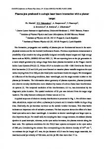
Figure 1 shows the part of the copper artwork coated with a thin layer of deposit and four areas of laser-light irradiation with different energy densities. Figure 2 shows the interaction zones on ceramic artwork after treatment with laser beams of different energy densities. The ruby laser used in the experiment was in the TEM00 mode, Q-switched. The pulse length was t = 30 ns, the wavelength of the laser light λ = 694.3 nm, and the coherent length lc = 1 m. The output beam of light was Ø = 16 mm, the output energy was E = 1 J with the Gaussian distribution, and the energy density ED = 5.00 · 103 J/m2 . The process of irradiation was performed under atmospheric conditions. Laser light was focused by a lens of f = 0.1 m. The artwork was placed normal to the laser-beam propagation. The laser energy density was changed by changing the distance between the artwork surface and the lens. The following parameters are shown in Table 1: the diameter of the interaction zone Ø, surface of interaction spots S, energy of incident laser beam E, and energy density for selected interaction zones on the surface of copper and ceramic artwork ED. Selected areas have similar values of energy density in order to compare the results of interaction.
382
Volume 31, Number 4, 2010
Journal of Russian Laser Research
Fig. 1. Interaction zones on copper artwork.
Fig. 2. Interaction zones on ceramic artwork.
The effects of laser light irradiation are studied using a JEOL JSM-6610LV scanning electron microscope (SEM) connected to an INCA350 energy-dispersive EDX unit. To accelerate, an electron voltage of 20 kV is chosen. The EDX unit was carried out the chemical analysis of the cleaning zone and the sediment composition; the area of the sediment covered the case. The ceramic surface of the sample before ion analysis was coated with a thin gold layer of 20 nm thickness and 19,320 kg/m3 density, in order to achieve better image structure. Table 1. Interaction Parameters. Interaction
Copper zones
Ceramic zones
parameters
3
4
4
5
Ø, mm
3.33
3.02
3.38
4.51
mm2
8.761
7.062
8.971
15.896
E, J
0.9
0.9
0.9
0.9
ED = E/S, 103 J/m2
102.82
61.63
100.36
56.60
S,
The results of the experiment described above should allow one to conduct a series of tests with similar artifacts using holographic interferometry.
3.
Results and Discussion
The interaction of laser beams with materials is a complex phenomenon that depends on many factors. The energy density of the laser beam, the time of irradiation or pulse length, the wavelength, and the energy distribution within the beam are related to laser characteristics. The reflection and absorption coefficients, surface shape, homogeneity, temperature coefficient, melting point, and boiling point are related to the material of the object. In nanosecond-laser divestment of inorganic encrustation using a Q-switched ruby laser, the prevailing processes are the selective explosive vaporization and the palliation induced by shock-wave generation. Macroscopic, visual analysis of the tested samples of cultural heritage objects (Figs. 1 and 2) show that the applied laser energy density caused more or less removal of deposits. Microscopic tests, conducted by SEM and EDX, allow one to determine the limit laser density energy that cleans the surface without
383
Journal of Russian Laser Research
Volume 31, Number 4, 2010
degradation. The interaction zones Nos. 1 and 2 on the copper samples and No. 1 on the ceramic one show the melting effects, which have to be avoided. The analysis is focused on the zone where there is no damage on the artwork surfaces. Table 2. Chemical Composition of Copper, Weight %. Number of IZ
C
O
S
Cl
Cu
Zn
Sum
3, CZ
7.29
4.78
0.00
0.00
87.93
0.00
100.00
3, DZ
21.76
8.33
0.00
0.29
69.62
0.00
100.00
4, CZ
6.05
4.99
0.00
0.00
88.17
0.79
100.00
4, DZ
22.87
8.10
0.00
0.24
68.30
0.50
100.00
a)
b)
c)
d)
Fig. 3. SEM and EDX of interaction zone No. 3 on the copper surface at laser energy density ED = 102.82·103 J/m2 . Notation: SEM, interaction zone No. 3 (a), SEM, center of interaction zone No. 3 (b), EDX, center of interaction zone No. 3 (c), and EDX, sediment in the center of interaction zone No. 3 (d).
Tables 2 and 3 give a quantitative analysis of the interaction zones Nos. 3 and 4, for artwork made of copper and Nos. 4 and 5 for artwork of neolithic pottery (IZ is the interaction zone, CZ is the clean zone, and DZ is the deposit zone). Figures 3 and 4 show interaction zones 3 and 4 for copper artwork and their spectra obtained using SEM and EDX analysis.
384
Volume 31, Number 4, 2010
Journal of Russian Laser Research
a)
b)
c)
d)
Fig. 4. SEM and EDX of interaction zone No. 4 on the copper surface at laser energy density ED = 61.63·103 J/m2 . Notation: SEM, interaction zone No. 4 (a), SEM, center of interaction zone No. 4 (b), EDX, center of interaction zone No. 4 (c), and EDX, sediment in interaction zone No. 4 (d).
Table 3. Chemical Composition of Neolithic Ceramic, Weight %. Number of IZ
C
Na
Mg
Al
Si
Cl
K
Ca
Mn
Fe
Cu
O
Sum
4 CZ
0.00 0.36 1.41 9.78 27.25 0.00 4.10 4.39 0.00 7.24 0.00 45.47 100.00
4 DZ
24.05 0.00 0.23 1.05 3.07 0.06 0.47 0.79 0.00 0.93 0.00 69.34 100.00
5 CZ
22.80 0.00 0.27 1.50 4.43 0.00 0.59 0.96 0.00 1.30 0.00 68.16 100.00
By visual inspection, SEM and EDX analysis confirmed that interaction zones Nos. 3 and 4 show incomplete cleaning of the surface of copper samples, but no melting effects. Results of chemical analysis of interaction zone No. 3, shown in Table 2, indicate that an energy density of 103 · 103 J/m2 reduced carbon content (7.29%) and oxygen (4.78%) content compared to the sediment zone (21.76% C and 8.33% O). In this zone, partial removal of the organic sediment layer occurred. This is confirmed by qualitative analysis shown in Fig. 3c and d. A similar phenomenon is present in interaction zone No. 4. The laser energy density was around 60 · 103 J/m2 , which is the maximum allowed laser energy density for diagnostic methods. Interaction zone No. 4 (shown in Fig. 5) was irradiated by laser light with energy density ED = 100.36 · 103 J/m2 .
385
Journal of Russian Laser Research
Volume 31, Number 4, 2010
Fig. 5. SEM of interaction zone No. 4 at laser energy density ED = 100.36 · 103 J/m2 .
a)
c)
b)
Fig. 6. EDX analysis of chemical composition of different parts of interaction zone No. 4 at laser energy density ED = 100.36 · 103 J/m2 . Central part of the zone irradiated with laser light of the great intensity (a), borders – the border area between the interaction zones irradiated with laser light and sediment (b), and sediment – area covered by organic sediment (c).
Figures 6a, b, and c show EDX spectra and quality analysis of different parts of interaction zone No. 4, namely, the central part of the zone irradiated with laser light of the great intensity (a), borders – the border area between the interaction zones irradiated with laser light and sediment (b), and sediment – area covered by organic sediment (c). The EDX spectrum, shown in Figs. 6b and c, contains elements characteristic of the organic deposits (C, O), as opposed to the zone without sediment (Fig. 6a). Analysis of the distribution of elements within zone No. 4 is performed using the mapping method. Figure 7 shows the mapping pictures.
386
Volume 31, Number 4, 2010
Journal of Russian Laser Research
Fig. 7. Mapping of elements within zone No. 4 of ceramic artwork. SEM (a), Al (b), Si (c), K (d), Fe (e), Mg (f), O (g), Ca (h), and C (i).
Fig. 8. SEM and EDX analysis of interaction zone No. 6 at ED = 56.60 · 103 J/m2 . SEM center of interaction zone No. 5 (left) and EDAX spectra of interaction zone No. 5 (right).
387
Journal of Russian Laser Research
Volume 31, Number 4, 2010
Generally, SEM and EDX analysis shows a partial cleaning of a sample surface of the ceramics. Results of chemical analysis of interaction zone No. 4, shown in Table 3, indicate that the zone irradiated by laser light with energy density of 100.36 · 103 J/m2 show no presence of carbon (0%) and reduced oxygen content (45.47%). In the border zone, 8.30% C and 54.44% O are present, while the zone of the sediment has 5.24% C and 69.34% O. Partial removal of the organic sediment layer has occurred, as indicated by the qualitative analysis presented in Figs. 6 and 7. At the border of zone No. 4, the amount of carbon and oxygen (8.30% C and 54.44% O) is much less, compared to the same part of interaction zone No. 1 (22.87% C and 68.15% O). In contrast, the content of these elements in the sediment is approximately the same as in the area outside of the laser irradiation. Figure 8 presents the SEM and EDX spectrum of central zone No. 5. The laser beam had ED = 56.60 · 103 J/m2 . Chemical analysis (Table 3) shows that in this zone there was no significant removal of organic layers. In the central part of zone No. 5, the amount of C and O is the same as in the sediment of zone No. 4. The results of chemical analysis are shown in Fig. 8. The ablation rate represents the single-pulse efficiency of laser removal at a given fluence. Knowledge of the dependence of the ablation rate on laser fluence is of practical importance for precise control of the removal of the encrustation [16]. Laser ablation starts to be observed above the ablation threshold. Above this value, the removal is almost linear up to the saturation fluence, indicating the point at which the efficiency is significantly reduced by dissipative phenomena such as, in particular, ionization and plasma formation. The maximum efficiency is achieved at the intermediate fluence. For example, in a linear ablation domain, the ablation rate for vaporization removal of black crust [16] using a Nd:YAG laser with different pulse duration between 125–950 ns is 1–2.5 J/cm2 (10 − 25 · 103 J/m2 ). These results are similar to the results obtained in our tests.
4.
Conclusions
We have presented the results of ruby-laser-light interaction with the surfaces of samples of copper and ceramics from the Neolithic Age. It is useful to determine the damage threshold for a chosen wavelength, although light in the visible part of the spectrum is commonly used in diagnostic methods. SEM and EDX analysis shows that the maximum allowable energy density is 100 · 103 J/m2 for ruby-laser light in diagnostic methods and in the cleaning of organic sediment deposited on the surface of the artwork without degradation of the surface and melting the copper and ceramic. The recommended safety laser energy density is 20 · 103 J/m2 for both cases. The characteristic fluences obtained are specific for the tested artifact materials in this experiment and can be recommended for use in the treatment of other related objects only with great caution. The unwanted, hypothetical focusing effect, due to concavity of the surface, can be five times greater without damaging the tested artefacts. This fluence (20 · 103 J/m2 ) is quite sufficient for holographic interferometric testing, but it is necessary to repeat the operation on the same zone to produce satisfactory cleaning results on the tested artefact surfaces.
Acknowledgments This work was financially supported by the Ministry of Science and Technological Development of Serbia under Project No. TR-19205A.
388
Volume 31, Number 4, 2010
Journal of Russian Laser Research
References 1. M. Bass, Laser Material Processing, North Holland, Amsterdam (1983). 2. I. Cooper, Laser Cleaning in Conservation: An Introduction, Butterworth-Heinemann, Oxford, UK (1998). 3. V. Tornari, Anal. Bioanal. Chem., 387, 761 (2007). 4. L. Lazarini, L. Marchesini, and J. F. Asmus, J. Vac. Sci. Technol., 10, 1039 (1973). 5. J. F. Asmus, Laser Focus, 12, 56 (1976). 6. J. Asmus, “Serendipity, punctuated,” Proceedings of LACONA VI [Lasers in the Conservation of Artworks], (Vienna, 2005), Springer-Verlag, Heidelberg (2006). 7. V. Verg`es-Belmin, “Comparison of three cleaning methods – microsandblasting, chemical pads and Q switched YAG laser – on a portal of the cathedral of Notre-Dame in Paris, France,” Proceedings of LACONA I [Lasers in the Conservation of Artworks], (Heraklion, Crete, Greece, 4–6 October, 1995), Verlag-Mayer (1997), p. 17. 8. K. Dickmann, S. Klein, and V. Zafiropulos, “Laser cleaning of marble: discolouration effects using various Nd:YAG laser wavelengths, (ω, 2ω, 3ω),” in: R. Salimbeni (ed.), Laser Techniques and Systems in Art Conservation, SPIE, 4402, 54 (2001). 9. J.F. Asmus, G. Guattari, L. Lazzarini, et al., Stud. Conserv., 18, 49 (1973). 10. V. Tornari, V. Zafiropulos, A. Bonarou, et al., J. Opt. Lasers Eng., 34, 309 (2000). 11. V. Tornari, A. Bonarou, V. Zafiropulos, et al., J. Cult. Heritage, 1, S325 (2000). 12. V. Tornari, A. Bonarou, V. Zafiropulos, et al., “Holographic interferometry sequential investigation of long-term photomechanical effects in the excimer laser restoration of artworks,” in: V. I. Vlad (ed.), ROMOPTO 2000: Sixth Conference on Optics, SPIE, 4430, 153 (2001). 13. J. Marczak, C. Jach, A. Sarzynski, and R. Ostrowski, “Experimental and Theoretical Indications on Laser Cleaning,” Proceedings of LACONA V [Lasers in the Conservation of Artworks], (Osnabruck, Germany, 15–18 September, 2003), Springer, Berlin/Heidelberg (2006), Pt. III [DOI: 10.1007/3-54027176-7 13]. 14. P. Targowski, B. Ruble, M. Gora, et al., Appl. Phys. A: Mater. Sci. Process., 92, No. 1 (2008) [DOI:10.1007/s00339-008-4446-x]. 15. P. Mottner, G. Wiedemann, G. Haber, et al., “Laser Cleaning of Metal Surface – Laboratory Investigations,” Proceedings of LACONA V [Lasers in the Conservation of Artworks], (Osnabruck, Germany, 15–18 September, 2003), Springer, Berlin/Heidelberg (2006), Pt. II [DOI: 10.1007/3-54027176-7 10]. 16. S. Siano, “Principles of Laser Cleaning in Conservation,” in: M. Schreiner and M. Strlic (eds.), Handbook on the Use of Lasers in Conservation and Conservation Science, COST G7 (2007). 17. M. Sreckovic, A. Kovacevic, S. Bojanic, et al., Laser Phys., 11, 336 (2001). 18. M. Sreckovic, Lj. Vulicevic, S. Bojanic, et al., J. Mater. Eng., 14, 327 (2003). 19. M. Sreckovic, S. Ristic, D. Druzijanic, et al., “Some aspects of the theory and praxis on laser interaction with the material for special construction, ” Proceedings of the 4th DAAAM International Conference on Advanced Technologies for Developing Countries (Slavonski Brod, Croatia, 21–24 September 2005), p. 321. 20. S. Polic-Radovanovic, “Application of lasers in the protection and diagnosis of material objects of cultural heritage,” PhD Thesis, Faculty of Mechanical Engineering, Belgrade Uni., Serbia (2007). 21. A. Milosavljevic, S. Petronic, M. Sreckovic, et al., Acta Phys. Pol. A, 15, 823 (2009).
389