Callicott, J.H., Mattay, V.S., Bertolino, A., Finn, K., Coppola, R., Frank, J.A., ⦠& Weinberger,. D.R. (1999). Physiological characteristics of capacity constraints in ...
RUNNING HEAD: N-BACK. META-ANALYSIS OF fMRI STUDIES IN CHILDREN
SUPPLEMENTARY MATERIAL FOR:
N-back Working Memory Task: Meta-analyses of Normative fMRI Studies with Children Zachary Yaple1 Marie Arsalidou2,3, * 1
Centre for Cognition and Decision Making, National Research University Higher School of
Economics, Moscow, Russian Federation 2
Department of Psychology, National Research University Higher School of Economics, Moscow,
Russian Federation 3
Department of Psychology, York University, Toronto, Canada
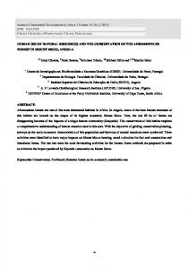
Methods The key terms: ‘fMRI’ AND ‘n-back’, specifying language as English, and document type as article were searched (http://www.webofknowledge.com) on the 7th of August 2017. This search was set to look for articles published from January 1st, 2011 to August 7th, 2017; eligible articles published before 2011 were taken from previously published meta-analyses using the n-back (Rottschy et al., 2012). Our literature search yielded 328 articles, which underwent a series of selection criteria (Figure S1). Specifically, in order to be included in the analyses, articles had to (a) report healthy participants, (b) use fMRI, (c) report whole-brain, within-group results using randomeffects analysis, (d) report stereotaxic coordinates in Talairach or Montreal Neurological Institute
RUNNING HEAD: N-BACK. META-ANALYSIS OF fMRI STUDIES IN CHILDREN
(MNI) space, and (e) include contrasts of the n-back task (e.g. 2 back > 1 back). Only those articles with adult participants (age range 18-65) were included in the analysis. Both examiners undertook this selection process separately, and then came to a final agreement. Data from 48 articles between 2012 and 2017 were included in the meta-analyses. Additional articles using the n-back task with adults were taken from a recent meta-analysis (Rottschy et al., 2012). Four articles, which included adults older than 65 years were not included (Wishart et al., 2006; Döhnel et al., 2008; Lim et al., 2008; McGeown et al., 2008), thus data from 46 articles from Rottschy et al. (2012) were included in our meta-analysis. The total number of articles was 94, with included data from 97 subject groups, 131 experiments (i.e., contrasts). Twenty-three articles reported more than one experiment (as indicated by a in Table S1) and three articles reported results for two separate subject groups (Goldstein et al., 2005; Schmidt et al., 2009; Yan et al., 2011 as indicated by b in Table S1). Table S1 shows article information, participant demographics and contrast included in the analysis. Activation likelihood Estimate (ALE) meta-analysis was computed using GingerALE 2.3.6 (http://brainmap.org/ale/; Eickhoff et al., 2017). All coordinates were transformed into the same space: MNI coordinates were converted to Talairach using the Lancaster et al., (2007) transformation algorithm. Resulting statistical maps were thresholded using a cluster level correction for multiple comparisons p = 0.05 at a cluster forming threshold set at p < 0.001 (Eickhoff et al., 2017). Single study and contrast study analyses were performed between adults and children. Contrast analyses are performed on images corrected for multiple comparisons, thus the threshold for contrasts between adults and children is set to p = 0.01 uncorrected, with 5000 permutations and minimum volume 50 mm3. Lastly, a Fisher-Freeman-Halton Exact Test was calculated to examine whether experiments were partial to task type (verbal, shape, visuospatial, etc.) or contrast type (e.g. 2-back minus baseline, 1-back minus baseline).
RUNNING HEAD: N-BACK. META-ANALYSIS OF fMRI STUDIES IN CHILDREN
Results N-back tasks for adults between 18 and 65 years, with an average age of 29.83 ±6.56 years (55.85% male; 85.45% right handed), yielded a total of 1648 foci. The Fisher-Freeman-Halton Exact Test (2x3) revealed no statistical significance in frequency across task modality (Verbal and Visuospatial n-back) and contrast type (2 back, 1-back, Linear Trend of load; p = 0.353, FisherFreeman-Halton Exact Test), indicating that the results were not biased towards a contrast type or task modality.
N-back: Adults N-back tasks in adults showed significant ALE scores mainly in large bilateral clusters in the prefrontal and parietal cortex (Table S2). Other regions included the insula, claustrum and cerebellum.
Contrast: Adults vs Children Table S3 shows a complete list of significant coordinates from the conjunction analysis and contrast between children and adults. A conjunction analysis shows significant ALE values in the superior and medial frontal gyri (Brodmann Area, BA 6) and in superior and parietal lobules (BA 7, 40). Other common regions include the insula and the cerebellum. Children did not have any suprathreshold clusters compared to adults. Compared to children, adults showed increased ALE values mainly in prefrontal regions (BA 9, 46, 10), the claustrum and parietal cortex (BA 7, 40).
RUNNING HEAD: N-BACK. META-ANALYSIS OF fMRI STUDIES IN CHILDREN
Keywords: TOPIC: fMRI AND “n back” AND LANGUAGE: (English) AND DOCUMENT TYPES (Article); TIMESPAN: 2011-‐Present; n = 328
Identification
Screening
No healthy contrasts n = 4
Age < 18: n = 14
No fMRI n = 35 No whole-‐brain coordinates n = 58
No n back contrast n = 10
No control group or within group contrast n = 65
Age > 65: n = 14
Eligibility
Removed n = 200
Eligible articles from previous meta-‐analysis (Rottschy et al., 2012) + n = 46
Eligible articles n = 128
No foci reported n = 80
Eligible articles n = 48
Included
Adults (18-‐65 yrs) n = 94; experiments = 131; foci = 1648
Figure S1. PRISMA flowchart for eligibility of articles included in the adult meta-analysis.
RUNNING HEAD: N-BACK. META-ANALYSIS OF fMRI STUDIES IN CHILDREN
Table S1. Information on source datasets included in the meta-analysis of adults Gender
Hand
Age range/ Mean,
Article
n
(M)
(R)
(STD)
Foci
Task Modality
Contrast
Allen et al., 2006
10
8
All
23-35
6
Verbal n-back
2-back > 0-back
Alonso-Lana et al., 2016 a
28
12
All
44.01(6.03)
1
Verbal n-back
2-back > 1-back
1
Verbal n-back
2-back > rest
3
Verbal n-back
1-back > rest
Beneventi et al., 2007
12
6
All
21-29
24
Face n-back
Linear trend of WM load
Binder & Urbanik, 2005 a c
12
7
All
20-29
19
Verbal n-back
2-back > 0-back
17
Shape n-back
2-back > 0-back
8
Verbal n-back
2-back and 1-back > 0-back
8
Verbal n-back
Linear trend of WM load
18
Visuospatial n-back
Linear trend of WM load
Cader et al., 2006 a
Callicott et al., 1999
16
9
6
6
All
NA
23-51
18-39
RUNNING HEAD: N-BACK. META-ANALYSIS OF fMRI STUDIES IN CHILDREN
Campanella et al., 2013
32
14
All
21.2 (~2.2)
6
Digit n-back
2-back > 0-back
Caseras et al., 2006
12
4
All
24-45
10
Verbal n-back
Linear trend of WM load
Cerasa et al., 2008
30
30
All
18-43
16
Visuospatial n-back
2-back > 0-back
Choo et al., 2005
14
9
All
21.8 (0.8)
8
Verbal n-back
Linear trend of WM load
Ciesielski et al., 2006
10
5
All
20.4-27.6
15
Categorical n-back
2-back > 0-back
Cohen et al., 1997
10
5
NA
18-34
9
Verbal n-back
Linear trend of WM load
D’Aiuto et al., 2015 a
17
10
NA
26.38 (6.78)
3
Verbal n-back
2-back > 0-back
1
Verbal n-back
1-back > 0-back
Deckersbach et al., 2008
17
17
All
25.6 (5.9)
12
Verbal n-back
2-back > rest
Dima et al., 2014 a
40
20
All
31.5 (10.4)
10
Verbal n-back
3-back > 0-back
8
Verbal n-back
2-back > 0-back
5
Verbal n-back
1-back > 0-back
Dores et al., 2014
10
6
All
27.1 (2.27)
20
Visuospatial n-back
2-back > rest
Drapier et al., 2008 a
20
10
NA
26-63
4
Verbal n-back
1-back > rest
6
Verbal n-back
2-back > rest
7
Verbal n-back
3-back > rest
RUNNING HEAD: N-BACK. META-ANALYSIS OF fMRI STUDIES IN CHILDREN
Druzgal & D’Esposito, 2001
9
5
All
21-27
12
Face n-back
Linear trend of WM load
Duggirala et al., 2016 a c
50
28
All
23.62 (3.17)
13
Categorical n-back
2-back > 0-back
15
Face n-back
2-back > 0-back
18
Verbal n-back
2-back > 0-back
El-Hage et al., 2011
90
45
All
19-56
5
Verbal n-back
Linear trend of WM load
Elzinga et al., 2007
14
14
NA
34.6 (10.9)
21
Verbal n-back
3, 2, 1-back > rest
Falkenberg et al., 2015
15
10
All
19-35
9
Verbal n-back
2, 1-back > 0 back
2013
41
24
All
40.27 (9.8)
2
Verbal n-back
2-back > rest
Forn et al., 2007
10
5
NA
NA
10
Verbal n-back
2-back > 0-back
Frangou et al., 2008 a
7
2
All
39 (5.88)
11
Verbal n-back
2-back > 0-back
5
Verbal n-back
Linear trend of WM load
Fernández-Corcuera et al.,
Fusar-Poli et al., 2011
15
9
All
25.18 (5.07)
8
Verbal n-back
Linear trend of WM load
Garrett et al., 2011
19
13
17
34.85 (12.54)
12
Verbal n-back
1-back > 0-back
Gillis et al., 2016
15
15
All
18-36
34
Categorical n-back
2-back > 0-back
Göbel et al., 2016
21
21
All
21-49
65
Verbal n-back
Linear trend of WM load
RUNNING HEAD: N-BACK. META-ANALYSIS OF fMRI STUDIES IN CHILDREN
Goldstein et al., 2005 b
7
7
All
32.1 (6.6)
9
Verbal n-back
3-back > 1-back
Goldstein et al., 2005 b
7
0
All
34.1 (12.2)
16
Verbal n-back
3-back > 1-back
Gropman et al., 2011
21
7
17
31.8 (2.7)
43
Verbal n-back
Linear trend of WM load
Harvey et al., 2005
10
5
All
18-42
10
Verbal n-back
3, 2, 1-back > rest
Honey et al., 2000
20
20
All
19-64
10
Verbal n-back
Linear trend of WM load
Honey et al., 2003
27
21
All
35.1 (9.9)
11
Verbal n-back
2-back > 0-back
Huang et al., 2015 a
18
6
All
36-55
10
Visuospatial n-back
2-back > 1-back
5
Visuospatial n-back
1-back > 0-back
Jogia et al., 2012
37
21
NA
18-63
5
Verbal n-back
3, 2, 1-back > rest
Johannsen et al., 2013
12
4
All
21.7-37.8
14
Verbal n-back
2-back > 0-back
Kasahara et al., 2011
9
4
All
19-53
9
Verbal n-back
Linear trend of WM load
Kim et al., 2006
12
9
11
21-46
8
Verbal n-back
2-back > rest
Koppelstaetter et al., 2008
15
15
All
25-47
16
Verbal n-back
2-back > 0-back
Korsnes et al., 2013
11
0
NA
18-45
9
Digit n-back
2-back > 1-back
Koshino et al., 2008
11
10
10
28.7 (10.9)
15
Face n-back
2, 1, 0-back > rest
Kumari et al., 2003
12
12
All
20-40
8
Digit n-back
2, 1-back > 0-back
RUNNING HEAD: N-BACK. META-ANALYSIS OF fMRI STUDIES IN CHILDREN
Kumari et al., 2006 a
13
13
All
18-55
16
Visuospatial n-back
0-back > rest
22
Visuospatial n-back
1-back > 0-back
18
Visuospatial n-back
2-back > 0-back
Lamp et al., 2016
16
5
All
18-27
17
Shape n-back
1-back > rest
Leung & Alain, 2011 a c
16
5
All
18-30
13
Categorical n-back
2-back > 1-back
13
Visuospatial n-back
2-back > 1-back
18
Verbal n-back
2-back > rest
10
Verbal n-back
1-back > rest
7
Verbal n-back
0-back > rest
Li et al., 2014 a
15
0
All
19-22
Loughead et al., 2009
33
18
All
33 (10.55)
13
Shape n-back
Linear trend of WM load
Luo et al., 2014
25
25
All
20-28
12
Face n-back
2-back > 0-back
Lythe et al., 2012
20
20
26.7 (6.7)
2
Verbal n-back
Linear trend of WM load
Manelis & Reder, 2014
16
5
All
24
18
Verbal n-back
Linear trend of WM load
Manktelow et al., 2014
21
13
All
18-60
22
Verbal n-back
2-back > 0-back
Marquand et al., 2008
20
7
All
43.7 (8.6)
19
Verbal n-back
2-back > 0-back
Matsuo et al., 2007 a
15
6
12
37.7 (12.1)
2
Visuospatial n-back
2-back > 0-back
RUNNING HEAD: N-BACK. META-ANALYSIS OF fMRI STUDIES IN CHILDREN
4
Visuospatial n-back
1-back > 0-back
Mattfeld et al., 2016
17
11
NA
28.7 (4.0)
6
Verbal n-back
Linear trend of WM load
McAllister et al., 1999 a
11
4
All
30.6 (11.2)
2
Verbal n-back
2-back > 0-back
5
Verbal n-back
1-back > 0-back
Monks et al., 2004
12
12
All
45.6 (3.52)
14
Verbal n-back
2-back > 0-back
Nebel et al., 2005 a
19
12
All
26-37
30
Verbal n-back
2-back > rest
10
Verbal n-back
1-back > rest
Nichols et al., 2017
41
88
All
30.8 (7.9)
7
Verbal n-back
3-back > 0-back
Norbury et al., 2014
15
10
All
23-61
6
Verbal n-back
3, 2, 1-back > 0-back
Oren et al., 2017
24
16
All
22-35
5
Digit n-back
Linear trend of WM load
Park et al., 2016
45
22
All
22.87 (~2.205)
41
Shape n-back
2-back > 0-back
Pomarol-Clortet et al., 2012
46
27
All
20-62
2
Verbal n-back
2-back > rest
Qin et al., 2009
27
27
All
18-25
14
Digit n-back
2-back > 0-back
Ragland et al., 2002 a c
11
6
All
21-53
10
Verbal n-back
2-back > 1-back
7
Verbal n-back
2-back > 0-back
6
Verbal n-back
1-back > 0-back
RUNNING HEAD: N-BACK. META-ANALYSIS OF fMRI STUDIES IN CHILDREN
Rama et al., 2001 a
8
0
All
21-25
6
Shape n-back
2-back > 1-back
9
Shape n-back
2-back > 0-back
5
Shape n-back
1-back > 0-back
32
Verbal n-back
2-back > 0-back
24
Verbal n-back
1-back > 0-back
Richter et al., 2013
34
26
NA
23.8 (~2.15)
25
Face n-back
2-back > 0-back
Reynolds et al., 2009
18
7
All
19-29
5
Verbal n-back
3-back > 1-back
Riccaiardi et al., 2006 a c
6
6
All
28 (1)
36
Tactile n-back
1-back > 0-back
28
Visuospatial n-back
1-back > 0-back
Rocca et al., 2014
52
24
All
22-52
16
Verbal n-back
Linear trend of WM load
Sabri et al., 2014
20
10
All
25 (5)
16
Verbal n-back
2-back > 1-back
Sánchez-Carrión et al., 2008 a
14
NA
All
24.2 (4.7)
18
Digit n-back
3-back > 0-back
16
Digit n-back
2-back > 0-back
Savini et al., 2012
12
12
All
19-32
9
Shape n-back
Linear trend of WM load
Scheuerecker et al., 2008 a
23
19
All
32.6 (9.9)
8
Verbal n-back
2-back > 0-back
15
Verbal n-back
2-back deg. > 0-back deg.
RUNNING HEAD: N-BACK. META-ANALYSIS OF fMRI STUDIES IN CHILDREN
18-58/34.36 Schmidt et al., 2009 b
25
25
All
(13.24)
8
Verbal n-back
Linear trend of WM load
18-58/33.13 Schmidt et al., 2009 b
25
0
All
(12.31)
6
Verbal n-back
Linear trend of WM load
Schmidt et al., 2012 a
32
NA
NA
24.6 (~3.6)
1
Verbal n-back
3-back > 2-back
16
Verbal n-back
3-back > 0-back
12
Verbal n-back
2-back > 0-back
Schneiders et al., 2011
48
22
All
19-31
22
Shape n-back
2-back > 0-back
Seo et al., 2011
22
0
All
38.27 (8.48)
18
Verbal n-back
2-back > 0-back
Shen et al., 1999
9
6
All
20-40
24
Visuospatial n-back
2-back > rest
Spreng et al., 2014
36
17
NA
22.3 (3.8)
18
Face n-back
2-back > rest
Stretton et al., 2012 a
15
4
11
19-58
5
Visuospatial n-back
2-back > 0-back
4
Visuospatial n-back
1-back > 0-back
Takeuchi et al., 2012
248 126
All
21.1 (1.8)
11
Verbal n-back
2-back > 0-back
Thomas et al., 2005
16
5
NA
21-50
9
Verbal n-back
2-back > 0-back
Thornton & Conway, 2013
16
6
All
22 (2.3)
16
Face n-back
Linear trend of WM load
RUNNING HEAD: N-BACK. META-ANALYSIS OF fMRI STUDIES IN CHILDREN
Veltman et al., 2003
21
7
NA
22.7 (3.6)
11
Verbal n-back
Linear trend of WM load
Veltman et al., 2005
10
3
All
22.9 (1.27)
20
Verbal n-back
Linear trend of WM load
Wesley et al., 2016
11
4
NA
28.8 (7.8)
3
Verbal n-back
1-back > 0-back
Winston et al., 2013
28
11
25
19-64
3
Visuospatial n-back
Linear trend of WM load
Wu et al., 2017
45
24
All
24.07 (4.83)
4
Digit n-back
2-back > 0-back
Yan et al., 2011 b
28
12
All
20.4 (1.4)
6
Visuospatial n-back
2-back > 0-back
Yan et al., 2011 b
28
12
All
20.9 (1.5)
8
Visuospatial n-back
2-back > 0-back
Verbal Yoo et al., 2004 a c
14
9
All
21-34
16
n-back
(visual) Verbal
2-back > 1-back n-back
23
(auditory)
2-back > 1-back
Yoo & Choi, 2005
10
8
All
20-30
22
Face n-back
2-back > rest
Zhou et al., 2014
18
9
All
24.94 (7.29)
5
Verbal n-back
2-back > 0-back
Ziemus et al., 2007
9
6
All
35-63
15
Verbal n-back
2-back > 0-back
RUNNING HEAD: N-BACK. META-ANALYSIS OF fMRI STUDIES IN CHILDREN
Note: n = sample size; M = Male; R = Right handed; STD = Standard deviation; NA = not available; a = for each experiment with within-group contrasts, foci was compiled into one experiment; b = experiment contained more than one group with a different set of foci; c = study includes multiple task modalities
RUNNING HEAD: N-BACK. META-ANALYSIS OF fMRI STUDIES IN CHILDREN
Table S2: Concordant brain regions related to the n-back task in adults
Cluster # Volume mm3 ALE Value x 1
2
3
4
27152
14560
13856
12824
y
z
Label
0.143 -30
20
4 L Insula BA 13
0.114 -42
2
0.084 -44
20
32 L Middle Frontal Gyrus BA 9
0.082 -30
-6
54 L Middle Frontal Gyrus BA 6
0.072 -34
46
18 L Middle Frontal Gyrus BA 10
0.036 -44
12
34 L Precentral Gyrus BA 6
8 L Precentral Gyrus BA 44
0.123 -34 -54
40 L Inferior Parietal Lobule BA 40
0.055 -12 -70
48 L Precuneus BA 7
0.133
38 -48
40 R Inferior Parietal Lobule BA 40
0.082
30 -58
42 R Superior Parietal Lobule BA 7
0.057
14 -70
50 R Precuneus BA 7
0.031
48 -38
50 R Inferior Parietal Lobule BA 40
0.103
40
34
30 R Superior Frontal Gyrus BA 9
0.042
48
10
36 R Middle Frontal Gyrus BA 9
0.041
48
14
24 R Inferior Frontal Gyrus BA 9 48 L Superior Frontal Gyrus BA 6
5
11488
0.129
0
12
6
6808
0.114
26
0
7
5416
0.153
30
20
8
3256
0.062
32 -58 -30 R Cerebellum Tuber
0.048
32 -62 -20 R Cerebellum Declive
56 R Sub-Gyral BA 6 4 R Claustrum
RUNNING HEAD: N-BACK. META-ANALYSIS OF fMRI STUDIES IN CHILDREN
9
3240
0.075 -30 -56 -32 L Cerebellar Tonsil
10
2848
0.068 -16
11
1136
0.042
10
2 -4
14 L Caudate Body 8 R Thalamus Ventral Anterior Nucleus
Note: L = Left; R = Right; BA = Brodmann area; Coordinates are reported in Talairach and all results are thresholded with cluster-level threshold was set to p = 0.05 whereas the cluster-forming threshold was set to p < 0.001.
RUNNING HEAD: N-BACK. META-ANALYSIS OF fMRI STUDIES IN CHILDREN
Table S3: Concordant brain regions related to the n-back task in adults versus children Conjunction: Adults AND Children Cluster # Volume mm3 1
2
4088
1488
ALE Value
x
y
z
Label
0.027
-2
14
48 L Superior Frontal Gyrus BA 6
0.023
-8
6
0.018
24
-62
42 R Superior Parietal Lobule BA 7
0.015
48
-44
48 R Inferior Parietal Lobule BA 40
0.014
32
-50
38 R Inferior Parietal Lobule BA 40
0.013
36
-52
44 R Inferior Parietal Lobule BA 40
0.012
42
-48
48 R Inferior Parietal Lobule BA 40 40 L Superior Parietal Lobule BA 7
50 L Medial Frontal Gyrus BA 6
3
1320
0.022
-30
-54
4
936
0.021
-24
-2
5
856
0.023
-38
0
6
648
0.021
30
18
7
520
0.016
-34
-60
-34 L Cerebellar Tonsil
0.014
-28
-64
-26 L Cerebellum Uvula
56 L Sub-Gyral BA 6 38 L Precentral Gyrus BA 6 10 R Insula BA 13
Adults > Children Cluster # Volume mm3
ALE Value
x
y
3.719 43.2
z
Label
1
4928
34.4 27.2 R Middle Frontal Gyrus BA 46
2
480
2.848
-28
14
3
368
2.948
-50
14
28 L Middle Frontal Gyrus BA 9
2.794
-44
12
34 L Middle Frontal Gyrus BA 9
2 L Claustrum
RUNNING HEAD: N-BACK. META-ANALYSIS OF fMRI STUDIES IN CHILDREN
4
272
2.605
-33
42
11 L Middle Frontal Gyrus BA 10
5
208
2.576
34
-4
52 R Middle Frontal Gyrus BA 6
6
136
2.652
38
-46
7
120
2.687
34
8
8
72
2.478
34
-66
38 R Inferior Parietal Lobule BA 40 48 R Middle Frontal Gyrus BA 6 48 R Superior Parietal Lobule BA 7
Children > Adults No suprathreshold cluster Note: L = Left; R = Right; BA = Brodmann area; Coordinates are reported in Talairach and all results are thresholded with cluster-level threshold was set to p = 0.05 whereas the cluster-forming threshold was set to p < 0.001.
RUNNING HEAD: N-BACK. META-ANALYSIS OF fMRI STUDIES IN CHILDREN
References Allen, P.P., Cleare, A.J., Lee, F., Fusar-Poli, P., Tunstall, N., Fu, C.H., … & McGuire, P.K. (2006). Effect of acute tryptophan depletion on pre-frontal engagement. Psychopharmacology (Berl). 187, 486-497. doi 10.1007/s00213-006-0444-x Alonso-Lana, S., Goikolea, J.M., Bonnin, C.M., Sarró, S., Segura, B., Amann, B.L., ... & Salvador, R. (2016). Structural and functional brain correlates of cognitive impairment in euthymic patients with bipolar disorder. PloS one, 11(7), p.e0158867. doi:10.1371/journal.pone.0158867 Beneventi, H., Barndon, R., Ersland, L., & Hugdahl, K. (2007). An fMRI study of working memory for schematic facial expressions. Scand J Psychol, 48, 81-86. 10.1111/j.14679450.2007.00536.x Binder, M., & Urbanik, A.S. (2006). Material-dependent activation in prefrontal cortex: working memory for verbals and texture patterns--initial observations. Radiology. 238, 256-263. https://doi.org/10.1148/radiol.2381041622 Cader, S., Cifelli, A., bu-Omar, Y., Palace, J., & Matthews, P.M. (2006). Reduced brain functional reserve and altered functional connectivity in patients with multiple sclerosis. Brain, 129, 527-537. doi:10.1093/brain/awh670 Callicott, J.H., Mattay, V.S., Bertolino, A., Finn, K., Coppola, R., Frank, J.A., … & Weinberger, D.R. (1999). Physiological characteristics of capacity constraints in working memory as revealed by functional MRI. Cereb Cortex, 9, 20-26.
RUNNING HEAD: N-BACK. META-ANALYSIS OF fMRI STUDIES IN CHILDREN
Campanella, S., Peigneux, P., Petit, G., Lallemand, F., Saeremans, M., Noël, X., … & Ward, R., (2013). Increased cortical activity in binge drinkers during working memory task: a preliminary assessment through a functional magnetic resonance imaging study. PLoS One, 8(4), p.e62260. doi:10.1371/journal.pone.0062260 Caseras, X., Mataix-Cols, D., Giampietro, V., Rimes, K.A., Brammer, M., Zelaya, F., … & Godfrey, E.L. (2006). Probing the working memory system in chronic fatigue syndrome: a functional magnetic resonance imaging study using the n-back task. Psychosom Med, 68, 947-955. doi 10.1097/01.psy.0000242770.50979.5f Cerasa, A., Gioia, M.C., Fera, F., Passamonti, L., Liguori, M., Lanza, P., … & Quattrone, A. (2008). Ventro-lateral prefrontal activity during working memory is modulated by MAO A genetic variation. Brain Research, 1201, 114-21. doi:10.1016/j.brainres.2008.01.048 Choo, W.C., Lee, W.W., Venkatraman, V., Sheu, F.S., & Chee, M.W. (2005). Dissociation of cortical regions modulated by both working memory load and sleep deprivation and by sleep deprivation alone. Neuroimage, 25, 579-587. doi:10.1016/j.neuroimage.2004.11.029 Ciesielski, K.T., Lesnik, P.G., Savoy, R.L., Grant, E.P., & Ahlfors, S.P. (2006). Developmental neural networks in children performing a Categorical N-Back Task. Neuroimage, 33, 980990. doi:10.1016/j.neuroimage.2006.07.028 Cohen, J.D., Perlstein, W.M., Braver, T.S., Nystrom, L.E., Noll, D.C., Jonides, J., Smith, E.E. (1997). Temporal dynamics of brain activation during a working memory task. Nature, 386, 604-608. D’Aiuto, L., Prasad, K.M., Upton, C.H., Viggiano, L., Milosevic, J., Raimondi, G., … & Moore, J.C. (2014). Persistent infection by HSV-1 is associated with changes in functional
RUNNING HEAD: N-BACK. META-ANALYSIS OF fMRI STUDIES IN CHILDREN
architecture of iPSC-derived neurons and brain activation patterns underlying working memory performance. Schizophrenia bulletin, 41, pp.123-132. doi:10.1093/schbul/sbu032 Deckersbach, T., Rauch, S.L., Buhlmann, U., Ostacher, M.J., Beucke, J.C., Nierenberg, A.A., … & Dougherty, D.D. (2008). An fMRI investigation of working memory and sadness in females with bipolar disorder: a brief report. Bipolar Disord, 10, 928-942. doi: 10.1111/j.13995618.2008.00633.x Dima, D., Jogia, J. & Frangou, S. (2014). Dynamic causal modeling of load-‐dependent modulation of effective connectivity within the verbal working memory network. Hum Brain Mapp, 35, 3025-35. doi: 10.1002/hbm.22382 Döhnel, K., Sommer, M., Ibach, B., Rothmayr, C., Meinhardt, J. & Hajak, G. (2008). Neural correlates of emotional working memory in patients with mild cognitive impairment. Neuropsychologia, 46, 37-48. Dores, A.R., Barbosa, F., Carvalho, I.P., Almeida, I., Guerreiro, S., Rocha, B.M., … & Castro-‐ Caldas, A. (2017). Study of behavioural and neural bases of visuo-‐spatial working memory with an fMRI paradigm based on an n-‐back task. J Neuropsychology, 11, 122-134. doi:10.1111/jnp.12076 Drapier, D., Surguladze, S., Marshall, N., Schulze, K., Fern, A., Hall, M.H., … & McDonald, C. (2008). Genetic liability for bipolar disorder is characterized by excess frontal activation in response to a working memory task. Biol Psychiatry, 64, 513-520. doi:10.1016/j.biopsych.2008.04.038 Druzgal, T.J., & D'Esposito, M. (2001). Activity in fusiform face area modulated as a function of working memory load. Brain Res Cogn Brain Res, 10, 355-364.
RUNNING HEAD: N-BACK. META-ANALYSIS OF fMRI STUDIES IN CHILDREN
Duggirala, S.X., Saharan, S., Raghunathan, P. & Mandal, P.K. (2016). Stimulus-dependent modulation of working memory for identity monitoring: A functional MRI study. Brain Cogn, 102, 55-64. http://dx.doi.org/10.1016/j.bandc.2015.12.006 Eickhoff, S. B., Laird, A. R., Fox, P. M., Lancaster, J. L., & Fox, P. T. (2017). Implementation errors in the GingerALE Software: Description and recommendations. Hum Brain Mapp, 38, 7-11. doi: 10.1002/hbm.23342 El-Hage, W., Phillips, M.L., Radua, J., Gohier, B., Zelaya, F.O., Collier, D.A. & Surguladze, S.A. (2013). Genetic modulation of neural response during working memory in healthy individuals: interaction of glucocorticoid receptor and dopaminergic genes. Mol Psychiatry, 18, 174-182. doi: 10.1038/mp.2011.145 Elzinga, B.M., Ardon, A.M., Heijnis, M.K., De Ruiter, M.B., Van, D.R., Veltman, D.J. (2007). Neural correlates of enhanced working-memory performance in dissociative disorder: a functional MRI study. Psychol Med, 37, 235-245. doi:10.1017/S0033291706008932 Falkenberg, I., Chaddock, C., Murray, R.M., McDonald, C., Modinos, G., Bramon, E., … & Allen, P. (2015). Failure to deactivate medial prefrontal cortex in people at high risk for psychosis. Eur Psychiatry, 30, 633-640. http://dx.doi.org/10.1016/j.eurpsy.2015.03.003 Fernández-Corcuera, P., Salvador, R., Monté, G.C., Sarró, S.S., Goikolea, J.M., Amann, B., … & Maristany, T. (2013). Bipolar depressed patients show both failure to activate and failure to de-activate during performance of a working memory task. J Affect Disord, 148,170-178. http://dx.doi.org/10.1016/j.jad.2012.04.009 Forn, C., Barros-Loscertales, A., Escudero, J., Benlloch, V., Campos, S., Antonia, P.M., Avila, C., 2007. Compensatory activations in patients with multiple sclerosis during preserved
RUNNING HEAD: N-BACK. META-ANALYSIS OF fMRI STUDIES IN CHILDREN
performance on the auditory N-back task. Human Brain Mapping 28, 424-430. doi: 10.1002/hbm.20284 Frangou, S., Kington, J., Raymont, V., & Shergill, S.S. (2008). Examining ventral and dorsal prefrontal function in bipolar disorder: a functional magnetic resonance imaging study. Eur Psychiatry, 23, 300-308. doi:10.1016/j.eurpsy.2007.05.002 Fusar-Poli, P., Broome, M.R., Woolley, J.B., Johns, L.C., Tabraham, P., Bramon, E., … & McGuire, P. (2011). Altered brain function directly related to structural abnormalities in people at ultra-high risk of psychosis: longitudinal VBM-fMRI study. J Psychiatr Res, 45, 190-198. doi:10.1016/j.jpsychires.2010.05.012 Garrett, A., Kelly, R., Gomez, R., Keller, J., Schatzberg, A.F. & Reiss, A.L. (2011). Aberrant brain activation during a working memory task in psychotic major depression. Am J Psychiatry, 168,173-182. doi: 10.1176/appi.ajp.2010.09121718 Gillis, M.M., Garcia, S. & Hampstead, B.M. (2016). Working memory contributes to the encoding of object location associations: Support for a 3-part model of object location memory. Behav Brain Res, 311, 192-200. http://dx.doi.org/doi:10.1016/j.bbr.2016.05.037 Göbel, A., Heldmann, M., Göttlich, M., Dirk, A.L., Brabant, G. & Münte, T.F. (2016). Effect of mild thyrotoxicosis on performance and brain activations in a working memory task. PloS one, 11, p.e0161552. doi:10.1371/journal.pone.0161552 Goldstein, J.M., Jerram, M., Poldrack, R., Anagnoson, R., Breiter, H.C., Makris, N., ... & Seidman, L.J. (2005). Sex differences in prefrontal cortical brain activity during fMRI of auditory verbal working memory. Neuropsychology, 19, 509-519. doi: 10.1037/0894-4105.19.4.509 Gropman, A.L., Shattuck, K., Prust, M.J., Seltzer, R.R., Breeden, A.L., Hailu, A., … &
RUNNING HEAD: N-BACK. META-ANALYSIS OF fMRI STUDIES IN CHILDREN
VanMeter, J. (2013). Altered neural activation in ornithine transcarbamylase deficiency during executive cognition: an fMRI study. Hum Brain Mapp, 34,753-761. doi: 10.1002/hbm.21470 Harvey, P.O., Fossati, P., Pochon, J.B., Levy, R., Lebastard, G., Lehericy, S., Allilaire, J.F., Dubois, B. (2005). Cognitive control and brain resources in major depression: an fMRI study using the n-back task. Neuroimage. 26, 860-869. doi:10.1016/j.neuroimage.2005.02.048 Honey, G.D., Bullmore, E.T., & Sharma, T. (2000). Prolonged reaction time to a verbal working memory task predicts increased power of posterior parietal cortical activation. Neuroimage, 12, 495-503. doi:10.1006/nimg.2000.0624 Honey, G.D., Sharma, T., Suckling, J., Giampietro, V., Soni, W., Williams, S.C., & Bullmore, E.T. (2003). The functional neuroanatomy of schizophrenic subsyndromes. Psychol Med, 33, 1007-1018. doi: 10.1017/S0033291703007864 Huang, R.R., Jia, B.H., Xie, L., Ma, S.H., Yin, J.J., Sun, Z.B., … & Luo, D.X. (2016). Spatial working memory impairment in primary onset middle-‐age type 2 diabetes mellitus: An ethology and BOLD-‐fMRI study. J Magn Reson Imaging, 43, 75-87. doi: 10.1002/jmri.24967 Jogia, J., Dima, D., Kumari, V. & Frangou, S. (2012). Frontopolar cortical inefficiency may underpin reward and working memory dysfunction in bipolar disorder. World J Biol Psychiatry, 13, 605-615. doi: 10.3109/15622975.2011.585662 Johannsen, L., Li, K.Z., Chechlacz, M., Bibi, A., Kourtzi, Z. & Wing, A.M. (2013). Functional neuroimaging of the interference between working memory and the control of periodic ankle
RUNNING HEAD: N-BACK. META-ANALYSIS OF fMRI STUDIES IN CHILDREN
movement timing. Neuropsychologia, 51, 2142-2153. doi: 10.1016/j.neuropsychologia.2013.07.009 Kasahara, M., Menon, D.K., Salmond, C.H., Outtrim, J.G., Tavares, J.V.T., Carpenter, T.A., … & Stamatakis, E.A. (2011). Traumatic brain injury alters the functional brain network mediating working memory. Brain Inj, 25, 1170-1187. doi: 10.3109/02699052.2011.608210 Kim, J., Whyte, J., Wang, J., Rao, H., Tang, K.Z., & Detre, J.A. (2006). Continuous ASL perfusion fMRI investigation of higher cognition: quantification of tonic CBF changes during sustained attention and working memory tasks. Neuroimage, 31, 376-385. doi:10.1016/j.neuroimage.2005.11.035 Koppelstaetter, F., Poeppel, T.D., Siedentopf, C.M., Ischebeck, A., Verius, M., Haala, I., ... & Krause, B.J. (2008). Does caffeine modulate verbal working memory processes? An fMRI study. Neuroimage, 39, 492-499. doi:10.1016/j.neuroimage.2007.08.037 Korsnes, M.S., Lövdahl, H., Andersson, S., Björnerud, A., Due-Tönnesen, P., Endestad, T. & Malt, U.F. (2013). Working memory in recurrent brief depression: An fMRI pilot study. J Affect Disord, 149, 383-392. http://dx.doi.org/10.1016/j.jad.2013.02.017 Koshino, H., Kana, R.K., Keller, T.A., Cherkassky, V.L., Minshew, N.J., & Just, M.A. (2008). fMRI investigation of working memory for faces in autism: visual coding and underconnectivity with frontal areas. Cereb Cortex, 18, 289-300. doi:10.1093/cercor/bhm054 Kumari, V., Aasen, I., Taylor, P., ffytche, D.H., Das, M., Barkataki, I., ... & Sharma, T. (2006). Neural dysfunction and violence in schizophrenia: an fMRI investigation. Schizophr Res, 84, 144-164. doi:10.1016/j.schres.2006.02.017
RUNNING HEAD: N-BACK. META-ANALYSIS OF fMRI STUDIES IN CHILDREN
Kumari, V., Gray, J.A., Ffytche, D.H., Mitterschiffthaler, M.T., Das, M., Zachariah, E., ... & Sharma, T. (2003). Cognitive effects of nicotine in humans: an fMRI study. Neuroimage, 19, 1002-1013. doi:10.1016/S1053-8119(03)00110-1 Lamp, G., Alexander, B., Laycock, R., Crewther, D.P. & Crewther, S.G. (2016). Mapping of the underlying neural mechanisms of maintenance and manipulation in visuo-spatial working memory using an n-back mental rotation task: a functional magnetic resonance imaging study. Front Behav Neurosci, 10. doi: 10.3389/fnbeh.2016.00087 Lancaster, J. L., Tordesillas-‐Gutiérrez, D., Martinez, M., Salinas, F., Evans, A., Zilles, K., ... & Fox, P. T. (2007). Bias between MNI and Talairach coordinates analyzed using the ICBM-‐152 brain template. Hum Brain Mapp, 28, 1194-1205. doi: 10.1002/hbm.20345 Leung, A.W. & Alain, C. (2011). Working memory load modulates the auditory “What” and “Where” neural networks. Neuroimage, 55,1260-1269. doi:10.1016/j.neuroimage.2010.12.055 Li, L., Men, W.W., Chang, Y.K., Fan, M.X., Ji, L. & Wei, G.X. (2014). Acute aerobic exercise increases cortical activity during working memory: a functional MRI study in female college students. PloS one, 9, p.e99222. doi:10.1371/journal.pone.0099222 Lim, H.K., Juh, R., Pae, C.U., Lee, B.T., Yoo, S.S., Ryu, S.H., … & Lee, C.U. (2008). Altered verbal working memory process in patients with Alzheimer’s disease. Neuropsychobiology, 57, 181-187. doi: 10.1159/000147471 Loughead, J., Wileyto, E.P., Valdez, J.N., Sanborn, P., Tang, K., Strasser, A.A., & Lerman, C. (2009). Effect of abstinence challenge on brain function and cognition in smokers differs by COMT genotype. Mol Psychiatry. 14, 820-826. doi: 10.1038/mp.2008.132
RUNNING HEAD: N-BACK. META-ANALYSIS OF fMRI STUDIES IN CHILDREN
Luo, Y., Qin, S., Fernandez, G., Zhang, Y., Klumpers, F. & Li, H. (2014). Emotion perception and executive control interact in the salience network during emotionally charged working memory processing. Hum Brain Mapp, 35, 5606-5616. doi: 10.1002/hbm.22573 Lythe, K.E., Williams, S.C., Anderson, C., Libri, V. & Mehta, M.A. (2012). Frontal and parietal activity after sleep deprivation is dependent on task difficulty and can be predicted by the fMRI response after normal sleep. Behav Brain Res, 233, 62-70. doi: 10.1016/j.bbr.2012.04.050 Manelis, A. & Reder, L.M. (2014). Effective connectivity among the working memory regions during preparation for and during performance of the n-back task. Front Hum Neurosci, 8. doi: 10.3389/fnhum.2014.00593 Manktelow, A.E., Menon, D.K., Sahakian, B.J. & Stamatakis, E.A. (2017). Working Memory after Traumatic Brain Injury: The Neural Basis of Improved Performance with Methylphenidate. Front Behav Neurosci, 11. doi: 10.3389/fnbeh.2017.00058 Marquand, A.F., Mourao-Miranda, J., Brammer, M.J., Cleare, A.J., & Fu, C.H. (2008). Neuroanatomy of verbal working memory as a diagnostic biomarker for depression. Neuroreport, 19, 1507-1511. doi: 10.1097/WNR.0b013e328310425e Mattfeld, A.T., Whitfield-Gabrieli, S., Biederman, J., Spencer, T., Brown, A., Fried, R. & Gabrieli, J.D. (2016). Dissociation of working memory impairments and attention-deficit/hyperactivity disorder in the brain. NeuroImage: Clinical, 10, 274-282. doi: 10.1016/j.nicl.2015.12.003 Matsuo, K., Glahn, D.C., Peluso, M.A., Hatch, J.P., Monkul, E.S., Najt, P., … & Soares, J.C. (2007). Prefrontal hyperactivation during working memory task in untreated individuals with major depressive disorder. Mol Psychiatry, 12, 158-166. doi:10.1038/sj.mp.4001894
RUNNING HEAD: N-BACK. META-ANALYSIS OF fMRI STUDIES IN CHILDREN
McAllister, T.W., Saykin, A.J., Flashman, L.A., Sparling, M.B., Johnson, S.C., Guerin, S.J., … & Yanofsky, N. (1999). Brain activation during working memory 1 month after mild traumatic brain injury: a functional MRI study. Neurology, 53, 1300-1308. McGeown, W.J., Shanks, M.F. & Venneri, A. (2008). Prolonged cholinergic enrichment influences regional cortical activation in early Alzheimer’s disease. Neuropsychiatr Dis Treat, 4, 465476. Monks, P.J., Thompson, J.M., Bullmore, E.T., Suckling, J., Brammer, M.J., Williams, S.C., … & Curtis, V.A. (2004). A functional MRI study of working memory task in euthymic bipolar disorder: evidence for task-specific dysfunction. Bipolar Disord, 6, 550-564. Nebel, K., Wiese, H., Stude, P., de, G.A., Diener, H.C., & Keidel, M. (2005). On the neural basis of focused and divided attention. Brain Res Cogn Brain Res, 25, 760-776. doi:10.1016/j.cogbrainres.2005.09.011 Nichols, T.T., Gates, K.M., Molenaar, P. & Wilson, S.J. (2014). Greater BOLD activity but more efficient connectivity is associated with better cognitive performance within a sample of nicotine-‐deprived smokers. Addict Biol, 19, 931-940. doi:10.1111/adb.12060 Norbury, R., Godlewska, B. & Cowen, P.J. (2014). When less is more: a functional magnetic resonance imaging study of verbal working memory in remitted depressed patients. Psychol Med, 44, 1197-1203. doi:10.1017/S0033291713001682 Oren, N., Ash, E.L., Tarrasch, R., Hendler, T., Giladi, N. & Shapira-Lichter, I. (2017). Neural patterns underlying the effect of negative distractors on working memory in older adults. Neurobiol Aging, 53, 93-102. http://dx.doi.org/10.1016/j.neurobiolaging.2017.01.020
RUNNING HEAD: N-BACK. META-ANALYSIS OF fMRI STUDIES IN CHILDREN
Park, J.W., Kim, Y.T., Yun, B.J., Jin, S.U., Lee, S.H., Ahn, S.H., … & Chang, Y. (2016). Stereoscopic 3D objects evoke stronger saliency for nonverbal working memory: An fMRI study. Int J Imag Syst Tech, 26, 76-84. doi: 10.1002/ima.22159 Pomarol-Clotet, E., Moro, N., Sarró, S., Goikolea, J.M., Vieta, E., Amann, B., … & Mckenna, P.J. (2012). Failure of de-activation in the medial frontal cortex in mania: evidence for default mode network dysfunction in the disorder. World J Biol Psychiatry, 13, 616-626. doi: 10.3109/15622975.2011.573808 Qin, S., Hermans, E.J., van Marle, H.J., Luo, J., & Fernandez, G. (2009). Acute psychological stress reduces working memory-related activity in the dorsolateral prefrontal cortex. Biol Psychiatry, 66, 25-32. doi:10.1016/j.biopsych.2009.03.006 Ragland, J.D., Turetsky, B.I., Gur, R.C., Gunning-Dixon, F., Turner, T., Schroeder, L., … & Gur, R.E. (2002). Working memory for complex figures: an fMRI comparison of verbal and fractal n-back tasks. Neuropsychology, 16, 370-379. Rama, P., Martinkauppi, S., Linnankoski, I., Koivisto, J., Aronen, H.J., & Carlson, S. (2001). Working memory of identification of emotional vocal expressions: an fMRI study. Neuroimage, 13, 1090-1101. doi:10.1006/nimg.2001.0777 Reynolds, J.R., West, R., & Braver, T. (2009). Distinct neural circuits support transient and sustained processes in prospective memory and working memory. Cereb Cortex, 19, 120821. doi:10.1093/cercor/bhn164 Ricciardi, E., Bonino, D., Gentili, C., Sani, L., Pietrini, P., & Vecchi, T. (2006). Neural correlates of spatial working memory in humans: a functional magnetic resonance imaging study
RUNNING HEAD: N-BACK. META-ANALYSIS OF fMRI STUDIES IN CHILDREN
comparing visual and tactile processes. Neuroscience, 139, 339-349. doi:10.1016/j.neuroscience.2005.08.045 Richter, S., Gorny, X., Machts, J., Behnisch, G., Wüstenberg, T., Herbort, M.C., Münte, T.F., Seidenbecher, C.I. & Schott, B.H. (2013). Effects of AKAP5 Pro100Leu genotype on working memory for emotional stimuli. PloS one, 8, p.e55613. doi:10.1371/journal.pone.0055613 Rocca, M.A., Valsasina, P., Hulst, H.E., Abdel-‐Aziz, K., Enzinger, C., Gallo, A., … & Barkhof, F. (2014). Functional correlates of cognitive dysfunction in multiple sclerosis: a multicenter fMRI Study. Hum Brain Mapp, 35, 5799-5814. doi: 10.1002/hbm.22586 Rottschy, C., Langner, R., Dogan, I., Reetz, K., Laird, A. R., Schulz, J. B., ... & Eickhoff, S. B. (2012). Modelling neural correlates of working memory: a coordinate-based meta-analysis. Neuroimage, 60, 830-846. doi: 10.1016/j.neuroimage.2011.11.050 Sabri, M., Humphries, C., Verber, M., Liebenthal, E., Binder, J.R., Mangalathu, J. & Desai, A. (2014). Neural effects of cognitive control load on auditory selective attention. Neuropsychologia, 61, pp.269-279. doi: 10.1016/j.neuropsychologia.2014.06.009 Sanchez-Carrion, R., Gomez, P.V., Junque, C., Fernandez-Espejo, D., Falcon, C., Bargallo, N., … & Bernabeu, M. (2008). Frontal hypoactivation on functional magnetic resonance imaging in working memory after severe diffuse traumatic brain injury. J Neurotrauma, 25, 479-494. doi: 10.1089/neu.2007.0417 Savini, N., Brunetti, M., Babiloni, C. & Ferretti, A. (2012). Working memory of somatosensory stimuli: an fMRI study. Int J Psychophysiol, 86, 220-228. doi: 10.1016/j.ijpsycho.2012.09.007
RUNNING HEAD: N-BACK. META-ANALYSIS OF fMRI STUDIES IN CHILDREN
Scheuerecker, J., Ufer, S., Zipse, M., Frodl, T., Koutsouleris, N., Zetzsche, T., ... & Meisenzahl, E.M. (2008). Cerebral changes and cognitive dysfunctions in medication-free schizophrenia an fMRI study. J Psychiatr Res, 42, 469-476. doi:10.1016/j.jpsychires.2007.04.001 Schmidt, C., Collette, F., Reichert, C.F., Maire, M., Vandewalle, G., Peigneux, P. & Cajochen, C. (2015). Pushing the limits: chronotype and time of day modulate working memorydependent cerebral activity. Front Neurol, 6. doi: 10.3389/fneur.2015.00199 Schmidt, H., Jogia, J., Fast, K., Christodoulou, T., Haldane, M., Kumari, V., & Frangou, S. (2009). No gender differences in brain activation during the N-back task: an fMRI study in healthy individuals. Hum Brain Mapp, 30, 3609-15. doi: 10.1002/hbm.20783 Schneiders, J.A., Opitz, B., Krick, C.M. & Mecklinger, A. (2011). Separating intra-modal and across-modal training effects in visual working memory: an fMRI investigation. Cereb Cortex, 21, 2555-64. doi:10.1093/cercor/bhr037 Seo, J., Kim, S.H., Kim, Y.T., Song, H.J., Lee, J.J., Kim, S.H., … & Lee, S.J. (2012). Working memory impairment in fibromyalgia patients associated with altered frontoparietal memory network. PloS one, 7, p.e37808. doi:10.1371/journal.pone.0037808 Shen, L., Hu, X., Yacoub, E., & Ugurbil, K. (1999). Neural correlates of visual form and visual spatial processing. Hum Brain Mapp, 8, 60-71. Spreng, R.N., DuPre, E., Selarka, D., Garcia, J., Gojkovic, S., Mildner, J., … & Turner, G.R. (2014). Goal-congruent default network activity facilitates cognitive control. J Neurosci, 34, 1410814. https://doi.org/10.1523/JNEUROSCI.2815-14.2014
RUNNING HEAD: N-BACK. META-ANALYSIS OF fMRI STUDIES IN CHILDREN
Stretton, J., Winston, G., Sidhu, M., Centeno, M., Vollmar, C., Bonelli, S., … &Thompson, P.J. (2012). Neural correlates of working memory in temporal lobe epilepsy—an fMRI study. Neuroimage, 60, 1696-1703. doi:10.1016/j.neuroimage.2012.01.126 Takeuchi, H., Taki, Y., Nouchi, R., Hashizume, H., Sassa, Y., Sekuguchi, A., … & Kawashima, R. (2014). Associations among imaging measures (2): The association between gray matter concentration and task-‐induced activation changes. Hum Brain Mapp, 35, 185-198.doi: 10.1002/hbm.22167 Thomas, R.J., Rosen, B.R., Stern, C.E., Weiss, J.W., & Kwong, K.K. (2005). Functional imaging of working memory in obstructive sleep-disordered breathing. J Appl Physiol, 98, 2226-34. doi:10.1152/japplphysiol.01225.2004 Thornton, M.A. & Conway, A.R. (2013). Working memory for social information: Chunking or domain-specific buffer? Neuroimage, 70, 233-239. doi: 10.1016/j.neuroimage.2012.12.063 Veltman, D.J., De Ruiter, M.B., Rombouts, S.A., Lazeron, R.H., Barkhof, F., Van, D.R., … & Phaf, R.H. (2005). Neurophysiological correlates of increased verbal working memory in highdissociative participants: a functional MRI study. Psychol Med, 35, 175-185. doi: 10.1017/S0033291704002971 Veltman, D.J., Rombouts, S.A., & Dolan, R.J. (2003). Maintenance versus manipulation in verbal working memory revisited: an fMRI study. Neuroimage, 18, 247-256. doi:10.1016/S10538119(02)00049-6 Wesley, M.J., Lile, J.A., Fillmore, M.T. & Porrino, L.J. (2017). Neurophysiological capacity in a working memory task differentiates dependent from nondependent heavy drinkers and controls. Drug Alcohol Depend, 175, 24-35. doi: 10.1016/j.drugalcdep.2017.01.029
RUNNING HEAD: N-BACK. META-ANALYSIS OF fMRI STUDIES IN CHILDREN
Winston, G.P., Stretton, J., Sidhu, M.K., Symms, M.R., Thompson, P.J. & Duncan, J.S. (2013). Structural correlates of impaired working memory in hippocampal sclerosis. Epilepsia, 54, 1143-53. doi: 10.1111/epi.12193 Wishart, H.A., Saykin, A.J., Rabin, L.A., Santulli, R.B., Flashman, L.A., Guerin, S.J., … & McAllister, T.W. (2006). Increased brain activation during working memory in cognitively intact adults with the APOE ε4 allele. Am J Geriatr Psychiatry, 163, 1603-10. doi: 10.1176/ajp.2006.163.9.1603 Wu, S., Wang, H., Chen, C., Zou, J., Huang, H., Li, P., … & Pandit, S. (2017). Task performance modulates functional connectivity involving the dorsolateral prefrontal cortex in patients with schizophrenia. Front Psychol, 8. doi: 10.3389/fpsyg.2017.00056 Yan, X., Zhang, J., Gong, Q. & Weng, X. (2011). Adaptive influence of long term high altitude residence on spatial working memory: an fMRI study. Brain Cogn, 77, 53-59. doi:10.1016/j.bandc.2011.06.002 Yoo, S.S., Choi, B.G., Juh, R.H., Park, J.M., Pae, C.U., Kim, J.J., … & Lee, C.U. (2005). Working memory processing of facial images in schizophrenia: fMRI investigation. Int J Neurosci, 115, 351-366. doi: 10.1080/00207450590520957 Yoo, S.S., Paralkar, G., & Panych, L.P. (2004). Neural substrates associated with the concurrent performance of dual working memory tasks. Int J Neurosci, 114, 613-631. doi: 10.1080/00207450490430561 Zhou, Y., Wang, Z., Zuo, X.N., Zhang, H., Wang, Y., Jiang, T. & Liu, Z. (2014). Hyper-coupling between working memory task-evoked activations and amplitude of spontaneous fluctuations in first-episode schizophrenia. Schizophr Res, 159, 80-89. doi: 10.1016/j.schres.2014.07.023
RUNNING HEAD: N-BACK. META-ANALYSIS OF fMRI STUDIES IN CHILDREN
Ziemus, B., Baumann, O., Luerding, R., Schlosser, R., Schuierer, G., Bogdahn, U., & Greenlee, M.W. (2007). Impaired working-memory after cerebellar infarcts paralleled by changes in BOLD signal of a cortico-cerebellar circuit. Neuropsychologia, 45, 2016-24. doi:10.1016/j.neuropsychologia.2007.02.012


![Running Head: [insert running head here] - ETD - Vanderbilt University](https://m.moam.info/img/260x300/running-head-insert-running-head-here-etd-vanderbi_59f11e271723dde995d05447.jpg)