www.nature.com/scientificreports
OPEN
Received: 18 December 2017 Accepted: 7 March 2018 Published: xx xx xxxx
Synthesis, fungicidal activity, structure-activity relationships (SARs) and density functional theory (DFT) studies of novel strobilurin analogues containing arylpyrazole rings Yuanyuan Liu1, Kunzhi Lv2, Yi Li3, Qiuli Nan1 & Jinyuan Xu3 A series of novel strobilurin analogues (1a-1f, 2a-2e, 3a-3e) containing arylpyrazole rings were synthesized and characterized by NMR spectroscopy. The structures of 1f, 2b and 3b were also determined by single crystal X-ray diffraction analysis. These analogues were collected together with other twenty-eight similar compounds 4a-4f, 5a-5h, 6a-6h and 7a-7f from our previous studies, for in vitro bioassays and thorough structure-activity relationships (SARs) studies. Most compounds exhibited excellent-to-good fungicidal activity against Rhizoctonia solani, especially 5c, 7a, 6c, and 3b with 98.94%, 83.40%, 71.40% and 65.87% inhibition rates at 0.1 μg mL−1, respectively, better than commercial pyraclostrobin. Comparative molecular field analysis (CoMFA) was employed to study three-dimensional quantitative structure-activity relationships (3D-QSARs). Density functional theory (DFT) calculation was also carried out to provide more information regarding SARs. The present work provided some hints for developing novel strobilurin fungicides. The resistance of pathogens has become one of the puzzling problems to crop protection, and the main solution is to develop novel fungicides with unique structures and mechanisms of action. Since the discovery of the strobilurin fungicide pyraclostrobin (Fig. 1), this novel fungicide class has occupied an important position due to its higher fungicidal activity, wider spectrum and lower toxicity toward mammalian cells1–3. Several representatives such as trifloxystrobin, kresoxim-methyl, metominostrobin and SYP-1620 have been commercialized (or marketed)4,5. Generally, the chemical structure of these strobilurins could be characterized by three parts: (i) a methyl (E)-β-methoxyiminoacetate or an isosteric methyl (E)-β-methoxyacrylate moiety as pharmacophore, ii) an aromatic bridge moiety, and (iii) a side chain. Combining the pharmacophore with a structurally diverse side chain is an effective way to get new strobilurin analogues, and the arylpyrazole structure of pyraclostrobin is such a side chain. In our previous work, the methoxyiminoacetate pharmacophore of trifloxystrobin was introduced into the arylpyrazole structure, and a series of strobilurin analogues 4a-4f, 5a-5h, 6a-6h and 7a-7f were synthesized (Fig. 1)6–8. However, their fungicidal activity and structure-activity relationships (SARs) have not been discussed together in detail. As one of the most important methods in the design of new drugs with computer-aided drug design (CADD), three-dimensional quantitative structure-activity relationships (3D-QSARs) play an important role in the bioactivity prediction and structure optimization. The comparative molecular field analysis (CoMFA) proposed by Cramer et al.9 has become the most standard and universal method for 3D-QSAR study because of its good predictivity and intuitive image. For example, Li et al.10 reported a CoMFA 3D-QSAR model about a series of 1
Department of Chemical and Pharmaceutical Engineering, Southeast University ChengXian College, Nanjing, 210088, P. R. China. 2Nanjing Sanhome Pharmaceutical Co Ltd, Nanjing, 210018, P. R. China. 3College of Food Science and Light Industry, Nanjing Tech University, Nanjing, 211816, P. R. China. Correspondence and requests for materials should be addressed to Yi.L. (email:
[email protected])
SCiENtiFiC REPOrTS | (2018) 8:7822 | DOI:10.1038/s41598-018-26154-5
1
www.nature.com/scientificreports/
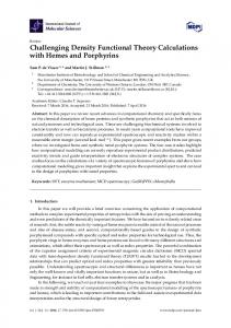
Figure 1. Design strategy of the target strobilurin analogues.
pyrazole derivatives against Rhizoctonia solani. Similarly Yang et al.11 reported fungicidal activity and 3D-QSAR study of phenylhydrazine substituted tetronic acid derivatives. Motivated by these findings, we conceived that establishing the 3D-QSAR model for our strobilurin analogues to predict the bioactivity and then optimize the structures might result in novel excellent fungicides. According to the frontier-orbital theory, Highest Occupied Molecular Orbital (HOMO) and Lowest Unoccupied Molecular Orbital (LUMO) are the two significant factors that affect the bioactivities12–14. They establish the correlation in various chemical and biochemical systems. Recently, Li et al.12,13 and Zhu et al.15,16 have reported studies on the frontier-orbital energies of some novel active molecules, which provide useful information about the biological mechanism and for further structural optimization. Thus, the study of the frontier-orbital energies may be helpful to the investigation of fungicidal activity. Taking all these into account, in this paper, in continuation of our studies on novel fungicidal strobilurin analogues, the methoxyiminoacetate pharmacophore of trifloxystrobin and methoxyacrylate pharmacophore of azoxystrobin were introduced into the halo-(un)substituted arylpyrazole structure, respectively, and a series of novel strobilurin analogues (1a-1f, 2a-2e, 3a-3e) were designed and synthesized (Fig. 1). The crystal structures of 1f, 2b and 3b were verified, to stimulate a better understanding of their binding nature. The fungicidal activity of these analogues and other twenty-eight similar compounds 4a-4f, 5a-5h, 6a-6h and 7a-7f from our previous studies were investigated together, with the aim of thorough understanding the structure-activity relationships (SARs) and developing novel fungicides. Their 3D-QSAR model and density functional theory (DFT) studies were also carried out to provide some guidance for further structure modification.
Results and Discussion
Synthesis. General synthetic routes for final compounds 1a-1f, 2a-2e and 3a-3e are shown in Fig. 2. Intermediates N-arylpyrazoles I were synthesized from arylhydrazines via addition-cyclization and oxidation, which could then afford 4-bromo-N-arylpyrazoles IV by bromination17. Intermediate benzyl bromide (E)-methyl 2-(2-(bromomethyl)phenyl)-2-(methoxyimino)acetate II was prepared from 1-(o-tolyl)ethanone via four steps including oxidation, esterification, oximation and bromination8. A previous report by Kim et al.18 described that intermediate (E)-methyl 3-methoxy-2-(o-tolyl)acrylate III-c could be synthesized from 1-bromo-2-methylbenzene and (E)-methyl 3-methoxyacrylate via Suzuki-Miyaura coupling reaction (Fig. 3). However, this approach required Grignard reagent and costly catalyst Pd(PPh3)4, which faced harsh reaction conditions and complicated processes. So in our procedure, readily accessible 2-(o-tolyl)acetic acid was used as starting material, and intermediate III-c could be obtained through three steps including esterification, condensation and methylation. The condensation of III-a with methyl formate was carried out under NaH alkaline condition, which gave III-b in 85% yield. A better yield (78%) of III-c was obtained in a molar ratio of III-b to dimethyl sulfate 1:1.2 equiv. in DMF as solvent, and with NaH as base. In our previous studies, several strobilurin analogues (4a-4f, 5a-5h, 6a-6h, Fig. 1) have been prepared by the substitution of N-arylpyrazoles with benzyl bromide in acetone, using potassium carbonate (K2CO3) as acid-binding agent7,8. Motivated by this reaction, in our procedure, N-arylpyrazoles IV and I were allowed to react with benzyl bromide II and III, respectively, in a molar 1:1.1 equiv. in boiling acetone in the presence of K2CO3, which afforded the target (E)-methyl 2-(2-(((4-bromo-1-aryl-1H-pyrazol-3-yl)oxy)methyl)phenyl)-2-(methoxyimino)acetate (1a-1f) and (E)-methyl 3-methoxy-2-(2-(((1-aryl-1H-pyrazol-3-yl)oxy)methyl) phenyl)acrylate (3a-3e) in 78–85% and 72–78% yields, respectively, as sole isolable products (Fig. 2). However, the target products (E)-methyl 2-(2-(((4-iodo-1-aryl-1H-pyrazol-3-yl)oxy)methyl)phenyl)-2-(methoxyimino) acetate (2a-2e) could not be obtained by the similar methods like 1a-1f via iodination first and then substitution (Fig. 4). Because the iodine was a better leaving group which made the bond ruptures more easily when the substitution took place. Therefore, compounds 5 were proposed to be synthesized firstly, which then underwent SCiENtiFiC REPOrTS | (2018) 8:7822 | DOI:10.1038/s41598-018-26154-5
2
www.nature.com/scientificreports/
Figure 2. General synthetic routes of target products 1a-1f, 2a-2e and 3a-3e.
Figure 3. Suzuki-Miyaura coupling reaction for the synthesis of III-c.
Figure 4. Synthesis of target products 2a-2e.
iodination to give products 2a-2e in 80–88% isolated yields with good functional-group tolerance. The iodination was carried out in a molar ratio of 5 to iodine monochloride (ICl) 1:2 equiv. in CHCl3 as solvent, and with K2CO3 as acid-binding agent. Other iodine reagents such as I2, N-iodosuccinimide (NIS), KI and NaI were also selected. However, the results were unsatisfactory. The regioselectivity of the reactions and the structures of the products 1a-1f, 2a-2e and 3a-3e were unequivocally determined by NMR spectroscopy and single-crystal X-ray diffraction analysis of (E)-methyl 2-(2-(((4-bromo-1-(4-fluoro-3-(trifluoromethyl)phenyl)-1H-pyrazol-3-yl)oxy) methyl)phenyl)-2-(methoxyimino)acetate (1f), (E)-methyl 2-(2-(((4-iodo-1-(4-iodophenyl)-1H-pyrazol-3-yl) oxy)methyl)phenyl)-2-(methoxyimino)acetate (2b) and (E)-methyl 2-(2-(((1-(4-chlorophenyl)-1H-pyrazol-3-yl) oxy)methyl)phenyl)-3-methoxyacrylate (3b).
Structure. The structures of 1a-1f, 2a-2e and 3a-3e were confirmed by their NMR spectra. In the 1H NMR
spectra, as a result of the deshielding effect of bromo and iodo groups, the CH of the pyrazole ring in 1a-1f and 2a-2e appeared as a singlet at low field δ 7.56–7.74 ppm and δ 7.65–7.81 ppm, respectively, whereas the two CH protons in 3a-3e appeared at δ 7.54–7.80 ppm and δ 5.73–5.83 ppm, respectively, as two doublets with coupling constants around 2.5 Hz. The CH2 in the oxy side chain appeared as a singlet at δ 5.08–5.26 ppm. The aromatic protons of all products resonated in the range of δ 7–8 ppm in 1H NMR spectra, and the 13C NMR signals were observed around δ 110–140 ppm. The chemical shifts of methoxy (OCH3) H-atoms appeared as two singlets around δ 3.67–4.09 ppm and δ 3.57–3.90 ppm, respectively. All products exhibited a carbonyl (C=O) 13C signal at the lowest field in the region of δ 162.2–168.1 ppm. The detailed crystal and structure refinement data of products 1f, 2b and 3b are listed in Table 1. Both 1f and 3b crystallize in a monoclinic space group P21/c, whereas 2b crystallizes in a monoclinic space group P21/n. In the crystal structures (Fig. 5), the bond lengths of C13-I1 (2.046(11) Å) and C18-I2 (2.070(12) Å) in 2b are longer than C9-Br (1.851(6) Å) in 1f, which represent a typical C-I bond length, and the values are similar to those SCiENtiFiC REPOrTS | (2018) 8:7822 | DOI:10.1038/s41598-018-26154-5
3
www.nature.com/scientificreports/ 1f
2b
3b
Empirical formula
C21H15BrF4N3O4
C20H17I2N3O4
C21H19ClN2O4
CCDC number
1433534
1433532
1433533
Formula weight
529.27
617.17
398.83
Temperature [K]
293(2)
293(2)
293(2)
Wavelength [Å]
0.71073
0.71073
0.71073
Crystal system
Monoclinic
Monoclinic
Monoclinic
Space group
P21/c
P21/n
P21/c
a [Å]
20.446 (4)
11.639 (2)
11.773(2)
b [Å]
13.551(3)
16.736(3)
7.8560(16)
c [Å]
7.0590(14)
12.343(3)
21.480(4)
α [°]
90.00
90.00
90.00
β [°]
90.10(3)
116.01(3)
93.27(3)
γ [°]
90.00
90.00
90.00
Volume [Å3]
1955.8(7)
2160.8(7)
1983.4(7)
Unit cell dimensions
Z
4
4
4
ρcalcd [g cm−3]
1.797
1.897
1.336
μ [mm−1]
2.177
2.941
0.222
F(000)
1060
1184
832
Crystal size [mm3]
0.20 × 0.10 × 0.10
0.20 × 0.10 × 0.10
0.30 × 0.20 × 0.10
θ range [°] for data collection
1.99 to 28.18
2.01 to 25.42
1.73 to 25.36
Index ranges
−24 ≤ h ≤ 0
0 ≤ h ≤ 14
0 ≤ h ≤ 14
−16 ≤ k ≤ 0
0 ≤ k ≤ 20
0 ≤ k ≤ 9
−9 ≤ l ≤ 9
−14 ≤ l ≤ 13
−25 ≤ l ≤ 25
Reflections collected
4077
4139
3824
Independent reflections
3967 [Rint = 0.0997]
3941 [Rint = 0.0990]
3637 [Rint = 0.0657]
Max. and min. transmission
0.8117/0.6699
0.7574/0.5908
0.9781/0.9364
Data/restraints/parameters
3967/2/298
3941/0/262
3637/1/253
Goodness-of-fit on F2
1.004
1.009
1.008
Final R indices [I > 2σ(I)]; R1, wR2
0.0772, 0.1344
0.0791, 0.1683
0.0771, 0.1690
R1, wR2 (all data)
0.1942, 0.1657
0.1544, 0.1963
0.1452, 0.1997
Largest diff. peak and hole [e·Å−3]
0.383 and −0.404
0.522 and −0.305
1.051 and −0.309
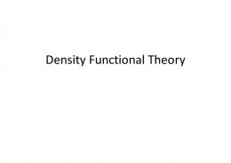
Table 1. Crystallographic data and structure refinement for compounds 1f, 2b and 3b. (2.071(3) and 2.070(4)) reported for other related derivatives19. The bond lengths of C8-O1 (1.312(7) Å) in 1f, C12-O4 (1.341(12) Å) in 2b and C9-O1 (1.337(5) Å) in 3b are longer than standard C=O double bond (1.229 Å), and belong to C-O single bond. The bond angle of C8-C9-C10 in 1f is 105.2(6)°, whereas the corresponding bond angles in 2b and 3b are 105.3(9)° (C12-C13-C14) and 104.3(4)° (C7-C8-C9), respectively. These values are similar to the typical angles of five-membered ring (108.0°). The bridge benzene ring A and the terminal benzene ring B are connected to the pyrazole ring C, twisted by 81.58° and 15.00° (1f), 59.58° and 21.11° (2b), 58.01° and 9.99° (3b), respectively, whereas they form a dihedral angle of 88.74°, 58.98° and 64.63°, respectively. The ester and methoxyimino (1f and 2b) or methoxyethene (3b) are almost coplanar, twisted by 76.21° and 68.47° (A in 1f), 59.43° and 2.72° (A in 2b), 80.78° and 89.15° (A in 3b), 14.52° and 23.67° (B in 1f), 21.51° and 61.79° (B in 2b), 80.84° and 78.88° (B in 3b), 22.58° and 29.97° (C in 1f), 8.82° and 22.45° (C in 2b), 84.90° and 86.31° (C in 3b), respectively, from the planes of the rings A, B and C. The intramolecular C5-H5A···F2 and C5-H5A···N2 H-bonds in 1f, C11-H11A···N3 H-bond in 2b, and C1-H1A···N2 H-bond in 3b result in the formation of four non-planar pseudo-rings D (C5/C4/C7/F2/H5A), E (N1/N2/H5A/C5/C6), F (N3/C12/O4/C11/H11A) and G (N1/N2/ H1A/C1/C6). In the crystal, six intermolecular H-bonds in 1f (C1-H1A···O3, C10-H10A···O3, C14-H14A···F1, C14-H14A···F4, C16-H16A···N2 and C21-H21B···Br), two intermolecular H-bonds in 2b (C14-H14A···O1 and C16-H16A···O1) and one intermolecular H-bond in 3b (C2-H2B···O3) reinforce the crystal packing (Fig. 5). These crystallographic data could provide a basis for elucidating the effect on their biological activities.
Fungicidal Activity and Structure-Activity Relationships (SARs). As an important method of drug
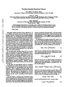
molecular design, the structure-activity relationships (SARs) can provide the guidance and enlightenment to the bioactivity prediction and structure optimization. In our previous studies, twenty-eight compounds 4a-4f, 5a-5h, 6a-6h and 7a-7f with similar strobilurin pharmacophores have been synthesized (Fig. 1). However, their SARs have not been discussed together. Here, we wish to report the SARs of all these forty-four strobilurin analogues (including sixteen compounds 1a-1f, 2a-2e and 3a-3e synthesized in this paper) from the following three aspects: (i) the effect of different pharmacophores and their positions; (ii) the effect of different substituents R on the terminal benzene ring; (iii) the effect of different substituents X on the pyrazole ring (Fig. 6). SCiENtiFiC REPOrTS | (2018) 8:7822 | DOI:10.1038/s41598-018-26154-5
4
www.nature.com/scientificreports/
Figure 5. X-ray crystal structures and packing diagrams of 1f, 2b and 3b.
Compounds 1a-1f, 2a-2e, 3a-3e, 4a-4f, 5a-5h, 6a-6h and 7a-7f were screened for bioactivity against two fungi, namely Rhizoctonia cerealis and Gibberella zeae, at the dosages of 10 μg mL−1, 1 μg mL−1 and 0.1 μg mL−1, respectively. As can be seen in Table 2 and Fig. 7, most compounds have excellent-to-good fungicidal activity against Rhizoctonia solani at 10 μg mL−1, especially 5c, 6c and 7a with 100% antifungal activity, as well as 3b, 5d, 5e, 6d, 7d and 7e with more than 80% antifungal activity. When the concentration was reduced to 0.1 μg mL−1, 5c, 7a, 6c, and 3b also had 98.94%, 83.40%, 71.40% and 65.87% inhibition rates, which were much better than commercial pyraclostrobin. This might imply that the introduction of suitable pharmacophores by taking the electronic effect and substituted positions into full consideration was important for improving the fungicidal activity. However, most compounds showed weak fungicidal activity against Gibberella zeae except 5c, 6b, 6c, 6e and 7d with moderate inhibitory activity at 10 μg mL−1. Therefore, the SARs based on the fungicidal activity against Rhizoctonia solani were discussed as follows. In terms of the o-substituted pharmacophores, the sequence of fungicidal activity against Rhizoctonia solani was methoxyiminoacetate moiety (5a-5h) > methoxyiminoacetamide moiety (6a-6h) > methoxyacrylate moiety (3a-3e) in general, irrespective of difference in substituent R on the terminal phenyl ring. For example, within the series of R = 4-Cl derivatives, methoxyiminoacetate-derivative 5c displayed a much higher fungicidal activity than the corresponding methoxyiminoacetamide-derivative 6c, while the methoxyacrylate-derivative 3b showed the lowest. Similar speculation could apply to the compounds 5e, 6e and 3d (R = 3-CF3). All the above three chloro-containing compounds 5c, 6c and 3b showed better activity than pyraclostrobin, which indicated the methoxycarbamate pharmacophore of pyraclostrobin might have no effective impact on the inhibition of Rhizoctonia solani. In addition, when changing the best o-substituted methoxyiminoacetate pharmacophore into the p-substituted, the results were unsatisfactory. Compounds 4a-4e showed much lower fungicidal activity than 5a-5h, which indicated the significant impact of pharmacophore position on the inhibition rates, and the o-substitution might be better. To examine the electronic effect of substituent R on the phenyl ring, the electron-donating CH3 and electron-withdrawing F, Cl, Br, I, CF3 were introduced. Compounds with electron-withdrawing substituents displayed higher fungicidal activity against Rhizoctonia solani than that with electron-donating substituents, as seen in the comparison of 1d (R = 4-Cl) vs. 1b (R = 3-CH3), 2b (R = 4-I) vs. 2a (R = 3-CH3), 3b (R = 4-Cl) vs. 3c (R = 2-CH3), 5c (R = 4-Cl) vs. 5b (R = 2-CH3), 6c (R = 4-Cl) vs. 6b (R = 2-CH3), and 7d (R = 4-Cl) vs. 7b (R = 3-CH3). According to the different electronic effect of electron-withdrawing substituent R, the sequence of fungicidal activity against Rhizoctonia solani is chloro-substituted > bromo-substituted, as seen in the comparison of 1d (R = 4-Cl) vs. 1a (R = 4-Br), 3b (R = 4-Cl) vs. 3e (R = 4-Br), 5c (R = 4-Cl) vs. 5 h (R = 4-Br), and 6c (R = 4-Cl) vs. 6 h (R = 4-Br), and within the series of R = 3-CF3 derivatives, the introduction of the fluoro group could make the fungicidal activity obvious improvement, as seen in the comparison of 1f (R = 3-CF3–4-F) vs. 1e (R = 3-CF3), 2e (R = 3-CF3–4-F) vs. 2d (R = 3-CF3), and 6d (R = 3-CF3-4-F) vs. 6e (R = 3-CF3). However, compounds 4c (R = 3-CF3) and 4f (R = 3-CF3-4-F), 5d (R = 3-CF3-4-F) and 5e (R = 3-CF3), 7e (R = 3-CF3) and 7f (R = 3-CF3-4-F) are three pairs of exceptions: 7f exhibited weaker fungicidal activity against Rhizoctonia solani as compared with 7e, whereas 4f and 5d showed better activity than 4c and 5e at 10 μg mL−1, respectively, and the results were just the opposite when the concentration was reduced to 1 μg mL−1 and 0.1 μg mL−1. These differences in fungicidal activity might be due to variations in combination of methoxyiminoacetate pharmacophore,
SCiENtiFiC REPOrTS | (2018) 8:7822 | DOI:10.1038/s41598-018-26154-5
5
www.nature.com/scientificreports/
Figure 6. The structure-activity relationships (SARs) of forty-four strobilurin analogues.
pyrazole ring and aromatic ring. Moreover, the positions of substituent R have little effect on the activity, as seen in the comparison of 5b (R = 2-CH3) vs. 5f (R = 3-CH3), and 6b (R = 2-CH3) vs. 6f (R = 3-CH3). According to the different positions of chloro group, the sequence of fungicidal activity is Cl-substituted phenyl ring > Cl-substituted pyrazole ring, as seen in the comparison of 5c vs. 7a-7f. However, compound 7a only containing a chloro on the pyrazole ring displayed nearly equal fungicidal activity to the best 5c, and with the increasing number of chloro group, the fungicidal activity was decreased, as seen in the comparison of 7d vs. 5c and 7a. These observations revealed that the mono-chlorination has an important influence on the fungicidal activity. To further investigate the effect of other halogen substituents X on the pyrazole ring, Br and I were introduced to the series of methoxyiminoacetate-derivatives, as compared with the non-substituted H and Cl. When the substituent R on the phenyl ring was the same, in most cases, the fungicidal trend of these four series against Rhizoctonia solani was H > Cl > Br > I. For example, within the series of R = 4-CF3 derivatives, compound 5e (X = H) had better fungicidal activity than 7e (X = Cl) and 1e (X = Br) at 10 μg mL−1 and 1 μg mL−1, respectively, whereas 2d (X = I) showed the weakest. Similar relationships could apply to the R = 3-CF3-4-F derivatives 5d (X = H), 7f (X = Cl), 1f (X = Br) and 2e (X = I), R = 4-OCF3 derivatives 5g (X = H), 7c (X = Cl), 1c (X = Br) and 2c (X = I), as well as R = 3-CH3 derivatives 5f (X = H), 7b (X = Cl), 1b (X = Br) and 2a (X = I). These results might indicate that the larger molecular volume (Br and I) was unfavorable for the intracellular uptake and transport in the fungus, and the non-substituted pyrazole ring might be the best. In summary, the SARs study revealed that the improvement of fungicidal activity required a reasonable combination of both methoxyiminoacetate pharmacophore and electron-withdrawing substituent R, and the type and size of substituent X on the pyrazole ring were critical. The chloro group had an effective impact on the fungicidal activity whether it on the terminal phenyl ring or pyrazole ring. The present work indicated that 5c (98.94% at 0.1 μg mL−1, methoxyiminoacetate pharmacophore, R = 4-Cl, X = H), 7a (83.40% at 0.1 μg mL−1, methoxyiminoacetate pharmacophore, R = H, X = Cl), 6c (71.40% at 0.1 μg mL−1, methoxyiminoacetamide pharmacophore, R = 4-Cl, X = H) and 3b (65.87% at 0.1 μg mL−1, methoxyacrylate pharmacophore, R = 4-Cl, X = H) could be used as potential lead compounds for further studies of novel fungicides.
Three-dimensional quantitative structure-activity relationships (3D-QSARs). In order to
obtain further insight into the structural requirements of novel strobilurin fungicides, we have performed a 3D-QSAR study of the above forty-four strobilurin analogues against Rhizoctonia solani using CoMFA technique. In CoMFA, it is assumed that the interaction between an analogue and its molecular target is preliminarily non-covalent and shape dependent in nature. The 3D-QSAR can be derived correlating the differences in steric and electrostatic fields surrounding a set of molecules to the fungicidal activity. The toxicity baselines and EC50 values were obtained from DPS data processing system, and the negative logarithm of EC50 (pEC50) was used as the biological activity in the 3D-QSAR study (Table 3). The lowest energy conformers were selected and minimized using Powell method to rms 0.001 kcal mol−1 Å−1. Alignment of the molecules was carried out using N-phenyl pyrazole ring as the common skeleton (Fig. 8, blue color), and the most active molecule 5c was used as a template molecule for database alignment according to the pEC50 values. Thirty-three molecules were randomly selected as the training set to establish the CoMFA model, and the remaining eleven molecules were used as the test set to test the predictive ability of the model (Table 3, “*”). The CoMFA model was generated using Partial Least-Squares (PLS) approach. The cross-validation with the Leave-One-Out (LOO) option and the SAMPLS program was carried out to obtain the optimal number of components (n) and cross-validated coefficient (q2). After n was determined, a non-cross validated analysis was performed without column filtering to obtain regression coefficient (R2) and its standard error (SEE), as well as F-test value (F) for the model evaluation.
SCiENtiFiC REPOrTS | (2018) 8:7822 | DOI:10.1038/s41598-018-26154-5
6
www.nature.com/scientificreports/ Inhibition [%][a] Rhizoctonia solani Structure
No.
10
1
Gibberella zeae 0.1
10
1
1a
57.97
54.73
45.68
30.28
12.31
0.1 0.32
1b
51.10
46.27
37.89
5.32
0.00
0.00
1c
51.33
47.28
40.69
12.69
9.17
0.00
1d
68.18
61.51
57.10
35.32
21.41
12.56
1e
51.85
45.32
37.24
17.45
7.52
0.32
1f
55.21
48.76
39.53
18.25
0.00
0.00
2a
42.02
32.18
25.37
4.79
0.00
0.00
2b
62.02
50.07
41.57
36.04
0.00
0.00
2c
48.21
44.22
34.93
0.00
0.00
0.00
2d
48.47
43.96
37.85
9.58
3.96
0.00
2e
54.05
52.99
45.55
13.96
10.00
8.54
3a
49.18
46.65
38.73
33.23
4.52
0.32
3b
85.78
80.86
65.87
48.56
19.49
10.54
3c
54.72
48.73
40.82
21.98
8.74
0.32
3d
52.83
40.57
32.41
22.15
9.87
0.18
3e
58.21
51.13
46.56
19.49
5.31
0.28
4a
47.73
38.31
24.67
10.34
8.91
8.62
4b
51.27
19.14
18.43
19.83
15.23
8.62
4c
44.85
35.68
29.38
23.85
10.63
1.44
4d
40.68
33.97
29.73
10.63
6.03
0.57
4e
53.04
33.97
17.02
15.80
9.20
8.33
4f
52.68
30.08
26.55
25.57
8.62
0.57
5a
54.02
48.98
44.51
37.65
28.34
18.85
5b
55.58
50.22
49.15
35.34
27.30
17.82
5c
100.00
100.00
98.94
62.53
51.32
34.77
5d
90.82
75.28
47.74
40.23
47.41
29.02
5e
85.17
81.29
48.09
36.49
34.20
33.33
5f
54.05
49.80
45.02
36.56
14.38
0.62
5g
59.36
52.19
44.49
42.50
31.04
0.00
5h
59.10
53.78
48.21
38.47
23.04
15.56
6a
53.97
42.84
37.56
48.34
39.31
17.72
6b
58.47
50.53
46.68
50.86
35.92
16.95
6c
100.00
94.70
71.40
56.15
44.48
33.62
6d
98.23
90.82
49.86
46.55
35.63
31.03
6e
66.53
47.68
35.72
50.63
48.54
0.00
6f
57.24
49.00
50.33
42.92
21.88
9.58
6g
54.65
43.72
35.57
46.25
36.25
5.00
6h
58.84
47.41
37.87
36.88
26.04
0.00
7a
100.00
98.23
83.40
39.37
32.76
26.15
7b
51.05
48.57
39.04
37.36
34.48
25.00
7c
55.86
47.03
31.50
34.48
33.91
31.61
7d
86.58
81.64
50.21
51.08
37.61
34.89
7e
81.64
74.93
51.98
33.33
33.05
33.05
7f
56.57
30.79
26.55
30.17
5.17
4.60
Continued SCiENtiFiC REPOrTS | (2018) 8:7822 | DOI:10.1038/s41598-018-26154-5
7
www.nature.com/scientificreports/ Inhibition [%][a] Rhizoctonia solani Structure
No.
10
61.51
1
38.91
Gibberella zeae 0.1
10
35.73
35.06
1
0.1
6.32
0.57
Table 2. Fungicidal activity of forty-four strobilurin analogues. a0, No activity, and 100, total kill.
Figure 7. The fungicidal activity of forty-four strobilurin analogues against Rhizoctonia solani.
The alignment gave a conventional R2 (R2ncv) of 0.977 with 2 components, a predictive R2 (R2pred) of 0.816 and an F value of 94.553. The model generated with a good internal predictive ability (q2 = 0.508) and a small standard error of estimation (SEE = 0.202) was selected as the best model to explain SARs and carry out further analysis. Observed and predicted fungicidal activity of the training and test sets were plotted in Fig. 9 and listed in Table 3. The results indicated that the observed and predicted data were in good agreement with each other, with a correlation coefficient R2 of 0.936. Figure 10a displayed the steric contour plot. The green contours describe regions where sterically favorable groups enhance activity (80% contribution), and yellow contours describe regions of unfavorable steric effects (20% contribution). The pharmacophore and its linked phenyl ring were surrounded by the sterically favorable green contours. The most active molecule 5c had a methoxyiminoacetate moiety substituted phenyl ring embedded in this green region. Other sterically-favorable green contours were observed near the terminal phenyl ring. This green contour was surrounded by the unfavorable yellow region. A substitution on the 2- or 4-position of the terminal phenyl ring was favored, whereas any substitution on the adjacent 3- or 5-position was unfavorable. This also suggested that the introduction of the chloro group to the 4-position was important for fungicidal activity. Figure 10b displayed the electrostatic contour plot. The blue contours describe regions where positively charged groups enhance activity (80% contribution), and red contours describe regions where negatively charged groups enhance the activity (20% contribution). In compound 5c, the red contours were found near the methoxyiminoacetate pharmacophore and the 4-position of the terminal phenyl ring, suggesting that a high electron density in this region increased the activity. A large negative-charge unfavorable blue contour was found to surround the side chain CH2 and the terminal phenyl ring. This indicated that substitutions in these regions with high electron density reduced activity and emphasized the necessity of positively charged groups. Overall, steric interactions (56.3% contribution) played a major role in the influence of fungicidal activity than electrostatic interactions (43.7% contribution).
Density functional theory (DFT) calculation. The frontier-orbital energies of a compound play an important role in bioactivities20. EHOMO is a rough measure of the electron-donating ability of a compound and, normally, increasing its value can improve the biological activity, whereas the ELUMO acts in reverse21,22. Energy gap between HOMO and LUMO characterizes the molecular chemical stability and it is a critical parameter in determining molecular electrical transport properties because it is a measure of electron conductivity. It also affects the bioactivity of a compound. Thus, the study of the frontier-orbital energies may be helpful to the investigation of fungicidal activity. Compounds 5c, 7a and pyraclostrobin which had wide difference in activity were selected for DFT comparison. The LUMO and HOMO maps of 5c, 7a and pyraclostrobin were shown in Fig. 11. Comparing the HOMO-LUMO gaps of the three molecules, the order was: pyraclostrobin >7a > 5c. The narrow HOMO-LUMO gap implies a high chemical reactivity because it is energetically favorable to add electrons to a low-lying LUMO or extract electrons from a high-lying HOMO, and so to form an activated complex in any potential reaction23. This suggested that compound 5c might possess a relatively high activity, which correlated well with the fungicidal activity results. In addition, the calculations indicated some similarities between 5c and 7a. In the HOMO,
SCiENtiFiC REPOrTS | (2018) 8:7822 | DOI:10.1038/s41598-018-26154-5
8
www.nature.com/scientificreports/
pEC50
Predicted pEC50
No.
Toxicity baselines
R2
0.670
6.174
6.832
5a*
Y = 0.1195 × +4.9791
0.9994
4.094
5.388
6.189
12.075
4.918
4.776
5b
Y = 0.0808 × +5.0415
0.9330
0.808
6.093
6.150
8.864
5.052
4.980
5c
Y = 1.3478 × +9.1015
0.8660
0.00225
8.648
8.659
0.9916
0.015
7.823
7.272
5d
Y = 0.6932 × +5.6521
0.9992
0.254
6.595
6.506
Y = 0.1859 × +4.8678
0.9977
10.038
4.998
4.240
5e
Y = 0.5458 × +5.6282
0.9242
0.163
6.788
6.714
1f
Y = 0.1983 × +4.9448
0.9945
3.581
5.446
5.427
5 f
Y = 0.1134 × +4.9905
0.9994
3.196
5.495
5.470
2a
Y = 0.2308 × +4.5577
0.9971
163.403
3.787
3.768
5 g
Y = 0.1877 × +5.0511
0.9998
1.189
5.925
5.874
2b*
Y = 0.2595 × +5.0316
0.9951
1.224
5.912
5.931
5 h
Y = 0.1375 × +5.0934
0.9998
0.471
6.679
6.814
2c
Y = 0.1712 × +4.8075
0.9728
23.161
4.635
4.724
6a*
Y = 0.2084 × +4.8674
0.9808
11.881
4.925
5.212
2d
Y = 0.1355 × +4.8334
0.9957
30.306
4.519
4.556
6b
Y = 0.1480 × +5.0480
0.9802
1.257
5.901
5.904
2e
Y = 0.1067 × +5.0216
0.9176
1.086
5.964
5.971
6c
Y = 2.2174 × +7.3938
0.9569
0.209
6.680
6.744
3a
Y = 0.1329 × +4.8697
0.9574
26.246
4.581
4.623
6d
Y = 1.0536 × +6.1433
0.9885
0.183
6.739
6.792
3b*
Y = 0.3308 × +5.784
0.9741
0.0108
7.967
7.663
6e
Y = 0.3965 × +5.0009
0.9918
2.300
5.638
5.639
3c*
Y = 0.1754 × +4.9515
0.9966
4.994
5.302
5.460
6 f
Y = 0.0871 × +5.0552
0.7815
0.614
6.212
6.184
3d*
Y = 0.2636 × +4.7920
0.9950
14.222
4.847
3.969
6 g*
Y = 0.2434 × +4.8629
0.9972
8.157
5.089
4.879
3e
Y = 0.1468 × +5.0498
0.9921
1.034
5.986
5.933
6 h
Y = 0.2662 × +4.9499
0.9988
3.481
5.458
5.378
4a*
Y = 0.3140 × +4.6536
0.9910
33.427
4.476
4.672
7a
Y = 2.0149 × +7.6913
0.9696
0.116
6.937
6.808
4b
Y = 0.4655 × +4.4200
0.8782
44.074
4.356
4.333
7b
Y = 0.1523 × +4.9041
0.9463
10.305
4.987
5.139
4c*
Y = 0.2064 × +4.6537
0.9962
109.776
3.960
4.074
7c
Y = 0.3146 × +4.8637
0.9859
5.604
5.252
5.233
4d
Y = 0.1482 × +4.6063
0.9935
2.996
2.881
7d
Y = 0.5507 × +5.6712
0.9402
0.139
6.857
6.808
4e
Y = 0.5148 × +4.5699
0.9996
17.124
4.766
4.825
7e*
Y = 0.4260 × +5.5412
0.9663
0.115
6.940
6.297
4 f
Y = 0.3469 × +4.6395
0.9273
24.248
4.615
4.673
7f
Y = 0.3960 × +4.6790
0.9299
13.307
4.874
4.950
pyraclostrobin
Y = 0.3292 × +4.8818
0.9187
5.896
5.229
5.254
No.
Toxicity baselines
R2
EC50 (μM)
1a
Y = 0.1548 × +5.0705
0.9653
1b
Y = 0.1680 × +4.8752
0.9873
1c
Y = 0.1344 × +4.9099
0.9902
1d
Y = 0.1469 × +5.3148
1e*
1009.92
EC50 (μM) pEC50
Predicted pEC50
Table 3. Experimental and theoretical fungicidal activity of forty-four strobilurin analogues against Rhizoctonia solani.
Figure 8. Superimposition of all molecules using database alignment.
Figure 9. Observed (X-axis) and predicted (Y-axis) biological activities of the training (blue dot) and test (red dot) sets. (Thirty-three molecules were randomly selected as the training set and eleven molecules (Table 3, “*”) were used as the test set to test the predictive ability of the model. The observed and predicted pEC50 could be obtained according to the location of each dot; the values were listed in Table 3).
SCiENtiFiC REPOrTS | (2018) 8:7822 | DOI:10.1038/s41598-018-26154-5
9
www.nature.com/scientificreports/
Figure 10. CoMFA steric (a) and electrostatic (b) contour plots.
Figure 11. Fungicidal activity and DFT comparison of 5c, 7a and pyraclostrobin. (The DFT calculations were carried out in the ground-state (in vacuo) with Gaussian 09 software by using B3LYP/6–31 G* method. The HOMO and LUMO maps were extracted from GuassView 5.0 program based on the optimized structures). the electrons were mainly delocalized on the terminal benzene ring and pyrazole ring (including CH2, O and Cl atoms). When electron transitions took place, some electrons in the HOMO would enter into the LUMO24; then, in the LUMO, the electrons were similarly delocalized on the bridge benzene ring and oxime ester moiety. The HOMO-LUMO gaps of 5c (0.160 a.u) and 7a (0.161 a.u) were very close to each other. The similar electron distributions and energy gaps between 5c and 7a might cause both with excellent fungicidal activity. However, pyraclostrobin exhibited quite different electron distributions as compared with 5c and 7a. Whether in the HOMO or LUMO, its electrons were mainly delocalized on the terminal benzene ring and pyrazole ring. In the LUMO maps, the general trend of electron delocalization was: pyraclostrobin >7a > 5c, which represented a negative correlation with their fungicidal activity. As reported, the frontier molecular orbitals are located on the main groups, the atoms of which can easily bind with the receptor12,13. Moreover, the different degrees of delocalization may affect the orbital interaction25. Therefore, it seemed that the high electron delocalization of pyraclostrobin in the LUMO might potentially make the orbital interactions limited, which might lead to a decrease in activity. Figure 12 is the molecular electrostatic potential (MEP) of 5c, 7a and pyraclostrobin. The MEP simultaneously displays molecular size, shape as well as positive, negative and neutral electrostatic potential regions in terms of color grading. From MEP, we can know the rich electron region and the lack electron region, where potential increases in the order of red < orange