Screening of Single-Chain Variable Fragments against TSP50 from a Phage Display Antibody Library and Their Expression as Soluble Proteins JINGYAN WEI,1,2 YANG LIU,1,3 SONGCHUAN YANG,1,4 JUNJIE XU,1 HANGTIAN KONG,1 BING HAN,1 YONGLI BAO,3 YIN WU,3 WEITIAN YIN,4 WEI LI,5 GANGLIN YAN,6 GUIMIN LUO,6 HAO-PENG XU,7 YUXIN LI,3 and BAI YANG2
A novel gene, testes-specific protease 50 (TSP50), is abnormally activated and differentially expressed in most patients with breast cancer, suggesting it as a novel biomarker for this disease. The possibility that TSP50 may be an oncogene is presently under investigation. In this study, the single-chain variable fragments (scFvs) against TSP50 were panned from a phage display antibody library using TSP50-specific peptide, pep-50, as a target antigen. After 4 rounds of panning, 3 clones (A1, A11, and C8) from the library were verified to show strong binding affinities for TSP50 by enzyme-linked immunosorbent assay (ELISA) and to contain the variable region genes of the light and heavy chains of scFv antibodies but different complementary determining regions by sequencing. The genes of scFv-A1 and scFv-A11 were cloned into expression vector pPELB and successfully expressed as a soluble protein in Escherichia coli Rosetta. The yields of expressions were about 4.0 to 5.0 mg of protein from 1 L of culture. The expressed proteins were purified by a 2-step procedure consisting of ion-exchange chromatography, followed by immobilized metal affinity chromatography. The purified proteins were shown a single band at the position of 31 KDa on sodium dodecyl sulfate–polyacrylamide gel electrophoresis. Sandwich ELISA demonstrated that the expressed scFv proteins were able to specifically react with pep-50, laying a foundation for the investigation of the function of TSP50 in the development and treatment of breast cancer. (Journal of Biomolecular Screening 2006:546-552) Key words: antibody library, phage display, single-chain variable fragments (scFvs), testes-specific protease 50 (TSP50)
INTRODUCTION 1
College of Pharmaceutical Science, Jilin University, 1266 Fujin Road, Changchun, 130021, China. 2
Key Laboratory for Supramolecular Structure and Materials, Jilin University, Changchun, 130023, China. 3
Institute of Genetics and Cytology, Northeast Normal University, Changchun, 130024, China. 4
The Third Clinical College, Jilin University, Changchun, 130031, China.
5
College of Life Science, Jilin University, Changchun, 130023, China.
6
Key Laboratory of Molecular Enzymology and Engineering of Ministry of Education, Jilin University, Changchun, 130023, China. 7
Biochemistry and Cell Biology Department, State University of New York at Stony Brook, USA. Received Sep 29, 2005, and in revised form Jan 22, 2006. Accepted for publication Jan 31, 2006. Journal of Biomolecular Screening 11(5); 2006 DOI: 10.1177/1087057106287901
546
www.sbsonline.org
B
of morbidity and mortality in women in the United States,1 China, and many other countries. Several candidate biomarker molecules correlated with breast tumors have been suggested and studied, such as KLK12, AZU-1, P7 antigen,2-4 and testes-specific protease 50 (TSP50).5 The TSP50 gene was isolated from a human testes cDNA library by using a genomic DNA probe, BR50. BR50 was isolated by a modified representational difference analysis technique due to its hypomethylated feature in a breast cancer biopsy. Northern blot analysis of RNA from multiple normal human tissues indicated that this gene was highly and specifically expressed in the testes, and it is a tissue-specific gene.5 TSP50 gene product shares a similar enzymatic structure with many serine proteases. Amino acid alignment with several serine proteases revealed that although TSP50 shared 2 critical catalytic triads, histidine and aspartic acid, the most critical catalytic site, serine, was substituted with threonine, which is different from traditional serine proteases; it could therefore be REAST CANCER IS A MAJOR CAUSE
© 2006 Society for Biomolecular Sciences
Screening of Single-Chain Variable Fragments classified as a novel protease.6 However, in addition to expression in normal testes, the TSP50 gene was abnormally activated and expressed in a vast majority of breast cancer patients tested.6 Earlier studies have suggested that the normal function of the TSP50 gene product might be involved in the human reproductive pathway but that its up-regulation could be linked to tumorigenesis in human breast cancer. The surprisingly high positive detection rate of the TSP50 protein in breast cancer could make it an attractive molecular marker for this disease.6 Based on the information above, we wished to acquire the gene fragments of single-chain variable fragments (scFvs) against TSP50 using phage display antibody library technology, clone them into the expression vector pPELB, and express them in Escherichia coli Rosetta. If the expressed proteins show a specific reaction with TSP50 molecules by immunological examination, it will be very beneficial to the investigation of the function of TSP50 in the development of breast cancer and its relevance to the treatment of breast cancer. This article is the 1st account of the selection of recombinant antibody fragments for TSP50 from a naive phage display library, that is, one constructed without prior exposure to antigen. The library was prepared without the involvement of laboratory animals, using a framework based on a pairing of heavy chain variable region (VH) and light chain variable region (VL) chains most common in the human immune response. If the scFv antibodies can be used as inhibitors or target carriers to treat breast cancer, the human antibody response against mouse protein can be avoided, enhancing therapeutic utility. MATERIALS AND METHODS Supplies Reagents and their suppliers are as follows: mouse anti-M13 phage, anti-hexa-histidine monoclonal antibody and Fast Flow Sepharose (Pharmacia Biotech, Uppsala, Sweden); Tryptone, yeast extract (Gibco, BRL, Gaithersburg, MD); Immunotubes (Maxisorp, Nunc); CM Fast Flow, Chelating Sepharose Fast Flow, and Sephadex G-25 (Pharmacia); tetramethylbenzidine (TMB), sodium dodecyl sulfate (SDS), phenyl methylene sulfonyl fluoride (PMSF), imidazole, isopropyl-β-D-thiogalactopyranoside (IPTG), N,N-methylene-diacrylamide, and acrylamide (Sigma, St. Louis, MO); 96-well flat-bottomed enzyme-linked immunosorbent assay (ELISA) plates (Becton Dickinson, Franklin Lakes, NJ); horseradish peroxidase (HRP)–conjugated goat antimouse and antirabbit immunoglobulin G (IgG; Santa Cruz Biotechnology, Santa Cruz, CA); restriction enzymes, NcoI and NotI (TaKaRa Bio Inc., Shiga, Japan); protein molecular weight markers (Bio-Rad, Hercules, CA; and Shanghai Biochemical Institute, China); and ELISA reader (ELX800; BioTek, Winooski, VT). All chemicals were of the highest purity available or otherwise of a quality noted in the text.
Journal of Biomolecular Screening 11(5); 2006
Phage antibody library, vector and bacterial strains Human synthetic VH + VL scFv library, helper phage M13K07, and E. coli TG1 used in this study were generous gifts from Fiona Sait, Centre for Protein Engineering, Medical Research Council (MRC), University of Cambridge, England. This scFv phagemid library contains synthetic V-gene (VH-VL) from lox library vectors7 recloned into the pHEN2 phagemid vector, and the library size is 1.47 × 10⁸ phagemid clones in E. coli TG1 cells and has a high proportion of functional antibody, with 96% of clones containing inserts (these are also commercially available from MRC and Pharmacia Biotech; see Web site). Vector pPELB and E. coli Rosetta were generous gifts from Dr. Sui Dexin, University of Michigan. Antigen used for screening The antigen, TSP50-specific peptide, pep-50, was synthesized at the Facility of Molecular Genetics, North Shore–Long Island Jewish Research Institute. The sequence of pep-50 was Ile-Trp-Arg-Asp-Val-Ile-Tyr-Ser-Val-Arg-Val-Gly–Ser-ProTrp-Ile-Asp-Gln-Met-Thr-Gln-Thr-Ala-Ser-Asp-Val-Pro-ValLeu-Gln-Val-Ile-Met-His-Ser-Arg-Tyr-Arg-Ala-Gln-ArgPhe-Trp-Ser-Phe-Val-Gly-Gln-Ala-Asn, corresponding to the 156 to 206 aa position, which was a nonhomologous hydrophilic region within the catalytic domain. Pep-50 is a critical catalytic site of TSP50 different from other traditional serine proteases and readily binds to antibodies when used as a target antigen. The activity of TSP50 can be inhibited when the prepared scFvs bind to pep-50, allowing investigation of the role of TSP50 in tumorigenesis in these studies. Jidong Shan et al. have prepared anti-TSP50 polyclonal antibody AT-50 using synthesized pep-50 as an antigen and applied it in their studies.6 Until now, full-length TSP50 has not been available; therefore, we used pep-50 as a target antigen substituting for the fulllength TSP50 to screen scFvs from the antibody library. Rescue of phage from the cells The whole of the bacterial library stock (about 1 × 10¹⁰ clones, stored at –80 °C) in 500 ml of 2 × TY culture medium containing 1% glucose and 100 µg/ml ampicillin was incubated with shaking at 37 °C to an OD600 of 0.4. M13K07 helper phage (1 × 10¹² pfu) was added to 50 ml of the culture, and the mixture was incubated without shaking for 30 min at 37 °C.8 Infected cells were pelleted and then resuspended in 100 ml 2 × TY broth containing 1% glucose and 100 µg/ml ampicillin and 50 µg/ml kanamycin and incubated overnight with shaking at 30 °C. PEG/NaCl (20% polyethylene glycol 6000, 2.5 M NaCl) in a 1/5 volume was used to precipitate phage particles from the culture supernatant as described previously.9 The pellet was resuspended in 2 ml phosphate-buffered saline (PBS)
www.sbsonline.org
547
Wei et al. and concentrated at 11,600g for 10 min in a microcentrifuge to remove most of the remaining bacterial debris. The phage supernatant was stored at 4 °C for short-term storage or in PBS, 15% glycerol for longer term storage at –70 °C. To determine the titer of the phage stock, 1 µl phage was diluted in 1 ml PBS, and 1 µl of this dilution was used to infect 1 ml of TG1 at an OD600 of 0.4 to 0.6 and incubated for 30 min at 37 °C. Fifty microliters of this, 50 µl of a 1:102 dilution, and 50 µl of a 1:104 dilution were plated on TYE plates containing 100 µg/ml ampicillin and 1% glucose and grown overnight at 37 °C. The titer of the phage stock was 5.6 × 10¹² pfu/ml determined according the number of TG1 clones on the TYE plates. Affinity selection of anti-TSP50 phage antibodies A 30 × 10 mm Immunotube (Maxisorp, Nunc) was coated with 1 ml pep-50 antigen, at 10 µg/ml in PBS (pH 8.5) and incubated overnight at 4 °C, washed 3 times with PBS, and blocked with 3% bovine serum albumin (BSA)–PBS at 37 °C for 2 h. The phage stock (5.6 × 10¹² pfu) was added to 1 ml 3% BSAPBS in the tube and incubated for 30 min at room temperature, rotating continuously on an under-and-over turntable, and then for a further 60 min at 37 °C without rotation. The tube was washed 20 times with PBS containing 0.1% Tween 20, and bound phage was eluted for 10 min using 1 ml 100 mM triethylamine as described above.10 During the elution, tubes were previously prepared with 0.6 ml 1 M Tris-HCl, pH 7.4, to add the eluted 1-ml phage for quick neutralization. Half of the eluted phage (0.8 ml) was added into 9.2-ml exponential-phase TG1 cell culture suspension in 2 × TY broth, and any remaining bound phage in the tube was eluted by adding 100 µl 1 M TrisHCl, pH 7.4, and 4 ml TG1 culture suspension. Both TG1 cultures were incubated for 30 min at 37 °C without shaking to allow for infection. The cells were pooled, concentrated by centrifugation, and plated on TYE agar11 containing 1% glucose and 100 µg/ml ampicillin. After incubation overnight at 30 °C, colonies were scraped into 5 ml of 2 × TY broth containing 1% glucose, 100 µg/ml ampicillin, and 20% glycerol and stored at –80 °C. Glycerol stock (100 µl) was then inoculated into 50 ml of 2 × TY broth containing 1% glucose and 100 µg/ml ampicillin and incubated with shaking at 37 °C to an OD600 of 0.4. Selected phages were then rescued from the culture as described above. Selection was repeated another 3 rounds with the following modifications: 1) pan 2, the Immunotube blocked with 3% glycine-PBS, and 2) pans 3 to 4 used pep-50 at 500 and 25 ng/ml, respectively; that is, the concentration of pep-50 used for coating was reduced from 10 µg/ml (pan 2) to 25 ng/ml (pan 4). Screening and selection of phage antibodies Individual colonies from pan 4 were grown in 96-well plates, and phage antibodies were rescued with M13K07
548
www.sbsonline.org
following the description above. The specificity of phage supernatant for binding to pep-50 was then determined using indirect ELISA. Indirect ELISA was performed to assess the binding of polyethylene glycol–precipitated phage at each round of panning, and phage clones binding to pep-50, but not to either BSA or glycine alone, were picked. The wells of a 96-well, flat-bottom ELISA plate were coated with 50 µl of pep-50 (5 µg/ml) overnight at 4 °C and then washed with PBS containing 0.1% Tween 20. The plates were blocked with 200 µl per well of 3% BSA-PBS at 37 °C for 2 h and then washed as described above. Phage precipitated at each round of panning (50 µl), used as the 1st antibody, were added to the wells in 3% BSA-PBS. The plates were then incubated at 37 °C for 2 h and washed 3 times with PBS containing 0.1% Tween 20 prior to adding 50 µl per well of a 1:1000 dilution of anti-M13 monoclonal antibody as the 2nd antibody and incubation at 37 °C for 1 h. Bound antibodies were detected with an HRP–goat antimouse IgG and TMB-H2O2 substrate. The reaction was terminated with 50 µl per well of 1 M H2SO4, and the optical density was read at 450 nm.11 Construction of the recombinant plasmids The antibody’s VH and VL genes from selected clones were amplified using PCR and primers corresponding both to the 5′ and 3′ sequences of the vector pHEN2(5′-CAG GAA ACA GCT ATG AC-3′ and 5′-GAA TTT TCT GTA TGA GG-3′; from TaKaRa Co.) and the phagemid DNA prepared from the selected clone as template. The products were sequenced by TaKaRa Co. in both directions using the same primers (5′-CAG GAA ACA GCT ATG AC-3′ and 5′-GAA TTT TCT GTA TGA GG-3′ from TaKaRa Co.).12,13 Phagemid DNAs from the clones found to have different complementary determining regions by sequencing were digested with NcoI and NotI, and then the scFv genes were cloned into the similarly digested soluble expression vector pPELB (a gift of Dr. Sui Dexin, University of Michigan). Expression of scFvs in E. coli Rosetta The recombinant plasmids were transformed into E. coli Rosetta by electroporation. Transformed E. coli Rosetta clones were incubated in 500 ml 2 × YT containing 100 µg/ml ampicillin and 34 µg/ml chloramphenicol at 37 °C. A total of 500 µl 1 M isopropyl thiogalactopyranoside (final concentration 1 mM IPTG) was added into the culture when the OD600 reached 0.6. Shaking was continued at 30 °C for a further 4 h.14 Cells were harvested by centrifugation at 6000 rpm at 4 °C for 10 min and either stored frozen at –70 °C or disrupted by sonication as described below and used directly for sodium dodecyl sulfate–polyacrylamide gel electrophoresis (SDS-PAGE) analysis as described previously.14
Journal of Biomolecular Screening 11(5); 2006
Screening of Single-Chain Variable Fragments Purification of the bacterial scFvs
Sandwich ELISA
The scFv proteins acquired above were purified as follows.14,15 Cells were resuspended in 50 mM PBS, pH 6.5, 1 mM PMSF, to a final ratio of approximately 5 ml lysis buffer per gram of cells. Cell suspensions were sonicated with middle pulse extension on ice, and supernatants were obtained by centrifugation at 12,000g at 4 °C for 15 min and filtered using a 0.45-µm filter. Ion-exchange chromatography was carried out on the column packaged with CM Fast Flow resin. The column was equilibrated with buffer A (50 mM PBS, pH 6.5). The sample was loaded onto the column overnight at 4 °C and eluted by a step NaCl gradient as follows. Weakly bound proteins were removed by adding 400 mM NaCl in buffer A. ScFvs were eluted by raising the concentration of NaCl to 1 M. The eluent containing scFvs was purified further with immobilized metal ion chelate affinity chromatography (IMAC) via the hexa-histidine tail on a 1-ml HiTrap chelating column using Ni2+ charged (AmershamPharmacia).15 The column was equilibrated with buffer B (50 mM PBS, pH 7.5, 300 mM NaCl). Sample (5-10 mg protein/ml gel) was loaded onto the column and eluted as follows. Nonspecifically and weakly bound proteins were removed by adding imidazole 80 mM in buffer B. ScFvs were eluted by raising the concentration of imidazole to 100 mM. The flow rate was 1 ml/min. After elution, EDTA was added to fractions at a final concentration of 1 mM to prevent the protein from precipitating on removal of the imidazole by dialysis. It is possible that the addition of EDTA scavenges leached Ni2+ from the HiTrap resin and prevents the His-tagged protein from divalent cationmediated cross-linking and precipitation. The concentration of the purified scFv proteins was determined with Lowry’s method.16 Purified antibody fragments were dialyzed against PBS and stored in 15% glycerol at –20 °C prior to use.
The wells of a 96-well ELISA plate were coated, respectively, with scFv-A1 and scFv-A11 at a concentration of 10 µg/ml in PBS overnight at 4 °C. The plates were blocked with 200 µl per well of 3% BSA in PBS at 37 °C for 2 h. Pep50 diluted in PBS-BSA to concentrations ranging from 200 to 0.001 µg/ml and 3% BSA in PBS as negative control was, respectively, added to the wells and incubated at 37 °C for 1 h. The wells were washed 3 times with PBS containing 0.1% Tween 20, and then rabbit anti-pep-50 polyclonal antibody was added and incubated at 37 °C for 1 h. Bound pep-50 was detected with an HRP–goat antirabbit IgG and TMB-H2O2 solution. The reaction was terminated with 50 µl per well of 1 M H2SO4, and the optical density was read at 450 nm.
Electrophoresis and Western blot SDS-PAGE in 15% gels using a Tris-glycine buffer was used to monitor the purification during the chromatographic procedures. ScFvs were confirmed by 15% SDS-PAGE and Western blot according to routine methods.17,18 Briefly, the proteins contained in the supernatant and in the chromatographic fractions were separated by electrophoresis as above and transferred onto a nitrocellulose membrane. The membrane was blocked with Tween-Tris buffered saline (TTBS; 100 mM Tris-HCl, 150 mM NaCl, 0.2% Tween 20, pH 7.5) containing 3% BSA overnight at 4 °C. This membrane was incubated for 1 h at 37 °C in TTBS and washed at room temperature by TTBS. The membrane was then incubated in 10 ml of TTBS containing 50 µg of monoclonal antibody against hexa-histidine for 1 h at 37 °C and washed in TTBS 4 times. The membrane was then incubated with HRP-labeled goat antimouse IgG for 1 h at 37 °C. After 4 washes with TTBS for 15 min, the bound antibodies were revealed using the TMB solution containing 0.03% H2O2.
Journal of Biomolecular Screening 11(5); 2006
RESULTS Selection of single-chain antibody against TSP50 ELISA of phage rescued from the original library (pan 1) and pans 2 to 4 indicated an enrichment of binders for pep-50 during panning but not for either BSA or glycine alone. The enrichment of phage antibodies to BSA was minimized by alternating BSA used to block Immunotube into glycine between pans 1 and 2. The stringency of selection for higher affinity pep-50 binders was increased further by decreasing the pep-50 concentration 20-fold between pans 2, 3, and 4.10 One hundred phage clones were randomly picked from the 4th round of panning and tested for specific binding to pep-50 by indirect ELISA. Three clones (A1, A11, and C8) showed strong binding only to pep-50; their affinities to pep-50 are 1.7 × 10², 1.3 × 10, and 1.1 L/mol, respectively. The results of DNA sequencing showed that they have 723 to 727 bp, consisting of 330 to 333 bp of light chain variable region gene, 341 to 344 bp of heavy chain variable region genes, and 45 bp of linker genes. However, they have different complementary determining regions. Two unique clones (A1 and A11) were selected according to their affinities to pep-50, and the results of sequencing and their molecular weights were about 31 to 32 kDa (predicted from their amino acid composition). Clone and expression of scFvs A1 and A11 in E. coli Rosetta The phagemid DNAs from the clones found to have different complementary determining regions by sequencing were digested with NotI/NcoI, and the scFv genes were cloned into expression vector pPELB, containing many polyclonal sites and leading sequence of making soluble expression, digested with the same 2 enzymes. The constructed expression vectors pPELB-A1 and pPELB-A11 were transformed into E. coli Rosetta. Before expression of induction, the recombinant
www.sbsonline.org
549
Wei et al.
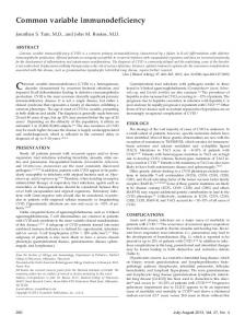
FIG. 2. The expression of soluble single-chain variable fragments (scFvs) under the preferred culture conditions. Lanes 1, 2, total soluble protein in the lysate of Rosetta transformed by pPELB-A1 and induced with isopropyl-β-D-thiogalactopyranoside (IPTG) in lane 2 and without IPTG induction in lane 1; lane M, protein molecular weight markers; lanes 3, 4, total soluble protein in the lysate of Rosetta transformed by pPELB-A11 and induced with IPTG in lane 3 and without IPTG induction in lane 4; lane 5, total soluble protein in the lysate of Rosetta transformed by pPELB and induced with IPTG.
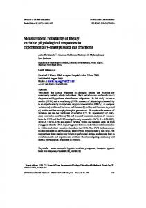
FIG. 1. Agarose (1.2%) gel electrophoresis of the digested A1pPELB and A11-pPELB with NotI/NcoI. Lane 1, the target fragment released from the A1-pPELB digested with NotI/NcoI; lane 2, the target fragment released from the A11-pPELB digested with NotI/NcoI.
plasmids in transformed bacterial cells were examined by digesting the plasmids extracted from the positive clones with the 2 enzymes of NotI and NcoI and sequencing. The result of DNA electrophoresis demonstrated that the target gene fragment of 750 bp was released by digestion (Fig. 1), and the results of DNA sequencing of the plasmids in the positive clones indicated that the coding sequences of VH, VL, linker, and leader were consistent with those before the recombination. Figure 2 demonstrates that scFv fragments were expressed as soluble protein in E. coli Rosetta. Characterization of the pep-50 binding scFvs The scFvs were purified by CM-Sepharose Fast Flow and IMAC. Their purity was tested by SDS-PAGE, followed by immunoblot experiments. Figure 3 shows electrophoretic analysis of the fractions resulting from the purification. The final products showed a single band, and their molecular weights were 31 and 31.4 kDa compared with the molecular weight of markers. The molecular mass accorded with predicted
550
www.sbsonline.org
molecular weight according to the sequencing results. The yields of soluble scFv-A1 and scFv-A11 after purification were about 4.0 and 5.0 mg of protein from 1 L of culture, respectively. Figure 4 shows the results of Western blotting of scFvs purified by CM-Sepharose Fast Flow and IMAC and the negative control Rosetta transformed with pPELB. The antibody used in this analysis detects the C-terminal His-tag fused to scFv gene. Binding of soluble scFvs to pep-50 The binding of the expressed single-chain antibody fragments to free pep-50 was determined by sandwich ELISA using scFv-A1 and scFv-A11 as the capture antibody and rabbit anti-pep-50 polyclonal antibody as the detecting antibody. The 2 antibody clones bound to free pep-50 with the lower detection limit of 4 µM scFv-A11 and 20 µM scFv-A1. In theory, the scFvs can react with the full-length TSP50, but as we have not acquired the full-length TSP50 to date, it should be explored in later studies. DISCUSSION In this study, phage display antibody library technology was applied to generate anti-TSP50 antibody fragments. The main advantage of this technique is that human antibodies can be produced much easier than human hybridomas can. These can be then developed for therapeutic use without the need to humanize mouse Mabs. Furthermore, by changing the
Journal of Biomolecular Screening 11(5); 2006
Screening of Single-Chain Variable Fragments
FIG. 3. Fifteen percent sodium dodecyl sulfate–polyacrylamide gel electrophoresis analysis of the fractions eluted from CM and immobilized metal ion chelate affinity chromatography (IMAC) columns. Lanes 1, 4, 25 µg of protein containing single-chain variable fragments (scFv)–A1 and scFv-A11 eluted from CM-Sepharose Fast Flow with 20 mM phosphate-buffered saline 1.0 mM NaCl, pH 6.5; lanes 2, 3, 8 µg of scFv-A1 and scFv-A11 eluted from IMAC with 100 mM imidazole; lane M, low-molecular-weight markers.
selection conditions, the antigen-binding properties of antibody phages can be influenced, for example, by selection for high-affinity antibodies or specificity for a predetermined epitope.19,20 The random pairing of V domains has the potential to generate artificial antibody specificities and could overcome the problem of the immune dominance of some antigens and epitopes in mice that limit the spectrum of specificities obtained with hybridomas.21 Furthermore, using this technique, the procedure was faster, easier, and considerably less expensive than the generation of polyclonal and monoclonal antibodies against the antigen. The antibody acquired in this study is scFv, which is the smallest antibody fragment containing a complete antigen-binding site. In comparison to the much larger Fab′, F(ab′)2, and IgG forms of monoclonal antibodies, scFvs have lower retention times in nontarget tissues. Unlike glycosylated whole antibodies, scFv antibodies can be easily produced in bacterial cells as functional antigen-binding molecules. A humanized monoclonal antibody against HER-2 (e.g., herceptin and scFv antibodies with biological function) has been developed for studies on the role of growth factor receptor pathways in breast cancer and leukemia. Some of them have been used as a therapeutic ally.22 They bind to the extracellular portion of transmembrane receptors, which is overexpressed in subsets of tumors, and inhibit cell proliferation in a dosedependent manner.22,23 The function of TSP50 in the development of breast cancer is still unknown. Thus, the scFvs selected in this study could be valuable reagents for the immunoassay of TSP50 as well as for the investigation of the function of TSP50 in the development of breast cancer. They were also eventually used as therapeutic agents for breast cancer.
Journal of Biomolecular Screening 11(5); 2006
FIG. 4. Western blot analysis of single-chain variable fragments (scFv) proteins purified by CM-Sepharose Fast Flow and immobilized metal ion chelate affinity chromatography (IMAC). The scFv proteins were purified, analyzed by 15% sodium dodecyl sulfate–polyacrylamide gel electrophoresis, transferred to nitrocellulose, and then detected by monoclonal antibody against hexa-histidine. The lysate of Rosetta transformed by pPELB was used as a negative control (lane 5). Lanes 1, 4, 25 µg of proteins containing scFv-A1 and scFv-A11 eluted from CM-Sepharose Fast Flow with 20 mM phosphate-buffered saline 1.0 M NaCl, pH 6.5; lanes 2, 3, 8 µg of scFv-A1 and scFv-A11 eluted from IMAC with 100 mM imidazole.
In conclusion, we have panned the scFv antibodies against TSP50 from a naive phage display antibody library using pep50 as the target antigen and successfully expressed the genes of scFvs in E. coli Rosetta. These antibodies have the ability to specifically recognize pep-50 with high affinity. If a gene modification to the scFvs can be accomplished using error-prone PCR, mutation strains, chain shuffling, or DNA shuffling,11,24 the antigen-scFv affinity can be further increased. Our results indicate that synthetic human antibody libraries can be useful tools for the selection of antibodies against a nonhomologous hydrophilic region within the catalytic domain of the protease. We are currently investigating the function of TSP50 in the development of breast cancer using selected scFvs and the clinical application of the scFv antibodies in the treatment of breast cancer. ACKNOWLEDGMENTS We thank Fiona Sait from the Medical Research Council, Cambridge, United Kingdom, for providing the phage library. We thank Dr. Sui Dexin from the University of Michigan for providing Vector pPELB and E. coli Rosetta. Jingyan Wei and Yang Liu contributed equally to this article. REFERENCES 1. 2.
Kaplan RM, Wingard DL: Trends in breast cancer incidence, survival, and mortality. Lancet 2000;356:592-593. Yousef GM, Magklara A, Diamandis EP: KLK12 is a novel serine protease and a new member of the human kallikrein gene family—differential expression in breast cancer. Genomics 2000;69:331-341.
www.sbsonline.org
551
Wei et al. 3.
4. 5.
6.
7.
8. 9.
10.
11.
12.
13.
14.
15.
552
Chen HM, Schmeichel KL, Mian IS, Lelièvre S, Petersen OW, Bissell MJ: AZU-1: a candidate breast tumor suppressor and biomarker for tumor progression. Mol Biol Cell 2000;11:1357-1367. Yang XW, Groshen S, Formenti SC, Davidson NE, Press MF: P7 antigen expression in human breast cancer. Clin Cancer Res 2003;9:201-206. Yuan LM, Shan JD, De Risi D, Broome J, Lovecchio J, Gal D, et al: Isolation of a novel gene, TSP50, by a hypomethylated DNA fragment in human breast cancer. Cancer Res1999;59:3215-3221. Shan JD, Yuan LM, Xiao QX, Chiorazzi N, Budman D, Teichberg S, et al: TSP50, a possible protease in human testes, is activated in breast cancer epithelial cells. Cancer Res 2002;62:290-294. Griffiths AD, Williams SC, Harley O, Tomlinson IM, Waterhouse P, Crosby WL, et al: Isolation of high affinity human antibodies directly from large synthetic repertoires. EMBO J 1994;13:3245-3260. Kristensen P, Winter G. Proteolytic selection for protein folding using filamentous bacteriophages. Fold Des 1998;3:321-328. Griffiths D, Malmqvist M, Marks JD, Bye JM, Embleton MJ, McCafferty J, et al: Human anti-self antibodies with high specificity from phage display libraries. EMBO J 1993;12:725-734. Marks JD, Hoogenboom HR, Bonnert TP, McCafferty J, Griffiths AD, Winter G: By-passing immunization: human antibodies displayed on phage. J Mol Biol 1991;222:581-597. McElhiney J, Lawton LA, Porter AJR. Detection and quantification of microcystins (cyanobacterial hepatotoxins) with recombinant antibody fragments isolated from a native human phage display library. FEMS Microbiol Lett 2000;193:83-88. Nissim A, Hoogenboom HR, Tomlinson IM, Flynn G, Midgley C, Lane D, et al: Antibody fragments from a single “pot” phage display library as immunochemical reagents. EMBO J 1994;13:692-694. Tomlinson IM, Walter G, Marks JD, Llewelyu MB, Winter G. The repertoire of human germline VH sequences reveals about fifty groups of VH segments with different hypervariable loops. J Mol Biol 1992;227:776-798. Sun Y, Li WJ, Ma JS, Mu Y, Du XB, Yan GL, et al: Soluble expression, purification and characterization of single-chain Fv catalytic antibody (sFv-2F3). Chem Res Chinese Universities 2004;20:317-322. Molloy P, Brydon L, Porter AJ, Harris WJ: Separation and concentration of bacteria with immobilised antibody fragments. J Appl Bacteriol 1995;78:359-365.
www.sbsonline.org
16.
Lowry DH. Protein measurement with the Folin phenol reagent. J Biol Chem 1951;193:265. 17. Kupsch JM, Tidman NH, Kang NV, Truman H, Hamilton S, Patel N, et al: Isolation of human-specific antibodies by selection of an antibody phage library on melanoma cells. Clin Cancer Res 1999;5:925-931. 18. Tordsson JM, Ohlsson LG, Abrahmsen LB, Karlstrom PJ, Lando PA, Brodin TN: Phage-selected primate antibodies fused to superantigens for immunotherapy of malignant melanoma. Cancer Immunol Immunother 2000;48:691-702. 19. Meulemans EV, Slobbe R, Wasterval P, Ramaekers FC, van Eys GJ: Selection of phage-displayed antibodies specific for a cytoskeletal antigen by competitive elution with a monoclonal antibody. J Mol Biol 1994;244:353-360. 20. Hawkins RE, Russell SJ, Winter G. Selection of phage antibodies by binding affinity: mimicking affinity maturation. J Mol Biol 1992;226:889-896. 21. Desai SA, Wang X, Noronha EJ, Kageshita T, Ferrone S: Characterization of human anti–high molecular weight–melanoma-associated antigen single-chain Fv fragments isolated from a phage display antibody library. Cancer Res 1998;58:2417-2425. 22. Shadidi M, Sioud M: An anti-leukemic single chain Fv antibody selected from a synthetic human phage antibody library. Biochem Biophys Res Commun 2001;280:548-552. 23. Reese DM, Slamon D: HER-2/neu signal transduction in human breast and ovarian cancer. Stem Cells 1997;15:1-8. 24. Hoogenboom HR: Designing and optimizing library selection strategies for generating high-affinity antibodies. Trends Biotechnol 1997;15:62-70.
Address reprint requests to: Jingyan Wei College of Pharmaceutical Science Jilin University 1266 Fujin Road Changchun, 130021, China E-mail:
[email protected]
Journal of Biomolecular Screening 11(5); 2006