Environmental Health and Toxicology • Original Article
Open Access Volume: 30, Article ID: e2015007, 7 pages http://dx.doi.org/10.5620/eht.e2015007
eISSN: 2233-6567
Screening of toxic potential of graphene family nanomaterials using in vitro and alternative in vivo toxicity testing systems Nivedita Chatterjee1, Ji Su Yang1, Kwangsik Park2, Seung Min Oh3, Jeonggue Park4, Jinhee Choi1 1
School of Environmental Engineering, Graduate School of Energy and Environmental System Engineering, University of Seoul, Seoul; 2College of Pharmacy, Dongduk Women’s University, Seoul; 3Fusion Technology Laboratory, Hoseo University, Asan; 4 Korea Environmental Institute, Seoul, Korea Objectives The widely promising applications of graphene nanomaterials raise considerable concerns regarding their environmental and human health risk assessment. The aim of the current study was to evaluate the toxicity profiling of graphene family nananomaterials (GFNs) in alternative in vitro and in vivo toxicity testing models. Methods The GFNs used in this study are graphene nanoplatelets ([GNPs]–pristine, carboxylate [COOH] and amide [NH2]) and graphene oxides (single layer [SLGO] and few layers [FLGO]). The human bronchial epithelial cells (Beas2B cells) as in vitro system and the nematode Caenorhabditis elegans as in vivo system were used to profile the toxicity response of GFNs. Cytotoxicity assays, colony formation assay for cellular toxicity and reproduction potentiality in C. elegans were used as end points to evaluate the GFNs’ toxicity. Results In general, GNPs exhibited higher toxicity than GOs in Beas2B cells, and among the GNPs the order of toxicity was pristine > NH2 > COOH. Although the order of toxicity of the GNPs was maintained in C. elegans reproductive toxicity, but GOs were found to be more toxic in the worms than GNPs. In both systems, SLGO exhibited profoundly greater dose dependency than FLGO. The possible reason of their differential toxicity lay in their distinctive physicochemical characteristics and agglomeration behavior in the exposure media. Conclusions The present study revealed that the toxicity of GFNs is dependent on the graphene nanomaterial’s physical forms, surface functionalizations, number of layers, dose, time of exposure and obviously, on the alternative model systems used for toxicity assessment.
Correspondence: Jinhee Choi 163 Siripdae-ro, Dongdaemun-gu, Seoul 130-743, Korea Tel: +82-2-6490-2869 Fax: + 82-2-6490-2859 E-mail:
[email protected] Received: March 14, 2015 Accepted: June 4, 2015 Published online: July 15, 2015 This article is available from: http://e-eht.org/
Keywords Alternative toxicity testing, Human bronchial epithelial cells, Caenorhabditis elegans , Graphene family nanomaterials
Introduction Graphene is typically composed of sp2-hybridized carbon atoms packed densely in a honeycomb crystal lattice arranged in a two-dimensional structure, resulting in a large surface area on
both sides of the planar axis. Graphene and related materials, including few-layer graphene, graphene nanosheets, graphene nanoplatelets (GNPs), ultrafine graphite, graphene oxide(GOs), and reduced GO etc., have been identified as graphene family nanomaterials (GFNs) [1-3]. The huge versatility of the GFNs
Copyright © 2015 The Korean Society of Environmental Health and Toxicology This is an Open Access article distributed under the terms of the Creative Commons Attribution Non-Commercial License (http://creativecommons.org/ licenses/by-nc/3.0/) which permits unrestricted non-commercial use, distribution, and reproduction in any medium, provided the original work is properly cited.
Page 1 of 7 http://e-eht.org/
Environmental Health and Toxicology 2015;30:e2015007
not only aiming for their potential future applications in the domain of electronics, photonics, composite materials, energy generation and storage, sensors and metrology, and biomedicine, specifically, biosensors, bioimaging and therapeutics, tissue engineering, and drug delivery etc., but also raised serious concerns about their environmental and human health impacts [1,3-5]. In recent years, several groups have devoted their studies to the elucidation of graphenes and related materials with in vitro and in vivo, but contradictory outcomes were reported. Therefore, at present, it is not possible to make any generalized conclusions about the biological interactions and toxic effects of GFNs. Moreover, GFNs are comprised of different forms of graphene nanomaterials that are endowed with different physicochemical characteristics; hence, not much has been understood about their toxicological profiles and more detailed studies are required [1,6]. In particular, toxicity data on GNPs which possess the potential to pose as nanohazards due to their specific platelet shape are still limited in comparison with related congeners [3,7] The aim of our present study was to evaluate and compare the toxic potentiality of different forms of GFNs in in vitro and alternative in vivo systems. We used five different commercially available forms of GFNs that consist of GNPs (with or without functionalization–pristine, carboxylate [COOH] and amide [NH 2 ]) and GOs (single layer [SLGO] and few layers [FLGO]). The human bronchial epithelial cells (Beas2B cells) and the nematode Caenorhabditis elegans were used as alternative in vitro and in vivo model systems, respectively, to profile the toxic responses of the GFNs.
Materials and Methods Graphene Family Nanomaterials The powdered GOs (SLGO and FLGO) and GNPs with different surface functionalization (pristine, COOH and NH2) were purchased from Cheap Tubes.com (http://www.cheaptubes.com/) and stocks prepared in distilled water at 1000 mg/ L with sonication.
Cell Culture and Graphene Family Nanomaterials Treatment Beas2B cells were cultured in Dulbecco’s modified Eagle medium/F12(DMEM/F12; GIBCO Life Science, Great Island, NY, USA), supplemented with 10% (v/v) fetal bovine serum and 1% (v/v) antibiotics, at 37°C in a 5% CO2 atmosphere. The GFNs were freshly prepared in cell culture medium DMEM/F12 at the desired concentrations with appropriate Page 2 of 7
amounts of each stock (1000 mg/L in distilled water) and sonicated for 10 minutes just before biological exposure.
Cytotoxicity and Cell Viability Assessment The cytotoxicities of all GFNs were determined with the EZCytox cell viability assay kit (Daeil Lab Service, Seoul, Korea) based on cleavage of the tetrazolium salt to water-soluble formazan by succinate-tetrazolium reductase, as described previously [8]. Approximately, 5 × 10³ cells/well were seeded in 96-well plates 24 hours prior to treatment and exposed to a range of concentrations (5 to 150 mg/L) of five different GFNs for the next 24 hours. Next, in a separate experiment, the cells (in 96well plates) were exposed to all GNPs (20 mg/L) and GOs (50 mg/L) for different time point (4 to 72 hours). After completion of the exposure time, 10 µL of EZ-Cytox reagent was added to each well including treated and control (without GFNs). Absorbance was detected at 450 nm after 2 hours of incubation at 3˚C. Appropriate blanks were used for each concentration to validate the absorbance. In addition, the cells (5 × 10⁴ cells/well in a 6-well plate) were also exposed to a fixed concentration (20 mg/L) of GFNs in complete and serum free DMEM/F12 medium for 24 hours. The cell viability was measured using the standard trypan blue (Invitrogen, Carlsband, CA, USA) staining method and the total numbers of stained and unstained cells counted using a hemocytometer.
Colony Formation and Morphology Changes The colony formation assays were carried out as described by Herzog et al. [9]. Exponentially growing cells were harvested and seeded in six-well culture plates at a density of 250 cells/ well. Each well contained 2 mL of cell culture medium. Cells were allowed to attach for approximately 24 hours. The cells were then washed with 2 mL of phosphate buffered saline (PBS) and treated with 2 mL of nanoparticles prepared in cell culture medium to final concentrations of 10 and 50 mg/L. Cells were exposed to nanoparticles over the time period they needed to form colonies, a colony being defined as at least 50 clones of one cell and incubated for 10 days. Before colonies were counted, the particle solutions were removed, the cells were washed with PBS and finally fixed and stained using a 0.1% Giemsa (Sigma-Aldrich, St Louis, MO, USA). Colony shape and cell morphology were detected under a light microscope.
Maintenance of C. elegans C. elegans were grown in Petri dishes on nematode growth medium and fed with Escherichia coli strain OP50 according to a standard protocol [10]. The worms were incubated at 20˚C and http://e-eht.org/
Nivedita Chatterjee, et al.
|
In vitro and in vivo graphene nanomaterials toxicity screening
young adults (3 days old) from age-synchronized cultures were used in all the experiments.
to 8 layer) but were of similar dimensional sizes.
Reproduction (72 Hours Assays) of C. elegans
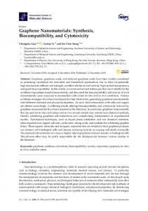
At first the viabilities of Beas2B cells were evaluated at various concentrations of GFNs for 24 hours. In general, a dose dependent decrease in viability was not markedly clear at lower doses (5 to 25 mg/L) but dose dependency became much more profound at higher doses (25 to 150 mg/L, Fig 1A). Unlikely, the GFNs induced decrease in viability at a dose of 20 mg/L was found to be clearly time dependent and become more distinct after 16 hours of exposure (Figure 1B). Furthermore, no significant difference in viability was observed between complete media and serum free media at a dose of 20 mg/L (Figure 1C). In general, GNPs were found to be more toxic than GOs. The order of sensitivity of Beas2B cells towards the GNPs were found as pristine > NH2 > COOH. The COOH and NH2 functionalization showed more or less similar cytotoxic effects on Beas2B cells, which only became different at the highest dose (150 mg/ L) and longest time (72 hours) point (Figure 1A, 1B). Interestingly, GOs mediated decreases in viabilities were not as profound as those induced by GNPs and both compounds showed more or less similar cytotoxicity, except at the highest dose (150 mg/L) and longest time (72 hours) point where SLGO become more toxic than FLGO (Figure 1A, 1B).
The effects of GFNs on the reproduction of wild type worms were investigated as described by Roh et al. [11]. After a young adult was exposed to GFNs at various concentrations (5 to 50 mg/L mixed in K-media) for 72 hours, the number of offspring at all stages beyond the egg were counted. Five replicates were conducted for reproduction assays.
Statistical Analysis The statistical significance of differences among/between treatments was determined using one way analysis of variance. This was followed by a post-hoc test (Tukey, p < 0.05). All statistical analyses were carried out using SPSS version 12.0 KO (SPSS Inc., Chicago, IL, USA) and graphs were prepared in SigmaPlot version 12.0 (Systat Software, Inc., Chicago, IL, USA).
Results Graphene Family Nanomaterials Characterization The details of layer number, thickness, dimensions, functionalization, methods of preparation etc. supplied by the manufacturer are summarized in Table 1. We used pristine and two other functionalized (COOH and NH2) GNPs of similar layer numbers, lateral dimensions, and average thickness, and two kinds of GOs which only differed in layer number - SLGO and FLGO (4
Effects on Cell Viability and Cytotoxicity
Colony Formation and Morphology Changes Significant dose dependent decreases in colony numbers were evident due to 10 days of exposure of GFNs (Figure 2A, 2B)
Table 1. Characterization of graphene nanomaterials (as supplied by the manufacturer) GOs
Properties Thickness X &Y dimensions (nm) No. of layers Purity (wt%) Method Solubility
Single-layered
Few-layered
0.7-1.2 nm 300-800 1 > 99.0 Modified hummers Water, NMP, DCB, DMF
4-8 layers 300-800 4-8 > 99.0 Modified hummers Water, NMP, DCB, DMF GNPs
Lateral dimensions (μm) Average thickness (nm) No. of layers Purity (wt%) Surface area (m2/g) Plasma process gas Primary functionality Other functionality Source material Form supplied
GNP-pristine
GNP-COOH
GNP-NH2
1-2 750 Argon None Atmospheric gas Natural graphite Dry powder
1-2 750 Proprietary oxygen based COOH COH, C= O, other oxygen Natural graphite Dry powder
1-2 750 Nitrogen NH2 N-H, O= C-N-H2, C= N Natural graphite Dry powder
GOs, graphene oxides; NMP, n-methyl-2-pyrrolidone; DCB, dichlorobenzene; DMF, dimethylformamide; GNPs, graphene nanoplatelets. http://e-eht.org/
Page 3 of 7
Environmental Health and Toxicology 2015;30:e2015007
GNP-pristine GNP-COOH GNP-NH2 SLGO FLGO
120
Viability (%)
100 80
Colony morphology Cell morphology
Colony morphology Cell morphology
60 40 20 0
0 5 10 25 50 75 100 GFNs (mg/L)
A GNP-pristine GNP-COOH GNP-NH2 SLGO FLGO
100 80
Viability (%)
150
GNP-pristine
GNP-pristine
GNP-NH2
GNP-NH2
60
A
40 50 mg/L 10 mg/L
20
4 8 16 24
48
72
Exposure time (hr)
B Serum free media Complete media
100
80 *
60
60
*
*
* ***
40
*** ***
20 0
80
Viability (%)
Colony forming units (% of control)
100
l tro Con
O SLG
2 O ne OH NH FLG -pristi -CO GNPP P GN GN
GFNs
B
40 20 0
l tro Con
O SLG
O e 2 FLG pristin COOH P-NH N P P G GN GN
GFNs
C
Figure 1. Cytotoxicity of GFNs to Beas2B cells. (A) The viability (%) determined by EZ-Cytox assay after 24 hours of exposure to GFNs in complete DMEM/F12 medium. (B) Cells exposed to GNPs (at 20 mg/L) and GOs (at 50 mg/L) at different time points and cytotoxicity determined by EZ-Cytox assay. (C) The viability (%) determined by trypan blue exclusion method after 24 hours of exposure to GNPs (at 20 mg/L) and GOs (at 50 mg/L) in complete DMEM/F12 media and serum free media. Data are presented as mean±standard error of mean. GFNs, graphene family nananomaterials; Beas2B, human bronchial epithelial; DMEM/F12, Dulbecco’s modified Eagle medium/F12; GNPs, graphene nanoplatelets; GOs, graphene oxides; SLGO, single layer GO; FLGO, few layers GO; COOH, carboxylate; NH 2, amide.
Page 4 of 7
Figure 2. The effects of GFNs exposure on colony formation in Beas2B cells for 10 days. (A) Colonies and cell morphology of Beas2B cells after 10-day exposure to GFNs at doses of 10 and 50 mg/L. (B) Effects on colony number. Data are presented as percent of control mean±standard error of mean. GFNs, graphene family nananomaterials; Beas2B, human bronchial epithelial; SLGO, single layer graphene oxide; FLGO, few layers graphene oxide; GNPs, graphene nanoplatelets; COOH, carboxylate; NH2, amide. *p pristine. In addition, a significant deformahttp://e-eht.org/
Nivedita Chatterjee, et al.
5 mg/L 10 mg/L 20 mg/L 50 mg/L
Reproduction potentiality (%)
100 80 *
60
*
*
*
*
*
40
*
***
***
20 0 O SLG
O FLG
2 ine COOH P-NH rist N p P G P GN GN GFNs
Figure 3. Effect of GFNs treatment on C. elegans. Percentage of reproductive potentiality of wild-type C. elegans due to GFNs exposure at different doses (5, 10, 20, and 50 mg/L) for 72 hours. Data are presented as mean±standard error of mean. GFNs, graphene family nananomaterials; SLGO, single layer graphene oxide; FLGO, few layers graphene oxide; GNPs, graphene nanoplatelets; COOH, carboxylate; NH2, amide. *p